In the last month of pregnancy, changes occur in the mother’s body that indicate the imminent birth of the baby. One of the symptoms of impending childbirth is a decrease in the level of the abdomen. You can understand that the baby is descending into the pelvis by the external signs and sensations of the mother. When will labor begin if the baby has dropped? Is it possible for labor to begin if the position of the abdomen remains unchanged?
Why and how does the stomach drop during pregnancy?
Many first-time mothers note that their belly droops at 33-34 weeks of pregnancy. As the baby grows, the uterus gradually moves. When the time is right, the baby takes a comfortable position in the pelvic area. The walls of the uterus soften, and it shortens and expands. After such changes, intensive preparation for the birth of the baby begins.
Certain changes also occur in the body of the expectant mother as the abdomen lowers.
| Name | Description |
| Uterus | In preparation for childbirth, the fundus of the uterus gradually moves into the pelvis. The abdominal muscles relax. There are no visual signs of the ongoing process. Only an obstetrician-gynecologist can determine changes at the next gynecological examination. In normal condition, the fundus of the uterus is located 5 cm above the vagina. Then there is a gradual expansion and further preparation for the passage of the baby through the birth canal. |
| Pelvic bones | The body of the expectant mother produces a large amount of the hormone relaxin. It helps the bones open up a little before giving birth. And after the abdomen subsides, the uterus moves to the pelvic area. Expansion of the pelvic bones before childbirth is normal. In some situations, if a pregnant woman has a narrow pelvis, this does not happen. |
| Child's Pose | The baby's head moves downwards, it fits tightly to the pelvic floor. The baby takes a certain position in order to move further along the birth canal. Previously, the baby's head was located in the abdominal cavity. As the body prepares for childbirth, the fetus descends to the pelvic area and awaits the onset of labor. |
The main reason for the movement of the abdomen in the last stages of pregnancy is the softening of the walls of the uterus and its stretching. Against the background of these changes, the fetus slowly falls down and rests its head on the pelvic bones.
Moving the abdomen even facilitates the birth process, since the fixed fetus in the small pelvis cannot turn over. Therefore, the onset of labor is facilitated by the correct positioning of the baby.
What to do if you have premature abdominal prolapse
Early descent of the fetal head into the pelvic cavity is fraught with squeezing. As a result, the baby may be born with a deformed skull: a flattened forehead or the back of the head sloping to one side. The degree and type of changes depend on the shape of the mother's pelvis.
Symptoms of premature prolapse:
- constant pressure on the bladder;
- frequent urge to urinate;
- sensation of a foreign body in the upper parts of the vagina.
A bandage during pregnancy reduces the load on the legs and supports the fetus.
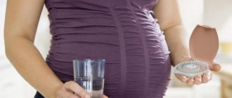
To accurately diagnose the problem, consult a doctor. If the diagnosis is confirmed, measures should be taken. The most effective methods of maintaining the fetus:
- Wearing a bandage. The device supports the baby's head in the correct position and prevents further advancement of the fetus along the birth canal. In addition, the bandage redistributes the load on the spine and speeds up the recovery process after childbirth.
- Gymnastics for pregnant women. Special poses help move the head upward and relieve discomfort. Lying on your back, lift your pelvis up and stay in this position for 5-10 seconds. Then kneel down and lift your pelvis above your chest level. Repeat the exercises every day for 3-4 approaches.
- Osteopathy. A skilled specialist will move the fetal head away from the entrance to the pelvis in one session.
If the risks of premature onset of labor are high, the doctor may place the pregnant woman in a hospital for preservation.
Determining the position of the fetus using the “abdominal map”
An interesting way to determine the baby’s position in detail is to draw up a “belly map” (“Belly Mapping”), offered by American midwife Gail Tully from Minnesota.
On a piece of paper we draw a circle - “belly”, and divide it into four segments. Above is the fundus of the uterus, below is the pubic bone. The woman’s sides will be mirrored on the map, that is, the left side will be reflected on the right side of the map. Mark the baby’s heart on the map, that is, the place where it can be heard most clearly. This is the upper part of his back. Therefore, it will be easy to find the head! Usually, a child’s back can also be determined by a particularly convex and flat area of the abdomen. Plot this line on the map too. And the large bulge in the upper abdomen is most likely the buttocks. Reclining or lying down, relaxed, observe the baby’s activity for two to three days, and note where two types of movements are felt: weak ones, most likely, mean movements of the arms, and strong shocks – the legs. Next, Gail Tully suggests taking a doll or soft toy approximately the size of a fetus and placing it on your stomach, trying to position it as marked on the map
You will be able to figure out exactly how your baby lies - not only in what presentation, but also how his body is rotated relative to the mother's back, which is also important for the course of labor. Most often, the baby's head is below, and the back is in the area of the mother's left side.
In her book, The Belly Mapping Workbook, Gail Tully explains in detail how to pinpoint your baby's position, using a whole bunch of technical terms. But, we believe, such information is more important to the doctor who delivers the child (besides, don’t forget, for such a detailed picture there is an ultrasound, which can even get a “portrait” of the child!).
The stomach does not drop - is it bad or not?
Knowing how to understand that the stomach is dropping, a woman looks forward to this moment and can be seriously puzzled if she does not notice changes in her body “on time”. What to do if the figure and sensations of a pregnant woman do not change in the later stages?
In fact, not all expectant mothers have a drooping belly before childbirth. This feature should be discussed with the supervising physician. We hasten to reassure you, there is no reason to panic - this is simply an individual feature of your body. It is likely that the desired changes will occur immediately before the baby is born. And the best part is that mom definitely won’t miss them. After all, she knows how to understand that the stomach drops before childbirth.
Photos of expectant mothers before and after the formation of the fetus can be viewed in our article. We wish you a pleasant pregnancy and an easy birth!
Nesting instinct on the eve of childbirth
On the eve of childbirth, many women receive another “news” from nature that the baby will be born very soon. An indomitable desire arises to prepare him a cozy “nest” and the expectant mother with great pleasure chooses tiny socks and baby vests for her future son or daughter, embroiders diapers, knits a hat and blanket, buys the softest and most beautiful bed linen for the crib, a comfortable stroller, a warm envelope, a lace blanket, a set of bibs and that nice little rattle! Another obligatory item of “building a nest” is often general cleaning (or even repairs). Having set up a children's corner, bought things and “cleaned” the house, the woman breathes a sigh of relief: she can give birth. And... it is at this moment that contractions often begin. “Nesting” is an incredibly happy time, because it reminds you that your dream of meeting your baby soon is about to become a reality!
Siblings' experience
Nena
— About two days before giving birth, she became more active - both times she began large cleanings throughout the entire apartment, which she had not had the strength to do for a long time.
I paid attention because I read here on the forum that this happens - preparing a nest. And there were no more harbingers
Ekaterina
— At 39 weeks 5 days I woke up with a terrible surge of strength and decided that it was simply vital to rip out all the floors in the house. Then I realized that it would start at night.
Enya
— I didn’t have any special feelings before giving birth, except that I, like a field mouse, had been dragging all the groceries home from the store for the last 10 days. Well, I couldn’t pass by the store. I take a full bag and drag it, then I realize that I can’t carry it, it’s hard, so I catch a taxi. Twice I came home by taxi with string bags...
Harbingers of labor - how to recognize them
Each female body is unique, so it is impossible to say unambiguously on what day the harbingers of childbirth will appear. Experts consider 37-38 weeks to be the beginning of the prenatal period. It is from this moment that the girl begins to feel changes in her body and body.
People around you can also see the harbingers of labor in first-time mothers. The most common is an altered gait.
Signs of labor in multiparous women
Harbingers are a number of symptoms that appear two (one) week before the upcoming event. They indicate the pregnant woman's readiness for childbirth. Most often, firstborns do not notice visible signs or do not pay attention to them
They do not provide a guideline or a clear idea of when exactly an important event will begin
Women who already have children are included in the list of those who give birth prematurely. Especially if the difference between the children is less than three years. Despite this, the harbingers should not be confused with the onset of labor. Signs can appear up to a week in advance and indicate that the body is preparing for an upcoming event.
Having noticed certain changes, a woman should prepare a bag for the maternity hospital and not go to long distances (suburban complexes, etc.).
What exactly do the harbingers of a second birth look like?
- The most obvious sign is a drooping belly. The fetus turns over and is now positioned with its head towards the entrance to the pelvis. This can happen several days before the start of the process.
- Removal of the mucus plug. A clot of brown or yellow mucus will come out of the uterus. This symptom is characterized by painful, pulling sensations in the pelvic area and lower abdomen. The “plug” protects the uterus and fetus from infections throughout pregnancy.
- Training contractions in second-time mothers can go almost unnoticed. This is due to the experience present and the greater readiness of the body. False contractions are not a reason to panic. They can appear at 32 weeks and periodically make themselves felt until the onset of prenatal periods.
- Weight loss is also a precursor to childbirth. A woman noticeably sheds kilograms from 36 weeks, or the numbers on the scale simply stop. This is due to the fact that the future baby has already gained weight. The female body, a couple of weeks before the onset of labor, gets rid of everything unnecessary, swelling in the legs, arms, and face goes away. During this period you can lose up to 3 kilograms. Losing weight does not pose any risks to the fetus or the expectant mother.
- Nesting syndrome occurs in most women. Over the course of a month (at 36-37 weeks), a woman’s hormonal levels stabilize and at this moment she feels a surge of strength. She wants to start general cleaning, rearranging furniture, ironing, getting rid of trash, and so on. All these processes bring untold pleasure to the expectant mother; she anticipates and looks forward to the imminent replenishment.
- Harbingers of imminent labor are the release of colostrum. It is colostrum that is the baby’s first nutrition, so if clear liquid begins to drip from the breast, you should prepare for an early feeding.
- Another harbinger of imminent labor in primiparas and others is a decrease in the number of movements. The reason for the decrease in activity lies in the fact that the baby becomes cramped. The fetus is also preparing for the upcoming event, and therefore conserves its strength - this is the second reason for the decrease in movements.
- Eating disorders and loose stools are also harbingers of the third birth, as well as the first and second. The body is actively cleansed, removing all unnecessary toxins and harmful elements. All efforts will be aimed at ensuring that the baby is born and the process is as easy as possible.
- Change in gait. Due to the different position of the fetus and large belly, girls walk with their backs tilted back. This allows you to not suffer so much from the load on the spine and maintain balance. This precursor is individual and directly depends on the size of the abdomen. If a girl is expecting twins, “proud gait” will definitely be present.
How to understand that your belly has dropped during pregnancy
A drooping abdomen is one of the harbingers, indicating that a pregnant woman will soon become a mother. Why does this happen, is this condition normal? To understand why abdominal prolapse occurs and what it is
It is worth paying attention to the anatomical features and structure of the woman’s pelvic bones
The bones of a woman’s pelvic joint are designed this way to, if necessary, allow the baby’s head to descend, putting pressure on the muscles, creating pressure and tone, causing labor. The anatomical features of the sacroiliac joint are formed in such a way as to keep the fetus in the desired position during early pregnancy. When the baby becomes heavier, his head puts pressure on the muscles, he sinks lower and lower.
However, this change in fetal descent occurs gradually. Initially, the muscles and bones of the pelvis should be ready for the lowering of the abdomen. Throughout pregnancy, the job of hormones was to preserve the child, providing him with the necessary conditions for growth and development. However, now that the baby is large enough to put pressure on the pelvic muscles, and the abdominal muscles have become difficult to support the overhanging belly, hormones work to soften the pelvic bones. Bone softening can be monitored by the following symptomatic manifestations:
- The bones in the sacral area ache. Especially if we talk about the period after a long walk in the later stages. However, pain can also be the cause of the formation of pathology. Therefore, it is worth informing a gynecologist about the changes, who will be able to determine the norm and establish the cause of such symptoms.
- The perineum hurts after a walk or when climbing stairs. Sometimes it feels like the baby is literally pressing on the muscles, creating a distinct pressure on the abdominal muscles; it becomes painful, but not so much that it is impossible to tolerate such a manifestation. If the feeling does not go away after 30-40 minutes, you should call an ambulance, explaining the symptoms.
- With a sharp turn, a woman may notice a slight cracking of the pelvic bones and upper leg bones (hips). There is no pain, but slight cracking or squeaking may occur.
Harbingers of change.
The first warning signs, including softening of the pelvic bones, can be missed even by a woman giving birth again. How to understand that the belly has dropped during a first-time pregnancy? As a rule, a woman learns that her stomach has already dropped from her friends and relatives, who from the outside note changes in appearance. The woman herself notes the following changes:
- It became easier to breathe. The protruding belly, which until that moment had only grown and rose above the pelvis, contributed to the fact that the internal organs changed their location. This applies to both the stomach and lungs. Shortness of breath even when going to the bathroom is a natural and unpleasant companion for any pregnant woman. After the stomach has dropped, the lungs have dropped, the amount of air is absorbed better, breathing, including at night, becomes easier.
- No heartburn. All for the same reason that shortness of breath goes away - the stomach drops a little, the sphincter does not become full after eating, and gastric juice does not flow back into the esophagus, as happened quite recently.
- A woman can place her palm between her chest and stomach. Prolapse is measured at home without knowledge in the field of obstetrics. If previously the palm did not fit between the chest and abdomen, then when lowered it is placed.
- The shape of the abdomen changes. As the baby descends into the pelvic area, the shape of the abdomen changes. If before it was more rounded, now there is an angularity, perhaps the lateral muscles holding the stomach are visible, but from the back of many women their interesting position is not visible.
All such signs are common to all pregnant women. However, it is they that allow us to say that soon the body will move into the final phase of pregnancy - childbirth.
The main signs of fetal descent into the pelvis
A woman notices a decrease in her abdomen by external signs and internal sensations. External signs:
- the stomach is lowered to the level of the navel so that a palm can be placed freely between it and the chest,
- the gait becomes heavy, the back straightens and leans back.
Internal sensations are expressed in relief of the condition of some parts of the body and increased tension in others. When the fetus drops, the expectant mother feels:
- Easier breathing. The uterus no longer puts pressure on the lung area, so shortness of breath stops.
- Unpleasant sensations in the ribs area disappear.
- When moving, the child does not make strong and painful pushes. It is difficult for a baby who has assumed the prenatal position to make sharp kicks and intense movements.
- Heartburn, heaviness in the stomach, and nausea go away.
- Problems with stool appear. The fetus puts pressure on the rectal area, which affects peristalsis.
- The frequency of urination increases. The expectant mother notes that the strength of the urge to urinate does not correspond to the fullness of the bladder.
- Painful sensations in the pubis, lower back, nagging pain in the upper legs, caused by the pressure of the fetus on the nerve processes.
To read: When can you start playing sports after a caesarean section: doctors’ recommendations
How can the fetus be positioned before birth?
There are several options for positioning the baby before birth. Not all of them are considered physiological: some can cause dangerous complications. If the presentation is incorrect, you will have to resort to surgery to prevent dangerous consequences and complications during childbirth. The position of the baby in the womb remains almost unchanged after 32-33 weeks.
Longitudinal position
The correct position of the fetus assumes that its tailbone and the back of the head are extended in one line, parallel to the axis of the mother's uterus. It is better if the back is turned towards the mother’s stomach: this reduces the likelihood of complications. The norm is also a position where the baby’s back is located closer to the mother’s back, and the duration of labor may increase. The head should be at the bottom, the pelvis at the top.
Oblique position of the fetus
The baby in the womb may lie diagonally to the mother's birth canal. This option is not considered normal, it can be dangerous and pose a risk of complications during childbirth. In most cases, doctors prescribe a caesarean section.
Transverse position of the fetus
Transverse presentation of the fetus is an indication for obstetrics through surgery. The child is positioned perpendicularly.
Breech presentation
Before birth, the baby may be in a position resembling a sitting position. After 32-33 weeks, the doctor makes a diagnosis and prescribes a caesarean section for obstetrics.
Causes of breech presentation
Malpresentation of a child is observed in the presence of various pathologies that provoke increased activity. Often diagnosed with microcephaly, anencephaly, hypoxia. Often children are born premature. Possible leg or breech presentation with polyhydramnios.
Women's pathologies can also become a cause. The baby often lies incorrectly if the pregnant woman has been subjected to severe or constant stress, is overly tense, or suffers from neuroses. If you have a history of difficult childbirth, multiple abortions and curettages, cervicitis, endometritis, then the likelihood of breech presentation increases.
Why is breech presentation dangerous?
If the position is incorrect, the baby often gets injured during childbirth. Possible injuries to the spine, craniocerebral, hip dysplasia. Hypoxia often develops during childbirth.
Childbirth is also more difficult for a mother whose child is in an incorrect position: ruptures of the vagina, vulva, perineum, and cervix occur more often. The pelvic bones are damaged.
Can labor begin if the belly has not dropped?
Abdominal prolapse is not a necessary symptom of impending childbirth. In some women, the uterus shrinks within a few hours of the start of labor. However, labor can also begin with a high abdominal position. The abdomen may not drop if the following factors are present:
- The fruit is positioned incorrectly. Sometimes the baby takes a transverse position. However, it does not sink into the pelvis.
- Polyhydramnios. With a large amount of amniotic fluid, lowering the baby's head becomes difficult.
- The baby's head is significantly larger than the mother's pelvis. This is possible if the fetus is very large or the mother’s pelvis is small.
- The fetal head at the entrance to the pelvis took on an incorrect tilt. With this arrangement, the likelihood of birth injuries increases, so a caesarean section is necessary.
- Congenital pathologies in a child. For example, hydrocephalus, which externally manifests itself in a pathologically large head size.
Norms and deadlines
There are no two similar births. But there are a number of general characteristics of the approach of the long-awaited action. During pregnancy, the baby turns and turns over. The position of the baby in the tummy changes before birth, the baby calms down, taking a decisive position. The mother notices at some point that the baby is submerged. This phenomenon is called relief. For women giving birth for the first time, this happens 2 to 4 weeks before the baby is born.
Everyone's tummy goes down differently. Clearly predict when labor activity will begin ( the process(es) of active interaction between a subject (living being) and an object (surrounding reality), during which the subject purposefully influences the object, satisfying
) it is forbidden. For some, symptoms appear in the middle of the 3rd trimester. For the rest, 2–3 days before the end of the gestation period.
Start pricing your belly at 32 – 34 weeks. The uterus will shift, which determines how long it will take to give birth. Prolapse occurs in pregnant women at different times.
The process is influenced by:
- body structure;
- progress of pregnancy, what kind;
- abdominal muscle tone;
- individual placement of the fetus.
When to give birth if the head has dropped into the pelvis? There are no problems with health, the first signs of descent ( the concept of criminal jargon, denoting a person occupying the lowest level in the prison hierarchy, a passive homosexual, or a person with whom homosexual intercourse was forcibly committed
) are observed a week before the onset of labor, on average at 37 weeks. There may be a deviation in one direction or the other for 7 days. This is not the first pregnancy, the process will begin within the next few days.
By what signs can you understand that your stomach has dropped?
A woman cannot always notice the difference in the height of the uterine fundus, since this distance can be insignificant (several cm) and not always visible to the eye. However, there are a number of other signs by which you can reliably determine that the stomach has dropped.
Subjective signs
Disappearance of heartburn.
It's no secret that most pregnant women, especially in the third trimester of pregnancy, suffer from heartburn.
This is due to the growth of the uterus and its pressure on all organs of the abdominal cavity, in particular on the stomach.
As a result of this pressure, stomach contents are increasingly refluxed into the esophagus, which causes the feeling of heartburn.
When the abdomen subsides before childbirth, the pressure on the stomach is significantly reduced. A woman may notice that she is practically not bothered by heartburn and she does not have to take antacid medications (Rennie, Rutacid, Gaviscon, etc.).
Reduced shortness of breath.
A decrease in shortness of breath occurs for the following reason: the prolapsed uterus stops putting so much pressure on the diaphragm. Thanks to this, breathing movements are performed effortlessly, and shortness of breath ceases to bother you.
Increased urination.
Due to the fact that the baby's head descends into the pelvis, excess pressure occurs on the bladder, which is located in anatomical proximity to the uterus. The pregnant woman notes that she has begun to feel the urge to urinate much more often.
Increased lower back pain.
This is due to the increased load on this part of the back during this period.
Objective signs
Decreased respiratory rate.
This is associated with easier breathing and decreased shortness of breath.
Decreased heart rate.
The lungs and heart are organs whose work greatly influences each other. Therefore, when shortness of breath disappears, the load on the heart also decreases, which is manifested by a slight decrease in heart rate.
Change in the height of the uterine fundus.
If you measure the distance from the symphysis pubis to the highest point of the uterus (fundus), you can determine that this distance has decreased by 3-4 cm.
The fetal head is pressed tightly against the entrance to the pelvis.
This sign can only be determined by an obstetrician; he places his hand on the fetal head and determines that it is fixed to the pelvic bones.
A shift in the center of gravity, which causes a woman’s gait to change.
What to do if the head drops prematurely
Bandage. It supports the uterus, and with it the fetal head, preventing it from falling too low. I recommend wearing a prenatal bandage to all pregnant women from the 22-24th week, as it allows you to properly distribute the load - transfer it from the abdominal muscles to the lower back. Thanks to this, the muscles of the anterior abdominal wall are not stretched too much and recover after childbirth much faster.
The bandage must also be worn after childbirth. If we are talking about the prolapse of internal organs, which caused the premature descent of the fetus, then, as mentioned above, after the birth of the child, the mother may experience some health problems. To minimize risks, I recommend wearing a bandage after childbirth, and after 2-3 months switching to shapewear.
Useful poses. Take a certain position during which the pelvis is higher than the chest. The most convenient option is the knee-chest position.
You can simply lie on your back and place a bolster or pillow under your pelvis. The fetus, under its own weight, will move towards the bottom of the uterus, the baby’s head will not be pressed against the pelvic bones so much.
Contact an osteopath. The doctor will use his hands to move the fetal head away from the entrance to the pelvis, and also restore the correct position of the pelvic bones. The manipulation is performed very carefully. But it is necessary to take action in the early stages - as soon as you notice or suspect signs of prolapse.
Physical activity. To ensure that the baby’s head is not fixed in the pelvis and is generally more mobile, maintain an active pregnancy. Of course, in moderation. For pleasure and without overwork.
Walking, water aerobics, pelvic stretching, yoga for pregnant women - I recommend practicing all these activities while carrying a baby. This will help the fetus and saturate the blood with oxygen. Plus, it also trains the baby’s vestibular apparatus: he will get used to different vestibular stimuli. In addition, these loads, especially stretching the pelvis, will prepare the woman for childbirth.
Prepare for conception and pregnancy. And in order to exclude the occurrence of disorders, deviations and unpleasant scenarios during pregnancy, I recommend that all women prepare for conceiving and bearing a baby and take a course of exercises to lift the internal organs. The fact is that these exercises cannot be performed during pregnancy, so it is important to do this before conception.
Other methods of maintaining pregnancy
Depending on the period at which the low location was detected, the condition of the cervix and the woman’s complaints, the following treatment options are possible:
- Bed rest. This is the most common and simple recommendation for women. If the position of the child is very low and there are structural changes in the cervix, strict bed rest is prescribed. You can only go up to the toilet, eat and shower. In a horizontal position, the presenting part of the fetus does not exert such strong pressure on the lower segment as in a vertical position. This helps maintain pregnancy.
- Installation of a pessary (abbreviated as RAP - unloading obstetric pessary). This ring for the cervix can be hard plastic, silicone, of various sizes (selected based on the anatomy of the woman’s genital organs).
- After installing the pessary, pressure from the presenting part of the fetus is applied not only to the cervix, but also to the entire surface of the product. This slows down structural changes in the cervix, helping to maintain pregnancy. Can be installed at any time up to the 32-35th week if necessary.
Suture on the cervix. Apply at a period of 16 to 20-22 weeks. It is used for isthmic-cervical insufficiency, in which case the presenting part of the fetus may also be low. It is a circular suture in the area of the internal pharynx, which literally prevents the cervix from opening even with pressure on it from the fetus. Apply only in hospital settings.
If, after examining a doctor and establishing the fact that the baby is low, the threat of premature birth is confirmed, in addition to the above, additional conservative therapy is carried out. It includes the following (sometimes several points at the same time):
- magnesium therapy: magnesium solution is dripped intravenously for up to 10-12 days;
- antispasmodic treatment: drugs such as “No-shpa”, “Papaverine”, “Drotaverine” are used according to an individual regimen;
- tocolytic therapy: Ginipral is used in the form of intravenous injections or tablets for oral administration; it relieves the tone of the uterus and thus helps maintain pregnancy.
We recommend reading about the causes of obstetric bleeding at different stages of pregnancy. From the article you will learn about the classification of obstetric hemorrhage, first aid for massive bleeding, treatment and prevention of obstetric hemorrhage. And here is more information about what abnormalities of the placenta and umbilical cord may be.
A low position of the baby in the uterine cavity can result in premature birth. Therefore, if a condition is detected or suspected, it is necessary to consult a doctor for qualified medical care, which will primarily be aimed at maintaining the pregnancy. Options and methods of therapy are selected individually, based on the timing and clinical situation.
Birth of a baby
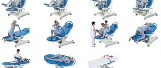
With each push, the head gradually passes through the pelvic cavity and begins to appear from the genital slit; doctors call this cutting in - the head appears from the genital slit only during pushing and through the eruption of the head (the head is constantly visible in the genital slit). This means the baby will be born soon. If there is a threat of perineal rupture, obstetricians often resort to dissection of the perineum - then they warn that they will do a perineotomy or episiotomy. This necessary measure helps prevent injuries to mother and baby. The perineotomy operation is a dissection of the perineum in the direction from the posterior commissure of the perineum to the rectal sphincter. Thus, the incision passes along the midline of the perineum. For an episiotomy, the incision is made on one side, through the labia majora (at an angle of 45° from the midline).
Immediately after birth, mucus is sucked out of the baby's nose and mouth using a rubber balloon so that it does not enter the lungs during his first breath. The condition of a newborn baby is assessed using the Apgar scale at 1 and 5 minutes. The following signs are taken into account: heartbeat, breathing, skin color, reflexes, muscle tone. The severity of each of the five signs is determined in points from 0 to 2. If the sum of points for all signs is from 7 to 10, then the condition of the newborn is satisfactory, 4-6 points - a condition of moderate severity, 1-3 points - severe.
After the baby is born, the obstetrician-gynecologist monitors for signs of placental separation. “It’s separated, we’re giving birth to the placenta” - this is what the doctor will say if, when pressing with the edge of the palm above the womb, the umbilical cord is not pulled inward, if the clamp previously placed on the umbilical cord near the genital slit has dropped slightly.
Of course, during childbirth and then after the baby is born, you will have to deal with a lot of new words and concepts. And the more you learn about them from reliable sources, the more reliably you will rid yourself of unreasonable fears.
Natalya Bulakh, obstetrician-gynecologist of the first category, Ph.D. honey. Sciences, MUZ Clinical Maternity Hospital, Astrakhan
A case of prenatal diagnosis of trisomy 18 in the second trimester of pregnancy
Ultrasound scanner HS70
Accurate and confident diagnosis.
Multifunctional ultrasound system for conducting studies with expert diagnostic accuracy.
Introduction
The incidence of trisomy 18 is approximately 1 case in 7000. It was first described by Edwards in 1960. The cytogenetically based form of the disease is simple trisomy 18. Children with trisomy 18 are more often born to older mothers. The average age of mothers at the birth of a child was 31.4, and of fathers 34.6 years, which is 5 years higher than in the control group. Girls with this disease are born approximately 2.5 times more often than boys.
The life expectancy of children with trisomy 18 is short: 60% die before the age of 3 months, about 10% survive up to 1 year. The main causes of death in the perinatal period are asphyxia associated with circulatory disorders, aspiration pneumonia and intestinal obstruction, older children die from pneumonia and urinary tract infections [1,2].
Children with trisomy 18 are born with severe prenatal hypoplasia. In 40% of cases, pregnancy is complicated by polyhydramnios. The phenotypic manifestations of the syndrome are diverse, and disturbances in the structure of the face and the musculoskeletal system are almost constant. There is a strawberry-shaped head (81%), choroid plexus cysts (50%), absence of the corpus callosum, expansion of the cistern magna, the lower jaw and the opening of the mouth are small, the auricles are deformed and low located. There is a flexor position of the hands (91%), shortening of the limbs, aplasia of the radial joint, in almost 80% of cases there is valgus deformation of the feet and their abnormal development: the arch sags, the heel protrudes sharply, swelling of the neck, diaphragmatic hernia, omphalocele, myelomeningocele. Of the defects of internal organs, the most constant are heart defects (impaired development of the interventricular septum, aplasia of the valve leaflets) and large vessels, which occur in 91-99% of cases. Congenital malformations of the digestive organs are observed in half of the cases of trisomy 18: Meckel's diverticulum, registered in every third child, incomplete intestinal rotation (16.5%) and esophageal atresia (85%). Almost half of the patients have congenital malformations of the genitourinary system. Fusion and duplication of the kidneys and ureters were noted in 14-27% of cases, cryptorchidism in 43.5% [1-4.5].
Case description
Patient S., 32 years old, during a screening ultrasound examination on a Medison SonoAce-1500 scanner during the 23rd week of pregnancy, based on specific signs, a preliminary diagnosis was made - trisomy 18 in the fetus. In the prenatal diagnostics room of the Clinical Maternity Hospital in Smolensk, in order to verify the diagnosis, cordocentesis was performed, followed by karyotyping in the laboratory of the Department of Biology of the Smolensk Medical Academy. This pregnancy, the fourth, proceeded without any special features. The previous three ended in artificial abortions without complications. My husband is 33 years old, somatically healthy. The spouses have not been genetically examined and have no occupational hazards.
Ultrasound examination revealed that fetometric indicators did not correspond to the gestational age, while the fetus had a brachycephalic shape of the head (Fig. 1), valgus deformity of the hands and feet with ectrodactyly of the left hand (Fig. 2, 3), hypertrophy of the right atrium (Fig. 4), low-set and deformed ears (Fig. 5), as well as an umbilical cord cyst (Fig. 6) against the background of polyhydramnios. Taking into account multiple markers of chromosomal pathology of the fetus, a cytogenetic study was performed. Fetal karyotype r = 47, xy, + 18. The pregnancy is terminated. During the pathological and anatomical study, the phenotypic features of the fetus corresponded to this karyotype and coincided with the ultrasound prenatal diagnosis. Right atrial hyperplasia is caused by hypoplasia of the tricuspid valve leaflets.
Rice. 1.
Fetal head.
Rice. 2.
Valgus deformity of the left foot.
Rice. 3.
Valgus deformity of the left hand with ectrodactyly.
Rice. 4.
Hypertrophied right atrium (arrow).
Rice. 5.
Deformed ear.
Rice. 6.
Umbilical cord cyst.
Conclusion
The described clinical observation is of interest to practitioners, as it confirms the possibility of diagnosing trisomy 18, which is one of the most common chromosomal diseases, using middle-class devices in the second trimester of pregnancy [1, 2, 4]. To determine pregnancy management tactics, we consider it advisable to carry out karyotyping in all cases of a pathognomonic echographic picture.
Literature
- Lazyuk G.I., Lurie I.V. Chromosomal diseases //Human teratology / Ed. G.I. Lazyuka M.: Medicine, 1991. - P. 358-364.
- Snyders RJM, Nicolaides KH. Assessing the risk of chromosomal disorders // Ultrasound markers of fetal chromosomal defects / Translation from English. - M.: Vidar, 1997. - P. 90-108.
- Medvedev M.V., Yudina E.V. Differential ultrasound diagnostics in obstetrics. - M.: Vidar, 1997. - P. 81, 114, 240, 257.
- Yudina E.V. What prenatal echography can do //Ultrasound. Diagnosis Obstetrics Gin. Pediatrician. - 2000. - T.8. - N1. — P. 18-22.
- Rusanova OK, Buntova O.V., Korbashova T.D. and others. Population frequency and diagnostic value of some ultrasound markers of congenital and hereditary diseases // Ultrasound. Diagnosis Obstetrics Gin. Pediatrician. - 1999. - T.7. - N 4. - P. 285-289.
Ultrasound scanner HS70
Accurate and confident diagnosis.
Multifunctional ultrasound system for conducting studies with expert diagnostic accuracy.
Choosing a method of delivery
A woman with a breech presentation of the fetus needs to go to the hospital at 38-39 weeks for examination and selection of rational tactics for labor management. The method of delivery is determined based on the number of births, the age of the mother, obstetric history, gestational age, readiness of the female body for childbirth, pelvic size and other factors. Breech presentation of the fetus is not an absolute indication for cesarean section, however, in cases where it is combined with various complicating factors, the issue is resolved in favor of surgical delivery.
Indications for elective caesarean section in full-term pregnancy are the age of the primigravida over 30 years; severe form of nephropathy; extragenital diseases that require switching off pushing; severe disturbance of fat metabolism; narrowing of the pelvis; estimated fetal weight over 3600 g in primiparous women and over 4000 g in multiparous women; fetal hypotrophy; signs of fetal hypoxia according to cardiotocography; disturbance of blood flow during Doppler measurements; Rhesus conflict; extension of the head of the 3rd degree according to ultrasound; unpreparedness of the birth canal at 40-41 weeks of gestation; post-maturity; foot presentation of the fetus; breech presentation of the first fetus in multiple pregnancies and other factors.
Childbirth is carried out through the natural birth canal if the expectant mother and fetus are in good condition, full-term pregnancy, normal pelvic size, average fetal size, with a bent or slightly straightened head, readiness of the birth canal, with a pure breech or mixed breech presentation.
It is best that when the fetus is breech, labor begins spontaneously. In the first stage of labor, the woman in labor must observe bed rest and lie on the side towards which the fetal back is facing in order to avoid complications (premature rupture of water, prolapse of the fetal leg or umbilical cord loops). Childbirth is carried out under monitor control of the fetal heartbeat and contractile activity of the uterus. In the second stage of labor, obstetric care is provided in the form of a benefit, the purpose of which is to preserve the fetal position (the legs are extended along the body and pressed to the chest by the fetal arms). First, the child is born to the navel, then to the lower edge of the angle of the shoulder blades, then to the arms and shoulder girdle, and then to the head. When a child is born, his head presses the umbilical cord up to the navel, and a lack of oxygen develops, so no more than 5-10 minutes should pass before the child is fully born, otherwise the consequences of oxygen starvation will be very negative. An incision is also made in the perineum to speed up the birth of the head and make it less traumatic.
Childbirth with foot presentation through the natural birth canal is carried out only in multiparous women with good labor activity, readiness of the birth canal, full-term pregnancy, medium size (weight up to 3500 g) and good condition of the fetus, a bent head, and the woman’s refusal to have a cesarean section. In this case, the obstetric care is as follows: cover the external genitalia with a sterile napkin and, with the palm facing the vulva, prevent the premature loss of the legs from the vagina. Holding the legs promotes full opening of the uterine os. During pushing, the fetus seems to squat down, resulting in a mixed breech presentation. The birthing legs are resisted until the uterine os is completely opened. After this, the fetus is usually born without difficulty.
The condition of children born in breech presentation through the birth canal requires special attention. Hypoxia suffered during childbirth can adversely affect the child’s nervous system; such pathology as dislocation of the hip joint is possible. A neonatologist and resuscitator must be present at the birth. If these precautions are observed, babies born in this way do not differ in development from other children.
Publications in the media
Breech presentation is classified based on the position of the legs and buttocks of the fetus • Pure breech presentation. The buttocks are presented, the legs of the fetus are bent at the hip joints, extended at the knee joints and extended along the body. Observed in 60–65% of cases of breech presentation • Mixed breech presentation. The legs are completely bent at the knee joints and pressed against the fetal abdomen. Observed in 25–35% of cases of breech presentation • Foot presentation. One or both feet or (extremely rarely) the fetal knees are presented •• A distinction is made between complete and incomplete breech presentations. With a complete breech presentation, both legs are presented; with an incomplete breech presentation, one leg or knees of the fetus are presented •• Observed in 5% of cases of breech presentation.
Statistical data . The incidence is 2.7–5.4% of all pregnancies. In 15–30% of cases it ends in the birth of a child with low body weight (less than 2500 g). Etiology • Pelvic narrowing, abnormal pelvic shape • Uterine malformations • Myomatous nodes in the lower segment of the uterus • Fetal malformations , eg anencephaly, hydrocephalus • Placenta previa • Uterine atony, a large number of births in history, uterine tumors, uterine scar, narrow pelvis • Low fetal weight or prematurity, multiple births • Excessive fetal mobility with polyhydramnios.
Clinical picture • High position of the uterine fundus, due to the location of the pelvic end of the fetus above the entrance to the pelvis • Using Leopold's techniques (see Normal pregnancy), it is determined that the fetal head (a round dense voting formation) is located in the fundus of the uterus, and the buttocks (large, irregularly shaped) non-ballot presenting part) - above the pelvic inlet • The fetal heartbeat is heard above the navel or at its level. Special examinations • Vaginal examination during labor •• With breech presentation, the presenting part is softer than with cephalic presentation. You can palpate the groove between the buttocks, the sacrum, the genitals of the fetus •• With a purely breech presentation, you can find the inguinal fold •• With a mixed breech presentation, the foot is palpated next to the buttocks. With the help of palpation of the sacrum, the position and appearance are clarified •• In case of leg presentation, in order not to mistakenly mistake the leg for a dropped handle (for example, in transverse positions), it is necessary to remember the distinctive features of the fetal limbs ••• The leg has a calcaneus, the fingers are straight, short, the thumb is not set back ••• The big toe of the foot cannot be pressed against the sole, unlike the thumb of the handle, which is easily pressed against the palm; you can “say hello” to the handle ••• The knee differs from the elbow by the movable patella, the foot meets the shin at a right angle •• By the location of the popliteal fossa, you can determine the position of the fetus. In the first position, the popliteal fossa faces to the left, in the second - to the right • Ultrasound easily diagnoses breech presentation.
Mechanism of childbirth • The first moment is the internal rotation of the buttocks, it begins when the buttocks pass from the wide part of the pelvic cavity to the narrow one. The rotation is performed in such a way that at the outlet of the pelvis, the transverse size of the buttocks is in the direct size of the pelvis. The front buttock fits under the pubic arch, forming a fixation point, while the rear buttock is installed under the coccyx. • The second point is lateral flexion of the fetal lumbar spine. Further forward movement of the fetus leads to greater lateral flexion of the fetal spine. In this case, the rear buttock rolls out above the perineum and after it, the front buttock is finally born from under the pubic joint. At this time, the shoulders enter with their transverse size into the same oblique size of the entrance to the pelvis through which the buttocks passed. • The third point is the internal rotation of the shoulders and external rotation of the body. This rotation is completed by placing the hangers at the straight exit size. In this case, the back is turned to the side, the front shoulder of the fetus fits under the pubic arch, and the rear one is installed in front of the tailbone above the perineum. • The fourth moment is lateral flexion of the cervicothoracic spine. The birth of the shoulder girdle and arms is associated with this moment. • The fifth moment is the internal rotation of the head (with the back of the head in front). The head enters with a small oblique size into the oblique size of the entrance to the pelvis, opposite to the one in which the shoulders passed. When transitioning from the wide to the narrow part of the pelvis, the head makes an internal rotation, as a result of which the sagittal (sagittal) suture appears in the direct size of the exit, and the suboccipital fossa is under the pubic joint, where a fixation point is formed. • The sixth moment is flexion of the head. The consequence of this is the eruption of the head: the chin, mouth, nose, crown, and back of the head sequentially roll out over the perineum. • In a pedicle presentation, the legs are born first. In front there is a leg facing the symphysis. The buttocks enter the small pelvis after the birth of the legs up to the knee. The further course of labor is the same as with breech presentation.
Pregnancy management tactics • From 35 weeks of pregnancy, it is recommended to engage in therapeutic corrective gymnastics • Some doctors recommend external obstetric rotation to transform a breech presentation into a cephalic presentation, however, obstetric rotation is unsafe - placental abruption, compression and entanglement of the umbilical cord, and premature birth may occur • In case of breech presentation, you should decide whether childbirth can be carried out naturally or whether it is necessary to resort to caesarean section. Currently, due to the risk of fetal trauma during vaginal delivery operations, most obstetricians consider it advisable to expand the indications for cesarean section for breech presentation of the fetus (80–90%). Labor management tactics • First stage of labor (dilatation period). Prevent premature rupture of the membranes. Strict bed rest is indicated. After the discharge of amniotic fluid, a vaginal examination is performed (clarification of the diagnosis, exclusion of umbilical cord prolapse). • Second stage of labor. After the birth of the fetus, the head presses the umbilical cord to the level of the navel. If labor does not end within 10 minutes, the fetus dies from asphyxia. Therefore, after the birth of the lower part of the body, prompt obstetric care is necessary. • Manual assistance for a purely breech presentation using the Tsovyanov method is used to prevent complications. The method is based on maintaining the normal position of the fetus. Technique. After eruption, the buttocks are grabbed as follows: the thumbs are placed on the legs pressed to the stomach, and the remaining fingers of both hands are placed along the sacrum to prevent premature loss of the legs. As the body is born, the arms are moved towards the genital opening of the woman in labor, continuing to press the outstretched legs to the stomach until the birth of the shoulder girdle. If after the birth of the shoulders the arms do not fall out on their own, the shoulder girdle is set in the direct size of the pelvis and the fetal body is tilted downwards (posteriorly). This creates the front handle. The body is then tilted upward (anteriorly), after which the posterior arm and legs (heels) of the fetus are born. At the birth of the head, the fetal body is also directed upward •• Manual aid for foot presentation using the Tsovyanov method. The method is based on holding the legs in the vagina until the throat is completely dilated. Technique. The woman's external genitalia is covered with a sterile napkin. A palm is placed on the vulva, delaying the birth of the legs, which leads to full opening of the pharynx. Thus, the fetus moves from a leg presentation to a mixed breech presentation. After complete opening of the pharynx, childbirth is carried out as in a breech presentation. •• Sometimes, during the use of manual aid according to Tsovyanov, premature loss of the legs still occurs. In such cases, a classic manual aid is used (a set of techniques aimed at freeing the arms and head of the fetus) •• If the birth of the head is delayed, it is released using the Moriso-Levre-Lachapelle maneuver - a method of extracting the fetal head in a breech presentation, in which the head is bent when inserted into the mouth the fetus with the index finger of one hand, followed by traction on the body with the other hand. Technique. The body of the fetus is placed astride the forearm of the hand, the second or third finger of the same hand is inserted into the vagina of the woman in labor, following its back wall, and then into the mouth of the fetus. With the second hand, grasp the fruit by the shoulders and release the head. • The third stage of labor with breech presentation is carried out as usual. Extraction of the fetus by the pelvic end is considered not an aid, but an obstetric operation, because in the process of manipulation all 4 stages of labor are artificially reproduced by applying drag force. The fruit is removed from the heels to the crown. • Indications for surgery •• The need for urgent vaginal delivery due to the severe condition of the woman in labor (for example, eclampsia) •• Acute fetal hypoxia and the absence of conditions for cesarean section •• Previous classic rotation of the fetus. • Conditions for carrying out •• Full dilatation of the cervix •• Discharge of amniotic fluid •• Matching the size of the fetus and the woman's pelvis. • Technique of operation •• First point. There are 2 methods: ••• Removing the fetus by the inguinal fold. The index finger of the hand grasps the front leg of the fetus by the inguinal fold; attraction is produced during pushing. To grasp the pelvic end, the thumbs are placed on the buttocks, the index fingers are placed on the groin fold, and the rest are placed on the hips of the fetus. The fetus is removed up to the umbilical ring ••• Removing the fetus by the stem. The leg is grabbed with the whole hand in the area of the knee joint and pulled down. The second leg is born independently •• The second moment. The fruit is removed to the level of the lower corner of the shoulder blades. This point is highlighted for two reasons ••• The release of the arms can be started only after the birth of the fetus to the level of the lower angle of the shoulder blades ••• After the birth of the fetus to the level of the navel, the head, entering the small pelvis, can pinch the umbilical cord, which threatens hypoxia •• Third and fourth points. The arms and head of the fetus are released as with classic manual assistance.
Caesarean section • Indications for cesarean section for breech presentation (in addition to absolute indications for any presentation) •• Combination of breech presentation with a burdened obstetric and gynecological history (infertility, stillbirth, birth of a child with trauma), uterine fibroids, uterine malformations, narrowing pelvis, gestosis, post-term pregnancy, primigravida age 30 years or more •• Scar on the uterus •• Large fetus •• Prolapse of umbilical cord loops •• Partial placenta previa. • Contraindications •• Intrauterine fetal death •• Deformity or extreme prematurity of the fetus •• Acute infectious disease in a woman. Caesarean section is undesirable during a long period without water (more than 12 hours). • Conditions for a cesarean section •• The fetus is alive and viable (not always feasible with absolute indications) •• The woman agrees to the operation (if there are no vital indications) •• The pregnant woman has no signs of infection. • Preparing the patient •• If Ht is less than 30%, infusion therapy is performed to compensate for fluid deficiency •• It is necessary to prepare for a possible transfusion of plasma and blood during surgery •• The woman’s bladder must be emptied. • Anesthesia can be inhalational (general) or regional (spinal or epidural). General anesthesia often leads to depression of the newborn’s vital functions, therefore, when performing general anesthesia, the time interval from the onset of anesthesia to the moment of extraction of the fetus should not exceed 10 minutes. • Progress of the operation •• Dissection of the abdominal wall •• Opening and separation of the vesico-uterine fold of the peritoneum, exposure of the myometrium •• Dissection of the myometrium (Kerr-Gusakov, Sellheim or Sanger incision) ••• Kerr-Gusakov incision (low transverse) currently used most widely. The incision is made in the lower segment of the uterus, which reduces the likelihood of rupture or divergence of the edges of the scar during subsequent pregnancies. Disadvantage - danger of damage to nearby vessels ••• Sanger incision (classical, or corporal, currently used rarely, according to indications) - a longitudinal incision on the anterior surface of the uterine fundus ••• Sellheim incision (low vertical) begins in the non-contractile part of the uterus and continue to the body of the uterus •• The child is carefully removed by hand •• The separated placenta with all membranes is removed •• The uterus is often removed from the abdominal cavity for the purpose of massaging the fundus, examining the appendages and visualizing the incision when applying sutures. To reduce blood loss, uterine contracting agents (oxytocin, methylergometrine, enzaprost, etc.) are injected into the uterine muscle. After separation of the placenta, manual examination of the uterine cavity is necessary to diagnose submucosal fibroids or to remove remnants of the membranes •• Single-row suturing of the uterus. The vesicouterine fold of the peritoneum is sutured with thin absorbable suture material, and the abdominal wall incision is sutured in the usual way. Observation • During childbirth, constant monitoring of the fetal heart rate should be carried out • Observation of the parturient woman is carried out as after other births. Prevention. Therapeutic corrective gymnastics for breech presentation of the fetus is indicated up to 35 weeks of pregnancy. Complications • Throwing back of the arms • Untimely rupture of amniotic fluid • Abnormalities of labor • Prolapse of the umbilical cord and small parts of the fetus • Spasm of the uterine pharynx with pinching of the torso or neck of the fetus • Extension of the head • Injuries to the head and soft tissues, brachial plexus and spinal cord of the fetus • Asphyxia of the fetus. Course and prognosis • Perinatal morbidity and mortality are higher in the case of breech birth • For fetal weights less than 1,500 g, the risk of cerebral hemorrhage, as well as perinatal mortality, is significantly higher in the case of vaginal delivery than after cesarean section.
ICD-10 • O32.1 Breech presentation of the fetus, requiring medical care for the mother • O64.1 Obstructed labor due to breech presentation • O80.1 Spontaneous birth in the breech presentation • O82 Singleton labor, delivery by cesarean section
Notes • Absolute indications for cesarean section •• Complete placenta previa •• Absolutely narrow pelvis •• Discrepancy between the size of the woman’s pelvis and the fetal head •• Incomplete placenta previa with unprepared birth canal and severe bleeding •• Premature abruption of a normally located placenta with unprepared birth canal and bleeding •• Tumors of the pelvic organs that prevent the birth of a child •• Severe scar changes in the cervix and vagina •• Threatening or incipient uterine rupture •• Severe gestosis with ineffective conservative treatment and unprepared birth canal •• Incompetent scar on the uterus •• Extragenital cancer and cervical cancer •• Serious extragenital pathology (for example, retinal detachment, complicated myopia, severe cardiovascular diseases).