How to determine a child's foot size by age
There are average indicators that allow you to approximately determine the parameters of a child’s foot length by age. But it is worth considering that each child is individual, and in some cases, the measurements taken from a particular child may not coincide with the generally accepted ones. Therefore, if you decide to determine the size of a child’s feet by age, the table will only tell you approximate numbers.
How to determine the size of a child's feet by centimeters
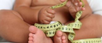
It is recommended to measure foot length while standing on the floor or other flat, smooth surface. A sheet of paper is placed under the leg, and the outline of the leg is outlined with a pen or pencil. It is better to measure both feet at once. If their length varies, then you should choose shoes based on the parameters of the larger foot. Half a centimeter is added to the resulting figures if you plan to purchase shoes for the summer season, and one and a half centimeters if for the winter.
For younger children who are not yet firmly on their feet, you can measure the length of the foot using a string or an ordinary student ruler. This simple tool will help you draw an imaginary line from the middle of the heel to the edge of the baby's big toe.
This line should not be straight, but slightly at an angle.
After the necessary measurements have been made to determine the size of the child’s feet in centimeters, you can use a special table that contains data on the correspondence of the metric foot length with a specific shoe size. And in our catalog you will find measurements of insoles for each model.
It is extremely important to choose the right size shoes for children. Buying boots or sandals with a large margin in length is also inadvisable, because wearing them will be uncomfortable.
Damage to growth areas
As you know, a child’s body is very vulnerable and undergoes constant change. Children grow up until the end of adolescence. Therefore, it is very important to monitor the normal development of the growth zones of the child’s body, in particular, bone tissue, namely the epiphyseal growth plates.
What are they? Growth plates, or as they are also called physis, are part of the growing tissue that is located at the ends of the tubular bones. These plates are only available for children and adolescents. At the end of the transition period, the child's plates ossify, and growth slows down significantly.
It should be noted that this period is somewhat different for girls and boys. This is due to the fact that the general maturation of the body in girls is faster and therefore ossification can occur by the age of thirteen. However, for boys this period is shifted to sixteen or seventeen years.
Each plate represents a part of the child's growing body and therefore their proper development is very important. They are perhaps the most vulnerable part of the child's skeleton. What can only cause sprains in an adult will lead to plate fractures in a child. It is even possible that the tendons remain untouched. As a rule, physes (plates) on the child’s body are often located in the area of the elbow, lower leg, ankle and forearm. But there may also be cases of damage to the lower and upper thighs and feet.
Let's look at the causes of damage to the epiphyseal plate. There can be many of them. So, the first thing that could be, of course, is mechanical damage to the limb caused by a fall or impact with any object. A child can receive such injuries at any time, even while under the supervision of his parents or older relatives. For example, no one is safe from falling from a swing or on a playground. However, what at first glance may seem like a minor injury is not always so in reality.
Further, the cause of damage to the plate may be chronic diseases. As a rule, this manifests itself already at an older age of the child. They can be provoked by playing sports, such as volleyball, basketball, gymnastics or athletics.
But, you should not immediately rush to limit your child’s physical activity in order to avoid such injury. Vigilant parents need to carefully monitor their child’s equipment and behavior after training. If you notice that your child is complaining of pain in the legs and arms, you should immediately consult a doctor.
If we talk about signs of damage to the growth plates, the pain will be local. It does not stop even when the body position changes or the physical load on the body decreases. You cannot force a child to move and play sports through pain. This can cause even more harm to the fragile little organism.
In addition to the above reasons, it is also possible to identify those that are not associated with mechanical effects on the body. These include bone infections, frostbite of the extremities, and harsh treatment of children.
This also includes the treatment of malignant tumors in children. That is, the use of chemotherapy, as well as radiation. It has already been scientifically proven that the use of such methods in the treatment of a disease such as cancer does not leave its mark on the entire body of an adult, and even more so a child. And, of course, in a child’s body, the most vulnerable parts of the body suffer first, which include the epiphyseal plates.
We should also consider cases of damage to a child’s growth plates due to neurological disorders and diseases from an early age.
An adult needs to closely monitor a child, regardless of his age. After all, as stated above, this growth development disorder can occur not only in the youngest children, but also in children ten and fifteen years old. That is, as the child grows, there is a danger of receiving damage of this kind.
If you notice that the child has stopped playing, he refers to pain in the arm or leg, there is deformation of the limbs, or the inability to move them, then you should immediately consult a doctor. Any delay can be dangerous for the further physical development of your son or daughter.
In any situation, if you receive an injury, you must contact a trauma surgeon. But if we are talking about small children, then it is necessary to undergo an examination under the supervision of a pediatric orthopedist-traumatologist. This must be done by such a doctor, since childhood diseases have their own characteristics that an ordinary surgeon may not notice or may not know.
When contacting a specialist, a child must be sent for magnetic resonance imaging after examination. Only in this case can you find out with certainty whether there is damage in the growth plates. Often, pediatric surgeons give directions for x-rays. However, such a survey does not provide an accurate conclusion on the topic of interest to us. Because x-rays show the area where the epiphyseal plates are located only in white. This fact does not allow us to understand if there are injuries or fractures.
Along with magnetic resonance imaging, the child may be prescribed a computed tomography, which is also acceptable.
In addition to establishing an accurate diagnosis, a highly qualified specialist must prescribe the correct treatment. And to do this, you need to determine the type of growth plate fractures.
It should be noted that initially in medicine there were five types of such fractures. However, not so long ago a new classification of such plate damage was developed, which was called the “Peterson qualification”.
So, let's take a closer look at the typology of growth plate fractures.
Types of fractures of the epiphysis plates (growth plates)
- The doctor diagnoses the first and most favorable type of fracture when the photographs show that the plate (physis) is not separated from the end of the tubular bone. That is, according to science, it is not separated from the pineal gland.
- The second type is a more severe case. With such a fracture, the epiphysis is separated along with the plate from the tubular bone itself. This is the most common fracture.
- The third type is characteristic of the lower leg and lower leg. It is extremely rare. It is characterized by the fact that the epiphysis, together with the plate, separates from the tubular bone, but only partially. That is, a fragment. In such a situation, only surgical intervention can help, since with this kind of fracture, blood circulation in this area may be impaired.
- The fourth type of fracture has complex consequences. Its essence lies in the fact that the fracture passes not only through the entire epiphysis and plate, but also rises higher, touching the metaphysis. This injury is of the nature of the shoulder and elbow.
- a type that is rare, characterized by the fact that as a result of bone crushing, the plate is compressed and ossification of the growth zone occurs.
- And the last, sixth type of fracture of the epiphysis plates is that the plate, epiphysis and metaphysis are completely absent. This fracture has the most unfavorable consequences, since bone growth in this area is always disrupted and sometimes even stops.
Almost any type of growth plate injury can be treated.
Let's consider the main methods of treatment for fractures of the epiphysis plates
- The first and most common method is the application of plaster (immobilization), which leads to immobilization of the injured surface. This method is sufficient for the treatment of the first and second types of growth plate fractures. However, when treating other types, plaster is also applied, but only after a number of other procedures.
- The second method involves surgical or manual restoration of the integrity of the bone tissue and epiphysis plate. This method is applicable for all types of damage to growth zones, except for the first, simplest and less painful. After applying this restoration, specialists must apply plaster. This is necessary to restore the integrity of the bone itself and the structure of the plate.
- The third method can be called restorative and preventive. This includes a physical therapy complex. It should be developed only by specialists in this field and is aimed at the speedy strengthening and healing of damaged tissue and limbs.
But even after completing all stages of treatment, constant monitoring of damaged growth zones is necessary. To do this, once every six months you need to undergo a regular examination by a pediatric orthopedist-traumatologist and x-rays, or for a more detailed view of the affected area, magnetic resonance imaging (computed tomography). As a rule, such observation continues for two years after fractures, or until the child’s growth plates have completely ossified.
If it does happen that the child has a fracture of the epiphysis plate (physis), then you should not immediately become despondent. Nowadays medicine does not stand still and the topics of child growth problems are constantly discussed, and a number of studies are carried out. In addition, doctors are currently making favorable prognoses for further development in cases of this type of injury.
Thus, it has already been established that with correct diagnosis and treatment of a growth plate fracture, 85 percent of children did not have any further disorders. Experts in orthopedic and traumatology say that the prognosis for the future depends on many factors.
Take a closer look at them.
Factors for healing bone tissue in damaged growth plates
The first, and perhaps one of the most important, is determining the severity of the injury and its location. Due to this type of damage, the flow of blood and lymph to the affected area may slow down. In addition, if you receive, for example, an open fracture, an infection can occur at the location of the growth zones and lead to infection of the bone tissue, which is the most severe case.
Next, consider a factor such as the child’s age. It has been established that the younger the child who has suffered damage to the growth plates, the more difficult it will be for him to recover. Since all tissues and organs are just beginning to form, and any impact on them can lead to disruption in the development of the entire organism.
Another important factor in a positive prognosis for the further development of a child’s growth is the doctor’s correct determination of the type of fracture. The most dangerous consequences are those of the fourth and fifth types, which require constant long-term observation and enhanced rehabilitation therapy.
After all the medical procedures have been carried out and doctors have assured them of a favorable prognosis for the future, many parents still notice stunted growth in their children, or incorrect, asymmetrical development of the limbs. We will immediately answer such questions. Given the nature of the injuries, this is a completely normal picture. Because the damaged area of the growth zone is already starting to work differently. The injured bone begins to grow slower than the rest. It may continue to grow on a different trajectory than similar healthy bone. In such a situation, you need to calm down and understand that the restoration of bone tissue and all its constituent elements, although it occurs quickly in children, requires special attention and control.
Currently, many scientific institutes are conducting research on the restoration and healing of bone tissue using genetic technologies.
This will avoid slowing the growth of the limbs, as well as their curvature. Author: K.M.N., Academician of the Russian Academy of Medical Sciences M.A. Bobyr
