Today it is difficult to find a person who is not aware of the dangers of ionizing radiation. However, despite the entire volume of available information, it is necessary to resort to diagnostic procedures performed using x-rays quite regularly. It would seem that what could be simpler than conducting a routine fluorographic examination or x-ray according to indications?
Such an indifferent attitude is typical for the average person, but is absolutely not typical for a pregnant or lactating woman, for whom the health of the child comes first. It is known that even minor exposure to radiation in the early stages of pregnancy can cause disruption in the development of the fetus, as well as unpredictable long-term consequences in the later stages. But what effect can radiation have on the body of a nursing mother, and is x-raying really harmful to the baby during breastfeeding? The answer to this and other questions can be found in the article.
The mechanism of the effect of ionizing radiation on the body
In order to track the relationship between the effect of x-rays on the mother’s body and the likelihood of a risk of negative impact on the child during breastfeeding (BF), it is necessary to obtain a minimal understanding of the ionization mechanism. Radiation, which is a stream of energy used to obtain x-rays and has the ability to pass through various obstacles, including the human body.
Due to the fact that the density of tissues and the location of internal organs create a heterogeneous barrier to radiation, overcoming obstacles leads to a partial loss of its intensity, which makes it possible to obtain an overexposed image in accordance with the anatomical features of the part of the body being examined. Thus, the rays that leave the body react with photographic paper, and the remaining rays react with the cells of the body.
The electrical interaction of radiation with the atoms of cells leads to irreversible damage, and the factor determining the amount of damage caused is the duration of exposure. The ability of radiation to split hydrogen atoms, which are in excess in the body in the form of water, leads to the formation of free radicals, which, in turn, cause a chemical reaction that disrupts the functional activity of surrounding cells at the molecular level.
Important! Due to the fact that the irradiation time at the time of taking an x-ray is several seconds, the damage caused to the body is minimal and is quickly eliminated by triggering the regeneration mechanism.
The effect of x-rays on the ability of lactation
Thus, the fact of the negative impact of X-rays on the body of a nursing mother is undeniable, but is ionizing radiation capable of directly affecting the reproduction of breast milk? Taking into account that the radiation dose received during X-ray diagnostics is so insignificant that it is unable to cause any serious damage to the tissues, thus depriving them of the ability to produce milk, the danger may lie in the milk itself, which is in the breast at the time of the procedure. picture.
Naturally, in this case we are not talking about an x-ray of an arm, leg or tooth, since the beam of rays used to take x-rays is quite narrow and is unable to have any effect on the body as a whole, and especially on the chest area. From this point of view, the following studies can be considered the most dangerous:
- mammography;
- fluorography;
- chest x-ray.
Important! The minimum threshold of radiation that can cause serious damage to the body is 1.5 3V (sievert), and the total number of diagnostic procedures performed using X-ray equipment is only 1.4 m3V (milisivet) per year.
It is permissible to take an X-ray of a tooth even during pregnancy, since it uses a very narrow beam of radiation.
When should a chest x-ray be taken?
Plain radiography of the chest organs is carried out:
- During an annual preventive health examination to identify risk factors and life-threatening diseases such as cancer or tuberculosis.
- For pain in the chest and heart area.
- With painful or bubbling breathing, sensations of lack of air.
- With loss of appetite and a sharp decrease in body weight.
- For swelling and high blood pressure.
- For injuries, to identify foreign objects in the esophagus, lungs, and respiratory tract.
- If you suspect lung diseases: pneumonia, abscess, pleurisy, silicosis, etc.
The effect of x-rays on the composition of breast milk
Another factor that elevates the effects of radiation to the rank of extremely dangerous phenomena is the ability to convert ordinary substances into radioactive ones. The essence of this process is the disruption of the stability of the nuclei of a non-radioactive substance due to prolonged exposure to ionizing radiation. As a result of such exposure, after the cessation of irradiation, the exposed substance continues to emit radiation for some time. In this case, the duration of radiation directly depends on the physical properties of substances.
There are three main options for the behavior of a substance exposed to radiation:
- death (for example, of a living cell);
- loss of stability (beginning of long-term or short-term radiation);
- modification (a substance changes its structure, but retains physical stability, that is, does not emit).
As you know, during breastfeeding the baby receives all the substances necessary for its full development. In addition to water, fats, proteins and carbohydrates, milk contains a complex of B vitamins, vitamins C, E and K, as well as a group of minerals:
- calcium Ca – 32 mg/100ml;
- potassium K – 51 mg/100ml;
- iron Fe – 0.03 mg/100 ml;
- magnesium Mg – 3 mg/100 ml;
- phosphorus P – 14 mg/100 ml;
- sodium Na – 17 mg/100 ml;
- zinc Zn – 0.17 mg/100 ml.
Important! Despite the fact that exposure to X-rays is extremely short-lived, the possibility of interaction of water, vitamins or minerals produced during lactation with ionizing radiation cannot be ruled out.
There is a possibility that X-ray radiation can affect the composition of breast milk. Unfortunately, there is no scientifically proven work on the changes that occur in breast milk after an x-ray, but the possibility of a physical reaction cannot be ruled out.
The least evil in this case will be the loss of useful qualities (in case of changes in physical properties), and the greatest will be the negative impact due to the acquisition of radioactive properties (loss of stability). It should also be taken into account that during feeding with milk exposed to x-ray radiation, even a short-term and mild negative effect will occur inside the child’s body, which will undoubtedly increase the harmful effects.
2.How is a chest x-ray done and how to prepare for it?
In most cases, a chest x-ray does not require prior preparation. During this procedure, it is necessary to remove outer clothing to the waist, as well as remove jewelry, removable dentures, glasses, and any metal objects, as they can affect the quality of the x-ray image.
Women should inform a specialist about the possibility of pregnancy, since X-ray radiation has a negative effect on the fetus.
Such exposure is especially dangerous for an unformed baby during the first trimester of pregnancy. In cases where the likelihood of complications for the mother if she refuses the study significantly exceeds the risk of exposure to radiation on the fetus, a chest x-ray is still performed, but the abdominal area is covered with a special apron that does not allow x-rays to pass through.
A chest x-ray is performed by a technologist or laboratory assistant - a person who monitors the procedure and coordinates the patient's actions. For a chest x-ray, you will need to press your chest against the x-ray machine plate, sit up straight, and hold your breath for a few seconds. If a second shot is required, you should turn sideways, raise your arms up and hold your breath again. The average duration of the procedure is 10 – 15 minutes. Although the image appears within 20 minutes after the examination, you will learn about its results after your doctor analyzes the image and writes a description of it.
Legal standards
Contrary to the existing likelihood of a negative impact of radiological research methods carried out during breastfeeding on the child’s health, the Ministry of Health of the RSFSR, in its recommendations dated 02/06/2004 No. 11-2/4-09, is silent about the need to limit diagnostics for preventive purposes.
Harm from X-rays
In the orders issued earlier (Order of the Ministry of Health of the USSR dated March 29, 1990 No. 129 and Order of the Ministry of Health of the RSFSR dated August 2, 1991 No. 132), examinations carried out using X-ray equipment in women during lactation are permissible only according to strict medical indications.
The loyal attitude of the Ministry of Health to performing x-rays during breastfeeding is also demonstrated by the regulations “On the environment of a newborn,” which sets out the rules obliging a woman to undergo fluorography for the purpose of prevention, immediately after the birth of a child.
This attitude of the state is understandable from the point of view of setting priorities, where the probable risk of harmful effects of X-ray radiation on a nursing woman is offset by the real risk of tuberculosis. The decisive factor determining the need for preventive examination is the increase in reported cases of tuberculosis.
Important! Nursing mothers who are at risk must undergo a fluorographic examination without fail.
For a woman from a socially prosperous background, sometimes it is enough to provide fluorographic images of all her closest relatives
Diagnostics with contrast
X-ray with contrast is another diagnostic method, the main purpose of which is to obtain detailed information about the condition of internal organs or the vascular system. Iodine-containing preparations are used as a contrast agent, which often cause a severe allergic reaction.
This diagnostic method is used exclusively according to indications, as a rule, in order to clarify the diagnosis. Taking into account that contrast is used mainly in computed tomography, in addition to radiation exposure, which is several times greater than conventional radiography, there is a risk of iodine-containing drugs passing into breast milk.
As a rule, x-rays are performed if the following pathologies are suspected:
- the presence of tumors in the chest or lungs;
- inflammatory lung diseases (pneumonia);
- tuberculosis.
Despite the fact that drugs in this group are characterized by a short half-life and fairly rapid elimination from the body, the possibility of penetration into breast milk cannot be completely excluded. Barium, used to obtain contrast images of the stomach, is not absorbed in the gastrointestinal tract, and therefore is not able to pass into breast milk. The use of radiocontrast agents makes it possible to detail information obtained using conventional x-rays.
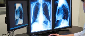
1.What is a chest x-ray and why is it needed?
Chest X-ray
- This is a flat image of all the tissues and organs that make up its composition.
This image is the result of a classic projection radiographic study - radiography
, which is used in the diagnosis of pathological changes in the chest cavity and nearby organs. X-rays allow you to see and evaluate the condition of the bones of the spine, abdominal cavity, soft tissues and organs.
In order to obtain accurate and complete information about the condition of the internal organs, doctors take pictures in two projections in one session: the first is from the back of the chest, and the second is taken from the side. If the results of this procedure are not enough to obtain a complete picture of the condition of the internal organs and bones, then doctors in addition prescribe other studies: echocardiography, ultrasound, computed tomography and magnetic resonance imaging.
Why do you need a chest x-ray?
A chest x-ray helps identify problems with internal organs that are not noticeable during a normal examination. It is appointed for the purpose of:
- diagnose diseases such as pneumonia, cystic fibrosis and lung cancer;
- Understand the causes of common symptoms: cough, shortness of breath and chest pain;
- identify pathologies of the cardiovascular system: heart failure, enlarged heart, etc.;
- diagnose damage to the lung tissue, as well as diseases accompanied by the accumulation of fluid in the lungs;
- identify chest injuries, rib fractures;
- identify foreign objects in the airways, lungs and stomach.
Alternative diagnostic methods
The danger of exposure to ionizing radiation, even small doses, should not be underestimated. But what to do if diagnostics are still necessary? In such cases, the question of whether it is possible to take an x-ray during breastfeeding is not entirely correct, since a diagnosis not specified in time can pose a serious threat to health. In some cases, you can use alternative diagnostic methods that do not pose any danger (ultrasound, MRI).
Moreover, if ultrasound cannot provide the necessary amount of information, then the only limitation to MRI is its cost. Another way to take an x-ray while breastfeeding and avoid possible negative consequences for the baby is to wean him off the breast during the diagnosis. To do this, immediately before the procedure, he should be fed, and subsequently fed with pre-expressed milk or formula. Within 12–24 hours, milk should be expressed and poured out, and after the specified time period has passed, you can return to your usual method of feeding.
After X-ray diagnostics, it is recommended to express milk for 24 hours
The modern approach to the use of diagnostic studies suggests neglecting the likely risk of negative consequences after an X-ray during hepatitis B, considering it very insignificant. If the decision on the advisability of diagnostics is made by the doctor, then only the mother has the right to decide whether the child can breastfeed immediately after the procedure.
X-ray and breastfeeding
Modern scientific research proves that there is no reason why a nursing mother should wean her baby off breast milk for some time due to X-rays.
Breastfeeding is NOT a CONTRAINDICATION for x-rays of any area of your body.
CT and MRI are also not contraindicated during breastfeeding.
If a contrast agent is administered, it is necessary to decide individually whether a particular contrast agent is compatible with breastfeeding. Mostly gadolinium-based contrast agents are used.
According to the website e-lactancia.org, X-ray examinations and gadolinium-based contrast agents are in the green (permitted) zone.
- X-ray does not affect the quality, quantity and taste of milk.
- There is no need to pump either before or after!
- There is no need to wean the baby from the breast after the X-ray examination. ⠀
X-ray is a short-term procedure (lasts a few seconds). The effect of radiation on the body stops immediately after the end of the procedure, the rays do not accumulate in it and radioactive substances are not formed.
Breast milk is not exposed to the negative effects of X-rays and its composition does not change.
Remember that any procedure must be justified and performed according to indications. Don't put off getting an X-ray if you need it! Late diagnosis worsens the prognosis of the disease!
More information: X-ray during pregnancy
Source:
Mohrbacher N., Stock J., La Leche League International, The Breastfeeding Answer Book, Third Revised Edition, 2008
Author: Tatyana Neeshpapa, @doctor_neeshpapa