X-ray examination is considered one of the most informative methods for examining internal organs. However, when it comes to children, parents are concerned about the question of at what age a child can be x-rayed. Concern for health is justified, because during the procedure the body is exposed to slight radiation. If an x-ray is unlikely to harm an adult, then in relation to a growing child’s body, caution will not be superfluous.
At what age and how often can children be x-rayed?
Modern medical equipment significantly limits the negative effects of radiation on the body. The radiation dose with digital radiography can be 10 times lower than with the conventional film method, as with digital fluorography. However, when X-rays are performed on children, it is important to understand at what age the procedure will be completely safe.
Pediatricians freely prescribe examinations for children who reach 15 years of age. It is believed that from this age the human body has developed as a whole and small doses of ionizing radiation do not pose a danger to it. At a younger age, examination can be carried out if there are indications and a doctor’s prescription in cases where the risks associated with the procedure do not exceed the harm from an incorrect diagnosis. Indeed, in most cases, improper treatment or its absence can lead to more severe consequences than a small dose of ionizing radiation. Therefore, at what age a child can be x-rayed is decided in each case by the attending physician and parents.
Indications for X-rays for children
A child can have an x-ray in the following cases:
- diseases of the chest organs;
- traumatic brain injuries;
- dislocations and fractures;
- entry of foreign objects into the respiratory system and gastrointestinal tract;
- suspicion of cancer;
- dental diagnostics.
These indications are relevant for a child over two years of age. For younger children, different rules apply when x-rays are prescribed.
When can an X-ray be taken on a newborn and infant?
For preventive purposes, X-ray examinations are not performed on children. Diagnostics can only be prescribed by a doctor in a situation where a quick and accurate diagnosis is needed, and the benefit of the examination outweighs the possible harm.
Such cases include:
- pathologies of the musculoskeletal system, injuries - birth and acquired;
- suspected pneumonia;
- intestinal obstruction;
- entry into the body of a foreign body.
Indications for radiography in children
X-rays can reveal a huge number of different pathologies of internal organs, soft tissues and the osteoarticular system. However, there are several diseases in which the question of at what age a child can be x-rayed recedes into the background, and the specialist prescribes an examination without hesitation. These are injuries to the spine, limbs or skull, foreign bodies in the gastrointestinal tract and respiratory system, pleurisy or pneumonia, heart disease or other anomalies in the development of internal organs.
X-rays are often required when inflammatory processes develop. A child cannot always describe symptoms (especially at an early age), so sometimes a doctor prescribes an examination to make a diagnosis and treatment.
With hip dysplasia, the question of at what age can children be x-rayed fades into the background. Without proper treatment, this congenital pathology can lead to impaired leg function, leaving the child disabled for life. To diagnose a dangerous pathology, a specialist may prescribe an x-ray at its first signs. Even at the age of 4-5 months.
Absolute contraindications
As mentioned earlier, the harm from radiography at a young age is incredibly great, and it is prescribed only in situations where there is a very serious reason for it. It is worth understanding that girls should never have the area where their reproductive organs are irradiated; boys, accordingly, should not have their testicles irradiated. Here are the absolute contraindications, in the presence of which it is strictly prohibited to conduct research:
- extremely close location of organs;
- the presence of any disturbances in the development of paired organs (for example, unevenness of this process);
- violation of an individual character;
- Training in the thyroid gland area is also prohibited.
An infant can be x-rayed from the first day of life, but there is some danger.
At what age can X-rays be taken?
It is important to remember that minors are not responsible for their health. Therefore, the question of whether it is worth conducting an examination and at what age you can have an x-ray for your child is decided by parents or legal representatives. If the doctor prescribed the procedure, it means that he decided that the risk was justified, and this opinion is worth listening to.
How often throughout the year?
The negative effects of ionizing radiation can accumulate in the body. Therefore, the question of whether it is possible to do an X-ray on a child and at what age it is permissible to conduct an examination, the frequency of procedures is not so important.
It all depends on the type of x-ray and what equipment is used to conduct the examination. For example, chest x-ray
can be carried out up to 5-6 times a year, dental examinations can be prescribed much more often.
Harm from X-rays
Ionizing rays pass through the cells of the body, “charging” them and affecting the structure of molecules. If the radiation was strong enough, changes can lead to the development of somatic diseases.
Ionizing radiation has the greatest impact on the bone marrow, causing blood diseases. However, for negative consequences to occur, the body must receive a large dose of radiation. Therefore, when answering the question about at what age a child can be x-rayed, you need to take into account the doses of radiation that he receives as a result of medical procedures and from natural sources.
In Russia, the upper limit of radiation exposure for preventive medical studies of citizens is set at the legislative level at 1 millisievert (mSv) per year. A radiation dose exceeding 50 mSv poses a health risk. Moreover, in just one hour of flight on an airplane, a person receives up to 0.1 mSv of radiation. From natural sources, a person is exposed to ionizing radiation of 1 to 10 mSv per year.
During medical examinations, the body receives from 0.001 mSv (with radiography of the bones of the extremities) to 10 mSv (with computed tomography of the whole body), and, for example, conventional fluorography carries a radiation dose equal to 0.5 mSv.
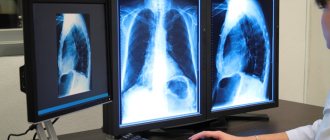
Description of the procedure
Let's first understand the principle of radiography. This method is based on the use of ionizing radiation, which is sometimes also called X-rays, or more precisely, on its basic physical properties. The fact is that such rays can easily penetrate the human body, but to some extent they linger there, on the basis of which photographs are taken. As you might guess, dense bones are much lighter in the image because they absorb X-rays quite well. For this reason, it will not be difficult for specialists to distinguish the location of the tubular bones from others. As for soft tissues, it is almost impossible to study them using X-rays, especially if it is performed without contrast (the use of a contrast agent cannot even be considered if we are talking about a newborn child).
X-ray of the abdominal and chest area of a newborn.
As for the advantages of radiology, among them experts always highlight the speed, low cost of the procedure, as well as sufficient effectiveness in many situations. Of course, there are more modern and safer diagnostic methods, for example, magnetic resonance imaging, but in many situations the overpayment is pointless, and if we are talking about the need for an X-ray for a newborn child, then this is almost impossible, because a person must be in the MRI machine for a long time (it can be difficult to explain this even to children, and a newborn baby will definitely not lie motionless). Using MRI, it is also impossible to study the condition of the long bones or, for example, the skull of a child’s head, because bone tissue is very poorly visualized.
Experts note that X-rays are practically not prescribed immediately after birth, because ionizing radiation can cause great harm, and the consequences can be extremely serious, because radiation will cause various pathological processes in a small body. For this reason, radiography of tubular bones or anything else in children under the age of 14 can only be performed if indicated. It is worth noting that only digital equipment should be used, because film devices have a much higher radiation dose.
How is an X-ray performed?
The tactics of the procedure depend on the area being examined and the examination method. Most often, the small patient takes a horizontal position on his back or side. In some cases, the child needs to take an upright position.
X-rays are taken for children in a matter of seconds, but it is important for parents to know at what age their child is able to remain still for short periods of time. In some cases, young patients need to be calmed or even “lulled to sleep.”
What can be a substitute for x-rays?
According to experts, if it is possible to avoid x-rays for a newborn, then it is best to use it. Nowadays, medicine, fortunately, is developed enough to have some choice among diagnostic procedures. Doctors recommend performing an ultrasound examination, that is, ultrasound. Thus, it becomes possible to identify many dangerous ailments without harmful ionizing radiation. This method helps to easily determine the presence or absence of dysplasia when it comes to the hip joint, and it can be used to identify dislocation or even subluxation of the hip without any problems. It is worth mentioning that ultrasound cannot always replace radiography, just as X-rays in some cases are less informative compared to ultrasound. For this reason, the diagnostic option should be selected only by a highly qualified specialist.
Unfortunately, it is not always possible to carry out safer research methods on infants (we are talking about procedures such as magnetic resonance or computed tomography), because they require a person to remain in a stationary position for a long time (it is impossible to explain this to a baby).
How are hip x-rays done for young children?
An X-ray of the hip joint is taken for a newborn lying down, the baby’s legs are specially stretched and straightened. Bending at the knees or pelvis is unacceptable and will not provide the necessary information. The legs also need to be moved slightly inward.
Before X-raying a newborn's hip joint, it is best to have him sedated. This will ensure that the clarity of the image does not deteriorate due to movement. The child's genitals are covered with a lead apron, which avoids future reproductive problems.
Technique
X-rays are taken only in specially equipped rooms on the territory of official medical institutions. The procedure is carried out under the supervision of a doctor. Sometimes a special device is used to secure the baby. The bandage and pads secure and at the same time protect from radiation.
The x-ray is performed in the presence of the parents (or one of them). To obtain clear images, the child must remain completely still, since a repeat session is highly undesirable. Babies cannot fulfill this basic condition on their own, so they are placed in a special bandage. It holds the baby in a fixed position.
If during the examination it is necessary to take a puncture or perform other procedures, the child is given anesthesia (immediately before the session). In this case, an anesthesiologist must be nearby during the session. Before the x-ray, the baby's body part that will be scanned is exposed. All metal objects (buttons, clasps, etc.) are removed from the rest of the clothing.
A protective lead apron is put on the body, which should not be exposed to radiation. The examination lasts from 20 minutes to half an hour. Afterwards, parents immediately receive black and white photographs with a transcript from the doctor. The results are recorded in the baby’s medical record. The photographs will be needed in the future, so it is advisable to save them.