A tragic phenomenon in medicine, a mystery without an answer - this is how sudden infant death syndrome is often called. And indeed, such an extremely mysterious and inexplicable phenomenon began to appear more and more often. Essentially, this is the death of a child under the age of 1 year without any signs of disease or abnormalities at autopsy. The baby simply dies for no apparent reason, unexpectedly and quietly. What is such a syndrome, why it can develop and how to deal with it - in the material of AiF.ru.
Risk group
Sudden infant death syndrome has no clear definition. The death of a child is often called unexplained. At the same time, there are no direct or indirect factors on the baby’s body or in the house that could serve as a catalyst - no infections, no bacteria, no genetic abnormalities, no other pathological problems. Doctors still cannot explain why exactly this problem develops. However, a number of studies on this topic have made it possible to draw some conclusions. For example, according to statistics, ADHD most often occurs in children under the age of 8 months, with the largest number of cases recorded at the age of 2-4 months. Of the total number of children killed, 60% were boys. As for time, the death of infants is noted at night - from midnight to 6 am.
Hello, Ambulance? 7 reasons to call the emergency room for your child
More details
Up to what age is Sudden Infant Death Syndrome possible?
SIDS is considered to be the cause of death in children from newborn to one year . But in fact, the risk of the syndrome decreases significantly with the appearance of the child’s ability to roll over, sit down, and stand up in the crib on his own, that is, after six months .
When a baby learns to turn, sit and crawl on his own, the risk of SIDS will decrease dramatically.
We study the reasons
There are no main and clear reasons for the development of SIDS. However, some doctors suspect that the catalyst is the imperfect functioning of the brain, when literally second-to-second glitches occur in it that can quickly unbalance the baby’s body. Others are confident that the leading role in the development of this pathology belongs to heart rhythm disturbances - children who have an extended QT interval on the ECG are susceptible to it. In addition, there is a theory according to which ADHD develops due to the fact that the position of a sleeping child on his stomach with his head turned to the side causes compression of the spinal artery, which causes hypoxia and apnea.
Today, doctors identify only concomitant factors that can provoke a corresponding reaction. Among them:
- Sleeping on your stomach (this factor is often called the main one)
- Excessive wrapping
- Using a mattress and pillows that are too soft
- Presence of unreasonable stops in breathing, incl. if such a situation was observed among the child’s brothers and sisters
- Mother's age is up to 20 years, when she is single and was not registered with a doctor
- Presence of illnesses in the mother during pregnancy
- Small gap between pregnancies (less than a year)
- Difficult course of childbirth
- Prematurity
- Artificial feeding
- Sleeping in separate rooms with parents
Also, such children often had sleep apnea. At the same time, it is worth understanding that an absolutely healthy baby may experience short pauses of 12-15 seconds in the first year, and this is a variant of the norm. If such breathing pauses take 20 seconds or more and are accompanied by pale skin, lethargy, and decreased muscle tone, then they indicate full-blown and life-threatening sleep apnea.
Early birds. How premature babies are cared for Read more
The article presents literature data indicating the complexity of the clinical and morphological diagnosis of sudden infant death syndrome (SIDS). The issues of prevention in SIDS risk groups and the basis for comparing clinical and morphological indicators that confirm or exclude the formulation of this diagnosis in the conclusions of clinicians, forensic experts and pathologists are reflected.
Sudden infant death
The article presents the literature showing the complexity of clinical and morphological diagnosis of sudden infant death syndrome (SIDS). It is reflected the issues of prevention in high-risk groups SIDS and comparison of clinical and morphological parameters, confirming or excluding the diagnosis in the conclusions of niyah clinicians, forensic experts and pathologists.
The term "sudden infant death syndrome" (SIDS) was first defined in 1969 at the II International Conference on Sudden Infant Death in Seattle as "the sudden, unexpected, nonviolent death of an infant aged 7 days to 1 year in which adequate explanation of the cause of death, data from anamnesis, clinic and pathological examination.” Terms such as “cot death” (or crib death) or “cradle death”, “sudden infant death syndrome” (SIDS), and “sudden infant death syndrome” (SIDS) are also used to refer to this pathological process. unexpected death of young children."
In 1991, SIDS was recognized by WHO as an independent diagnosis and, according to ICD-10, has code R 95.0 (sudden death of an infant). The incidence of SIDS is on average 1 case per 500 live births, reaching up to 3 cases per 1000 newborns in different countries of the world. According to WHO, the share of this syndrome in the structure of infant mortality in these countries ranges from 15 to 33%. The lowest rates are observed in the Scandinavian countries, Austria, Japan, Israel, and Northern Ireland. SIDS occurs only in children of the 1st year of life, in 90% of cases it occurs in the first 6 months with a peak mortality rate between the 2nd and 4th months of life. Boys predominate among those who suddenly died (60-75%). Most children are found dead at night (from 0 to 6 hours), lying on their stomach, with their head turned to the side. Death is often the first and last medical manifestation of the syndrome in question. In this case, an apparently healthy child is put to bed and found dead a few hours later. Less often, there are precursors in the form of excitement, lethargy, weakness 1-2 days before death, but there are no signs that would allow parents or a doctor to suspect the possibility of a catastrophe [1, 2, 5, 8, 10]. SIDS does not include cases of sudden death of children under 7 days of age and over one year of age.
Sudden infant death more than 100 years ago was explained by three theories: accidental suffocation (bed linen or pillow), Asthma thymicum (compression of the trachea by an enlarged thymus or goitrous thyroid gland), and Status thymicolymphaticus. Currently, these theories have significantly lost their practical significance.
Modern pathophysiological theories trying to explain sudden infant death come down to the following: 1) apnea hypothesis; 2) long QT syndrome; 3) decreased brainstem perfusion; 4) disturbance of the awakening reaction and “grasping” breathing; 5) congenital myocardial hypertrophy.
Other hypotheses come from an excess of endogenous drugs - endorphins, a defect in beta-oxidation of fatty acids, and a delay in the maturation of cardiorespiratory control by the central nervous system. To explain the thanatogenesis of SIDS, attention is paid to respiratory mechanisms in the form of various forms of apnea, including obstructive forms, and cardiac mechanisms in the form of heart rhythm disturbances due to changes in the conduction system and an imbalance in its autonomic regulation, as well as long QT interval syndrome [6, 7, 8, 12]. Biochemical changes are also noted with an increase in the total level of proteins, amino acids and insulin in the blood. An increase in the latter, combined with long breaks in feeding at night, leads to severe hypoglycemia, considered as a direct cause of “death in the crib” [4]. The mechanisms of thanatogenesis in SIDS also include the syndrome of immune-endocrine dysfunction with an increase in the thymus (thymomegaly) due to the cortical layer, a decrease in the mass of the adrenal glands due to a decrease in the width of the zona fasciculata and the volume of cells, a decrease in the mass of the pituitary gland with a decrease in the number of basophilic cells and the volume of their nuclei, which usually combined with hypofunction of these organs [1, 10]. A number of studies indicate that Status thymicolymphaticus does not exist as a special condition; others indicate its presence in SIDS in 40.4% of cases [2, 7, 12]. The described changes in the thymus and adrenal glands are defined as endocrinopathy and appear in ICD-10 under the term “polyglandular dysfunction” (E 31.0).
Mutations of genes associated with a high probability of sudden death were identified - beta-myosin heavy chains, myosin binding protein C, alpha-tropomyosin and troponin T. Identification of mutations in these genes made it possible to use implantation of an electrical pacemaker or cardioverter for preventive purposes, which reduced the number of children with sudden death (A.D. Tsaregorodtsev, 2010).
Thus, no single hypothesis can explain the causes of SIDS. It is likely that many factors must combine to cause sudden death of an infant. Most researchers are of the opinion about the polyetiological, multifactorial nature of SIDS, which develops against the background of increased sensitivity associated with the immaturity of the nervous, endocrine and immune systems [1, 8, 10, 13]. However, most often the true cause of death remains unclear.
In 1986, the concept of “obvious life-threatening episode” (ALE) in infants was introduced for the first time at a conference of the American Institutes of Health. It has a number of synonyms (near-miss, abortive SIDS, etc.) and refers to infants who have experienced life-threatening episodes and survived. At the same time, AFE should not be understood as a diagnosis, because it only gives a description of a situation, the cause of which at the moment remains unclear. The incidence of AFE is estimated to be about 0.6% and occurs with increasing frequency from the first week of life, reaching its peak in the first month of life. During the first 4 months, 60% of all cases of AFE are registered, which can be expected during the first year of life [4].
Some of the possible triggers for life-threatening manifestations are: central obstructive or mixed apnea on polysomnographic studies, seizures, upper and/or lower respiratory tract infections, gastroesophageal reflux, metabolic diseases, or circadian rhythm dysregulation.
A comprehensive analysis of a number of demographic indicators, the mother’s obstetric and gynecological history and characteristics of the early postpartum period of children revealed a number of factors (Table 1), a large number of which in one child significantly increases the risk of developing sudden infant death syndrome. Among them are the following:
Social factors:
1. Bad habits of parents, especially mothers.
2. Unfavorable living conditions.
3. Low educational level of parents.
4. Family hereditary burden.
Biological factors:
1. Death of another child in the family from SIDS, especially one of the twins.
2. A burdened obstetric and gynecological history of the mother.
3. Aggravated pregnancy.
4. Neonatal pathology (birth of a child with a very large or small weight, asphyxia, attacks of apnea during sleep).
5. Frequent infections and others.
Table 1.
Characteristics of risk factors for the development of SIDS (M.A. Shkolnikova, L.A. Kravtsova, 2004)
| Criterion | Gradation | Point |
| 1 | 2 | 3 |
| Social factors | ||
| Mother's education | average | 2 |
| low/secondary or elementary | 5 | |
| Father's education | average | 6 |
| low/secondary or elementary | 7 | |
| Footage per person in an apartment | less than 3 m2 | 4 |
| 3-7 m2 | 2 | |
| Single-parent family | 5 | |
| Maternal smoking during pregnancy | 10 | |
| Maternal smoking after childbirth | 3 | |
| Mother's alcoholism | 31 | |
| Father's alcoholism | 3 | |
| Perinatal risk factors | ||
| Ordinal number of births | 2nd | 1 |
| 3rd | 5 | |
| 4 or more | 32 | |
| The interval between these data and previous births is less than 14 months. | 12 | |
| Number of previous pregnancies -3 or more | 3 | |
| Age at first pregnancy | less than 17 years old | 3 |
| 18-21 | 1 | |
| Number of previous medical abortions | 1-2 | 1 |
| 3 or more | 3 | |
| Cases of previous sudden death of children | 20 | |
| Deadlines for registration at the antenatal clinic | later than 16 weeks | 2 |
| was not a member | 30 | |
| Maternal hypotension during pregnancy | 6 | |
| Multiple pregnancy | 6 | |
| Apgar score (at 5 minutes) | 6-7 points | 3 |
| 5 or less | 8 | |
| Baby's weight at birth (kg) | 4 or more | 6 |
| 2-2,5 | 6 | |
| less than 2 | 12 | |
| Presence of signs of morphofunctional immaturity | 10 | |
If the total score is more than 70, the child is considered to be at risk for developing SIDS, and if the score is more than 100, the child is considered to be at high risk.
When identifying perinatal risk factors, already at the first visit to a newborn, the following questions should be discussed with parents:
- special child care;
- careful visual observation;
- tactile stimulation in case of sudden respiratory arrest;
- prevention of rickets, ARVI and anemia.
In cases where a child is classified as a high-risk group for SIDS, it is necessary to recommend monitoring breathing with respiratory monitors of the “BABY-SENS” type; the latter can be used not only in the hospital, but also at home. It is believed that the duration of monitoring can be 4-6 months, i.e. during a critical period for the development of SIDS. The use of a monitor creates confidence in the doctor and parents that severe respiratory dysfunction will be registered in a timely manner.
A special approach is required for siblings of children who died from SIDS, as well as children who have suffered from acute thyroidectomy. This group of infants is indicated for an inpatient examination with hardware monitoring of cardiorespiratory activity, screening for congenital anomalies and excluding the secondary nature of GE. It is advisable to hold a consultation with a cardiologist and neurologist to determine the scope of necessary examinations.
It is considered quite effective for the prevention of SIDS if parents follow the following general recommendations [3, 9]:
- The baby's head should be open while sleeping
- The baby should not be swaddled tightly or left in bed with a pacifier.
- Limit or not use duvets, pillows, duvets, soft toys and soft bumpers in the crib
- It is recommended to put the child to sleep on his back or side with a bolster to prevent him from turning over and ending up lying face down
- Use a firm, fitted mattress
- It is necessary to protect the baby from second-hand smoke
- Be sure to regularly ventilate rooms
- Eliminate noise, loud music, etc.
- Provide tactile stimulation
For a doctor, from a deontological point of view, the form of presenting information to parents that the child is at risk for SIDS is difficult. Avoid medical terminology (SIDS) and be very careful when communicating that your baby may have breathing or heart problems. In conversations, “alarming” symptoms should be discussed: the child gives short-term pauses in breathing, chokes, “sniffles,” “snores,” “grunts,” screams “until he’s blue in the face,” and suddenly turns pale.
Treatment recommendations for the prevention of SIDS are currently under development. Considering the large role of post-hypoxic disorders of adaptation of the cardiovascular system, among the medications allowed in the neonatal period, levocarnitine (Elkar, pantocalcin, etc.) may be the drug of choice. The course of treatment is 2-3 weeks at a dose of 50 mg/kg/day. This recommendation is proposed based on the fact that levocarnitine helps restore energy supply processes in the myocardium. Other medications that improve mitochondrial processes include coenzyme Q preparations (kudesan Q10) in a dose of 5 drops 2 times a day.
Children with social risk factors for SIDS, in addition to the medical measures listed above, need enhanced social and legal protection, since the problem of SIDS also closely overlaps with two others - alcoholism and smoking of parents, especially the mother. The success of SIDS prevention also largely depends on knowledge of the clinical and morphological manifestations of fetal alcohol syndrome (fetal alcoholism, alcoholic embryofetopathy), which combines dysmorphic signs of varying severity and combination, deviations in the psychosomatic status of the child, the pathogenesis of which is based on the toxic effect of ethanol and products of its breakdown on the body of the embryo and fetus. In accordance with ICD - 10, alcoholic embryofetopathy is a legitimate diagnosis and in its Russified version is called “fetal alcohol syndrome” (code Q86.0). In this regard, it is necessary to remember about the withdrawal syndrome, which manifests itself already in the early neonatal period [11]. In addition to what has been noted, this contingent of infants represents a social risk group and is often at risk of abuse. Thus, death in these children can also occur from external causes.
I.A. Kelmanson [4] proposed a retrospective algorithm that can serve as an auxiliary diagnostic method and help in the interdisciplinary analysis of cases of death of children suspected of SIDS. The table, which includes 6 clinical and 12 morphological signs, makes it possible to distinguish between cases of SIDS and sudden death from life-threatening diseases (Table 2).
Table 2.
Calculation table for recognizing cases of SIDS (I.A. Kelmanson, 2006)
| Signs | Gradations of characteristics | Points |
| Clinical data | ||
| 1. Examination of the child by a pediatrician within 2 weeks before death | No | 0 |
| 2 days before death and later | 2 | |
| one day before death or earlier | 10 | |
| 2. Clinical diagnosis 2 weeks before death | ARVI | 7 |
| exanthema infections | 10 | |
| intestinal infections | 6 | |
| pneumonia | 8 | |
| 3. An emergency call from a pediatrician to a child a day before death | 13 | |
| 4. Symptoms and signs the day before death | catarrhal phenomena | 4 |
| diarrhea | 5 | |
| vomiting and regurgitation | 4 | |
| unmotivated anxiety | 4 | |
| scream | 3 | |
| lack of appetite | 6 | |
| lethargy | 6 | |
| convulsions | 7 | |
| rash | 9 | |
| 5. The child’s temperature the day before death | normal or no measurement required | 0 |
| less than 37.5°C | 6 | |
| 37.5°C and above | 13 | |
| 6. Prescribing medications to a child the day before death | antipyretic | 10 |
| antibiotics and/or sulfonamides | 15 | |
| anticonvulsants | 7 | |
| analeptics | 14 | |
| Autopsy data | ||
| 1. Signs of low nutrition | 4 | |
| 2. Gray skin tone | 2 | |
| 3. Weakly expressed cadaveric spots | 2 | |
| 4. Coagulated blood in the cavities of the heart and large vessels | 4 | |
| 5. Brain hemorrhages | 5 | |
| 6. Signs of pneumonia | none | 0 |
| unilateral involving one segment | 2 | |
| diffuse or bilateral involvement | 8 | |
| 7. Nature of pneumonic exudate | absent | 0 |
| serous | 4 | |
| purulent or hemorrhagic | 13 | |
| 8. Tonsillitis | 6 | |
| 9. Enteritis/colitis | 9 | |
| 10. Accidental transformation of the thymus | 4 | |
| 11. Hemorrhages in the adrenal glands | 4 | |
| 12. Seeding of pathogenic pathogens from blood | 4 | |
The interpretation of recognition results depending on the amount of points scored is as follows: the score is less than 5 - the probability of SIDS is very high, and the probability of sudden death as a result of a life-threatening disease is very small; from 5 to 24 - the probability of SIDS is high, the probability of a life-threatening disease is low; 25-44 - the likelihood of SIDS is low, the likelihood of a life-threatening disease is high; 45 and above - the likelihood of SIDS is very low, the likelihood of a life-threatening disease is very high.
Pathological findings in SIDS are few. The morphological picture is characterized by signs of “acute death”. During an external examination, it is usually noted that the child is of normal fatness, the lips and nail plates are cyanotic, there may be mucous and bloody discharge from the nose and mouth, the anus is soiled, and there are no signs of violent death.
The autopsy is recommended to begin with the body cavities, then open the spine and end with the opening of the skull. During internal examination, attention is drawn to the liquid state of the dark red color of cadaveric blood with an enlarged right ventricle of the heart. In more than half of the cases, pinpoint hemorrhages are found in the pleura, pericardium and epicardium. As a rule, an empty rectum and bladder are detected, and the presence of a large amount of curdled milk or other food contents in the stomach. The thymus is most often enlarged, has a lobular structure, surrounded by a thin capsule with a well-developed vascular network. Under the capsule, especially below the level of the collarbones, pinpoint hemorrhages may be detected. All lymphoid organs are either normal size or enlarged. The adrenal glands are predominantly reduced in size or correspond to the norm; “adenomas”—foci of cortical hyperplasia—can be found in their cortex and capsule. The mass of the pituitary gland is reduced [1, 12].
Microscopic signs are variable and may include the detection of isolated foci of fibrinoid necrosis of the larynx and trachea or focal intraepithelial inflammation of these organs. The lungs show small areas of atelectasis and emphysema, severe edema, as well as focal interstitial lymphoid infiltrates, often associated with the bronchi (bronchus-associated lymphoid tissue), focal intra-alveolar hemorrhages and focal acute or subacute bronchiolitis, sometimes bronchospasm. Particular attention is paid to hypertrophy of the right ventricle of the heart and thickening of the walls of the arteries of the pulmonary circulation due to hyperplasia of the muscle layer. Foci of extramedullary hematopoiesis are often found in the liver, and around the adrenal glands there are foci of persistent brown fat, with a decrease in the width of the zona fasciculata and the volume of epinephrocytes in the organ. The cortex and medulla in the thymus are well differentiated without signs of accidental transformation. In this case, thymomegaly is often observed due to an increase in the cells of the cortical layer. In the brain stem, venous congestion, edema, and sometimes traces of perinatal damage in the form of slight hydrocephalus, brown staining of the membranes with hemosiderin and signs of gliosis are found. In the pituitary gland, a decrease in the number of basophilic cells and the volume of their nuclei is observed, which is usually combined with its hypofunction. In parenchymal organs, dystrophic changes of varying severity are detected [7, 8, 13].
Intrathoracic petechiae are a characteristic finding in most cases of SIDS and they tend to be more numerous than in deaths from other causes, including death from mechanical asphyxia. The location and distribution of petechiae suggest that negative intrathoracic pressure plays a role in their origin. Upper airway obstruction is the final mechanism in most cases of SIDS. However, a study by the National Institute of Pediatric Pathology and Human Diseases (USA, 1979) was able to confirm only three reliably common findings in SIDS, which are likely markers of tissue hypoxia - persistence of brown fat around the adrenal glands, erythroblastosis of the liver and gliosis of the brain stem [13].
When autopsying children under 1 year of age, difficulties in interpreting the data obtained are determined by a number of reasons:
- a small number of dissectors, including histologists, who are well versed in acute pediatric pathology;
- quite rare virological testing;
- lack of detailed histological examination of the airways (turbinates, cells of the ethmoid bone);
- lack of blood and urine sampling for laboratory tests, including determination of alcohol in them.
The latter circumstance is due to the fact that the possibility of a child being poisoned by alcohol, including those with fetal alcohol syndrome, cannot be ruled out. If fluids are collected, it is necessary to recalculate the child’s weight.
First of all, it is necessary to carefully exclude possible other diseases or circumstances that could lead to death. Among them, in addition to those already mentioned, there may also be burning, poisoning, Quincke's edema or laryngeal phlegmon, etc.
In suspicious cases, it is necessary to determine the biochemical composition of the blood (the content of glucose, insulin, protein, sodium, potassium, chlorine, calcium, phosphorus, magnesium, residual nitrogen in the blood serum), since it is known, for example, that in cases of SIDS hypoglycemia is observed.
Recognizing the legitimacy of the diagnosis of SIDS, taking into account international statistics, it should nevertheless be noted that in clinical practice there has recently been a tendency towards overdiagnosis of this syndrome. Diagnosis of SIDS is a responsible decision for a doctor of any specialty, and is largely based on the method of excluding other diseases and clinical and anatomical signs of life-threatening conditions, and in autopsy practice also detecting signs of hypoxia in the form of dark liquid blood, diapedetic hemorrhages, and degeneration of parenchymal organs. Moreover, in some children who died suddenly, in 15-25% of cases, an obvious cause of death is revealed at autopsy, associated with a severe but not clinically diagnosed disease (meningococcal infection, bronchopneumonia, congenital heart defects, cerebral aneurysm, myocarditis, etc. ), which cannot be considered as SIDS.
Since the diagnosis of SIDS is made by exclusion, a detailed analysis of the case is required, including examination of the place and circumstances of death, analysis of medical records with a detailed study of the child’s developmental history, autopsy data and laboratory tests.
EM. Shakirova, G.M. Kharin, A.Z. Shakirova
Kazan State Medical Academy
Kazan State Medical University
Shakirova Elza Mustafovna - Candidate of Medical Sciences, Associate Professor of the Department of Pediatrics with a course of outpatient pediatrics at KSMA
Literature:
1. Bochkareva A.K. The role of immune-endocrine deficiency in the genesis of sudden death syndrome in infants // Pediatrics, 1998. - No. 3. - P. 69-73.
2. Grigoriev K.N. Sudden death syndrome in infants // Medical assistance, 2001. - No. 5. - P. 33-37.
3. Information letter of the Ministry of Health of the Russian Federation No. 13-16/23 dated 03/05/04 “On methods of preventing sudden death syndrome in children of the first year of life.” - M., 2004. - 3 p.
4. Kelmanson I.A. Sleep and breathing of young children. - St. Petersburg: Elbi-SPb, 2006. - 392 p.
5. Nepomnyashchaya V.A. Epidemiology and prevention of sudden death syndrome in children: Author's abstract. dis... cand. honey. Sci. - Donetsk, 2005. - 20 p.
6. Nisevich L.L., Talalaev A.G., Yatsyk G.V. and others. Acute respiratory viral infection and sudden death syndrome in young children // Pulmonology, 2002. - No. 5. - P. 6-9.
7. Pathological anatomy of diseases of the fetus and child. Guide for doctors / ed. THOSE. Ivanovskaya, L.V. Leonova. — 2nd ed., revised. and additional - M.: Medicine, 1989. - P. 384-387.
8. Pediatrics: Guide / R.E. Bergman, V.K. Vaughan. - M.: Medicine, 1994. - Book 8. - P. 401-408.
9. We had a child...: 300 questions from young parents / V.P. Alferov, V.S. Marinichev, T.A. Marinicheva. - L.: JV "SMART", 1991. - 143 p.
10. Tsibel B.N., Bochkareva A.K. Functional morphology of the adenohypophysis, thymus and adrenal cortex in sudden infant death syndrome // Archives of Pathology, 1998. - T. 60. - No. 2. - P. 23-27.
11. Shabalov N.P. Neonatology: textbook in 2 volumes. - M.: MEDpress-inform, 2006. - 636 p.
12. Shkolnikova M.A., Kravtsova L.A. Sudden infant death syndrome. - M.: Medpraktika, 2004. - 32 p.
13. Histopathology Atlas for the Sudden Infant Death Syndrome / M. Valdes-Dapena, PA McFeeley, HJ Hoffman et al. - Washington, 1993. - 339 p.
Why morning?
Most often, sudden infant death occurs early in the morning. And this is quite understandable, because... In any person - be it an adult or an infant - a part of the nervous system called the parasympathetic is activated at night - it is responsible for lowering the respiratory rate and heart rate. Also, in the morning, the level of glucorticoids in the blood decreases, which also causes a decrease in the body’s reserve capabilities.
If parents manage to notice the baby’s breathing stops during sleep, the situation can be corrected. First of all, the respiratory center should be stimulated. You need to take the child in your arms and stir him up - there is no need to be afraid of waking him up in such a situation, the main thing here is to save his life. After breathing has appeared, you should gently massage your arms, legs, feet and earlobes. It is also advised to vigorously run your finger along the spine.
If the baby does not wake up and breathing is not restored, it is necessary to begin the resuscitation procedure and call emergency help.
Article on the topic
Five minutes to save. What to do if a child chokes?
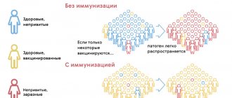
Sudden infant death syndrome and vaccinations
The start of vaccination coincides with the peak of infant mortality due to SIDS. This fact began to arouse suspicion among anti-vaccination mothers. Still would. If some consider childhood vaccination the source of all troubles and health problems, why not, out of ignorance, blame it for the deaths of babies?
But statistics and research results convince us of the opposite: vaccinated children die in their sleep less often than their unvaccinated peers. In addition, the risk of dying from cardiac or respiratory arrest during sleep during an infectious disease in unvaccinated children is much higher.
SIDS has nothing to do with routine vaccination
Prevention rules
The main danger of sudden infant death syndrome is precisely its suddenness - it is impossible to predict it. At the same time, doctors have identified measures that can act as preventative measures. For example, you should ensure that your baby sleeps on his back - today there are many devices for this that securely fix the baby and prevent him from rolling over. You will also have to exclude soft bedding sets. An excellent solution would be a hard mattress and a sleeping bag - it will serve as a blanket without completely covering the child. There should be no soft toys in the bed. Despite the fact that one of the risk factors is sleeping separately from your parents, you should still not choose to sleep together. Ideally, the baby will sleep in his own crib or cradle next to the parent's bed.
You should dress your baby correctly before going to bed - do not choose clothes that are too warm, and the temperature in the room should not be higher than 20 degrees. If there was feeding before bedtime, you must wait for the baby to burp.
Particular attention should be paid to the child in the following situations:
- Having a high temperature, especially during sleep
- Refusal to eat
- Decreased physical activity
- Presence of respiratory infection
- Baby's sleep after prolonged hysterics and crying
- Sleeping in new conditions (for example, at a party)
It’s worth being fully prepared and carefully monitoring your baby, then there is an opportunity to prevent the development of ADHD and save the baby’s life.
When is infant death syndrome a homicide?
Many child deaths have completely understandable causes. In most cases, the death of infants is caused by the intentional or unintentional careless behavior of their parents. When the autopsy and the expert commission discover violent factors, the diagnosis: “SIDS” changes to the verdict: “Murder.”
Intentional strangulation. There are cases where a baby was deliberately strangled by one of his own parents. Angry at the prolonged loud crying, the adult covered the helpless baby with a heavy pillow, cutting off the supply of oxygen.
Death due to shaking. At the moments when adults shake a child by the shoulders, trying to calm him down in this way, they do not even imagine that their baby is on the verge of death. Young children's necks are still so weak that even a few sudden, violent movements of the head can cause serious brain damage. The consequences of such shaking are often loss of consciousness, coma and death.
Suffocation in a dream. Occurs unintentionally when mother and baby sleep together. Women who take sleeping pills, tend to sleep deeply, or drink alcohol should not place their baby next to them. People say about such cases: “I slept with the child.”
A mother may accidentally cut off her baby's breathing by squeezing him in his sleep.