Ultrasound is one of the safest, most informative and accessible examination methods in modern pediatrics. If necessary, ultrasound diagnostics can be performed on the child immediately after birth. The method allows you to detect many diseases even before symptoms appear, so it is recommended to adhere to the timing of screening. If your child needs an ultrasound, we suggest you sign up at the Miracle Doctor clinic. We have the latest equipment and offer comprehensive medical services for the whole family.
Advantages of ultrasound over other examination methods
The safety of ultrasound does not involve radiation exposure to the body and has no absolute contraindications. If necessary, the examination can be carried out several times in a row in a short period of time.
Painless ultrasound is a non-invasive diagnostic method that does not require the use of painkillers. The procedure is quick and does not cause any discomfort to small patients.
Efficiency
With the help of ultrasound, you can literally examine the brain, internal organs, joint-muscular apparatus, that is, all the main structures of the body, in just half an hour. A high-tech diagnostic method allows you to detect even minor deviations from the norm.
When should an ultrasound be performed on a healthy child?
Children of the first year of life
It is advisable to carry out the first ultrasound examinations for early detection of possible pathologies and disorders of internal systems and organs at the age of one and a half to two months. The examination includes ultrasound of the kidneys, hip joints, abdominal cavity and neurosonography - ultrasound examination of the brain through the large fontanel.
Before classes in the sports section
To exclude anomalies in the development of internal organs, which serve as a contraindication for playing certain sports.
Before visiting school
It is advisable to perform an ultrasound of the child for preventive purposes and examine the kidneys, abdominal cavity and thyroid gland; Adolescent girls receive an additional ultrasound of the pelvic organs.
Ultrasound equipment allows you to examine the structure and study the condition of internal organs, detect malformations, circulatory disorders, track the appearance and development of neoplasms, etc.
The huge advantage of the diagnostic procedure is that many diseases are diagnosed at an early stage, which greatly facilitates treatment.
Indications for performing an ultrasound scan on a child
- early diagnosis of diseases of internal organs;
- assessment of the condition and development of the brain;
- diagnosis of heart diseases;
- checking the hip joints to exclude dislocations and dysplasia;
- monitoring the size of the thymus gland;
- planned examination at a certain age (1 month, six months, a year, before entering kindergarten and school).
In addition, an ultrasound scan is necessary for a child if any disease or developmental disorder is suspected. Timely diagnosis will allow you to detect pathology in the early stages and begin treatment on time.
An ultrasound of which organs should be performed on a newborn?
Ultrasound examinations are mandatory in the Russian Federation for a one-month-old baby:
- Brain. It is called neurosonography and is carried out through the large fontanel on the head. The task of diagnosis is to identify congenital developmental defects or pathological changes due to difficult childbirth.
- Abdominal and pelvic organs, including kidneys, liver and spleen, pancreas, gall and bladder, ureters.
- Hip joints to ensure the absence of dysplasia, dislocations, subluxations of the femurs.
- Hearts. Echocardiography or ultrasound of the heart of a newborn is mandatory to exclude various pathologies.
In some cases, especially with vomiting, an ultrasound scan of the pylorus is performed. This allows us to exclude hypertrophic pyloric stenosis.
Types of ultrasound that can be performed in our clinic
Screening for children in the first year of life. Ultrasound examination of the brain, heart, abdominal organs, kidneys and hip joints is among the mandatory diagnostic procedures for babies at 1 month. Next, up to a year, if indicated, the child will also have to undergo a series of examinations that will help detect the most common diseases.
Neurosonography. In terms of information content, ultrasound of the brain in children of the first year of life can be compared with CT and MRI, and the procedure has no restrictions or contraindications. It is especially important to undergo this examination for infants with birth injuries, hypoxia and other pathologies.
Doppler ultrasound of the vessels of the neck and head. The method is indicated for assessing blood supply to the brain, checking muscle tone and the functioning of large blood vessels. Doppler ultrasound is required for low body weight, birth injuries, convulsive syndrome and other pathologies.
Ultrasound of the paranasal sinuses. Ultrasonography is performed if sinusitis, sinusitis and other ENT diseases are suspected. You can perform an ultrasound of the paranasal sinuses on your child with virtually no restrictions.
Ultrasound of the thymus. This is the most important organ of the immune system, which needs to be checked if the child often suffers from colds, suffers from allergies and bronchial asthma, and also demonstrates high fatigue and low tone.
Ultrasound of the heart. EchoCG is performed for early diagnosis of diseases of the cardiovascular system. In addition, the doctor may order an examination of the child if arrhythmia, high or low blood pressure is detected, or changes are detected on the ECG.
Ultrasound of the abdominal organs. As a rule, diagnostics are prescribed to infants at 1 month for early detection of diseases of the liver, gallbladder, pancreas and spleen. Further examination is required if any symptoms of diseases of the listed organs appear. At a medical clinic in Moscow, abdominal ultrasound is performed for children of any age.
Ultrasound of the kidneys and bladder. It is recommended to undergo an examination of the urinary system if there are relatives in the family with corresponding diseases, as well as if characteristic symptoms appear (rare or frequent urination, urinary incontinence, pain in the lumbar region, etc.).
Ultrasound of the pelvic organs. Diagnostics are carried out to assess the development of reproductive organs in children of different ages. This is necessary to prevent diseases that can later cause infertility.
Ultrasound of the hip joints. The examination allows you to detect dysplasia, dislocations and other pathologies. An ultrasound of the hip joints is required for children with breech presentation, birth injuries, large body weight and other indications.
Ultrasound of blood vessels. It is recommended to undergo an examination if you complain of dizziness, regular headaches, noticeable deterioration in memory and concentration. Ultrasound will detect circulatory disorders and vascular pathologies (narrowing, blockage, stratification, etc.).
Ultrasound of lymph nodes. It is not always possible to detect enlarged lymph nodes in a child during a clinical examination. If the doctor suspects relevant pathologies (latent infections, lymphadenitis, etc.), he may prescribe sonography.
Ultrasound of soft tissues. Diagnostics makes it possible to detect neoplasms, inflammatory processes, areas of hemorrhage, foreign bodies and other pathologies in the head, neck, torso and extremities.
Preparing for an ultrasound examination
No preliminary preparation is required for the study:
- brain;
- thyroid gland;
- hearts;
- mammary glands;
- genitals in boys;
- hip joints.
Ultrasound of the abdominal organs (ultrasound in abdominis cavitatem) (liver, gall bladder, pancreas, spleen) is performed strictly on an empty stomach. For babies of the first year of life, a break in eating is allowed for about 3 hours, from 1 year - at least 6 hours. At the same time, for children who are not breastfed, 3 days before the examination it is recommended to introduce a diet that reduces the process of gas formation in the intestines.
Ultrasound of the urinary system (ultrasound de urinae ratio), as well as the pelvic organs in girls, is performed with a full bladder. To do this, you can make sure that the child does not urinate for 3-4 hours, or 40 minutes before the examination, invite him to drink from 250 to 500 ml of water (at the rate of 5-10 ml per 1 kg of weight).
Scheduled ultrasound for a child at three months
Neurosonography. Even if the first study does not reveal any pathology, ultrasound of the brain is repeated at the age of three months, because it is by this time that all the consequences of the hypoxia suffered by the child are visualized.
Ultrasound of the hip joints. At three months of age, it is recommended to re-perform an ultrasound of the hip joints; at this time, in addition to assessing the angles, attention is paid to the process of formation of ossification nuclei.
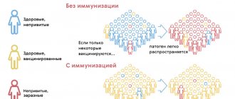
Ultrasound of the thymus. Vaccinations begin at the same time. In addition to tests, when preparing for vaccination, it is advisable to conduct an ultrasound of the thymus gland (thymus). Together with a blood test, this study provides the pediatrician with information about the state of the immune system, and can prevent a number of complications and make the vaccination campaign more effective.
How is ultrasound performed on children?
During the procedure, an adult accompanying person stays next to the child. The doctor suggests which part of the body needs to be freed from clothing, spreads a disposable diaper on the couch and helps the child take the desired position. Next, the specialist applies a water-based hypoallergenic gel to the patient’s skin and the ultrasound machine’s sensor. This transparent, colorless substance has no pungent odor and is easily removed with wipes.
After this, the doctor begins to carry out diagnostics by gently moving the sensor over the treated area.
An ultrasound scan takes from 10 to 40 minutes, depending on the type and scope of the examination. The diagnostic results are recorded in a computer program and given to visitors immediately after the procedure is completed.
Interpretation of diagnostic results
The ultrasound protocol contains all the basic information about the area being examined:
- sizes and contours of internal organs;
- arrangement of organs relative to each other;
- tissue structure;
- blood circulation features;
- assessment of the functionality of various body systems, etc.
In addition, ultrasound allows you to determine various individual developmental characteristics of a child, as well as identify pathologies that require treatment. Conclusions about the presence of diseases based on the examination protocol should be made by the attending physician.
Ultrasound of the kidneys and bladder.
One of the main diagnostic methods that allows us to identify a wide range of pathologies of the urinary system in children. An ultrasound of the kidneys will show in half an hour a painless and safe diagnosis whether any change in the structure of this organ has occurred.
Indications for the study:
- children under 1 year of age
- pain in the lower abdomen and lumbar region
- changes in urination (the child begins to urinate frequently)
- complaints of painful urination
- change in urine color
- bad urine tests
- increased body temperature for no apparent reason
- back and abdominal injuries
- defects and abnormalities of kidney development, urolithiasis in close relatives (heredity)
How to prepare?
No additional preparation is required for a kidney ultrasound.
An ultrasound of the bladder is performed on a full bladder, so children are given liquid to drink 1-2 hours before the examination (depending on age, from 0.25 to 1.5 l).
During an ultrasound of the urinary system (kidneys + bladder), the kidneys are examined first against the background of a full bladder, then against the background of an empty one.
Ultrasound of the genitals
in boys , this is one of the ultrasound diagnostic techniques that allows you to visualize the organs of the scrotum, including the testicles. The diagnosis is made with almost 99 percent probability. Inflammatory diseases and complications of injuries are also well identified. Moreover, the method is absolutely painless.
Indications for the study:
- epididymitis, orchitis, orchiepididymitis
- suspected tumor
- varicocele (varicose veins of the inguinal canal), as well as torsion of the spermatic cord
- hydrocele (dropsy)
- injuries
- non-inflammatory cysts, abscesses and other necrotic processes in the scrotum.
- cryptorchidism
How to prepare?
No preparation required.
Is ultrasound harmful for children?
It is important to know that ultrasound diagnostics is not based on penetrating radiation and does not harm the baby’s body. Ultrasound can be used both in the womb and from the first hours of a child’s life!
Why at the Perinatal Health Center?
In our center, small patients are examined using an expert-class device – “VOLUSON-E8”, which is equipped with all the necessary sensors and analyzers, printing devices and CD recording devices.
It should be noted that the result of an ultrasound examination depends not only on the class of equipment, but also on the competence of the doctor conducting the examination, on his clinical thinking, ability to use various techniques, a good spatial understanding of the anatomy of a small organism and competent interpretation of the resulting image.
At the end of the ultrasound examination, the expert doctor will give you a study protocol with ultrasound images (if necessary) and explain the detected changes. You will, of course, discuss recommendations for treatment and further tactics with your doctor.