Causes
To penetrate the scrotum during the intrauterine stage of fetal development, the testicle makes its way through the abdominal cavity and passes through the abdominal wall along the inguinal canal. In the embryonic period, the testicle is located first retroperitoneally, and then intraperitoneally. During the first three months of intrauterine life, the gonad is located in the lumbar region, on the side of the spine, adjacent to the kidney. By the end of the 3rd month of intrauterine life, the gonad is located near the internal opening of the inguinal canal. Only at the 8th month does the testicle penetrate the inguinal canal and from here descend into the scrotum. The gonad descends into the scrotum following the guide ligament - gubernaculum testit. The canal through which the testicle enters the scrotum is represented by the vaginal process of the peritoneum, which is obliterated (closed) by the time the baby is born.
The process of lowering the testicle into the scrotum occurs under the influence of several main factors:
- downward traction due to the gonad conductor - Gunter's cord;
- difference in the growth rate of the body and the growth of the spermatic cord;
- increased intra-abdominal pressure;
- bowel movements, the lower section of which, during the final descent of the testicles, is filled with meconium;
- appendage development.
The passage of the testicle through the inguinal canal largely depends on the concentration of androgens (male sex hormones) produced by the embryonic gonad. The greatest role is given to luteinizing hormone, which is actively produced by the pituitary gland of the fetus in the last trimester of pregnancy. Thus, a decrease in the concentration of male sex hormones that are produced in the fetus or a deficiency of gonadotropic hormones in the mother (it has been established that gonadotropic hormones affect the descent of the testicles by regulating the production of androgens) can significantly affect the process of testicular descent. Any mechanical obstacles along the route can also lead to its deviation.
Most common complications
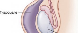
- Dropsy of the membranes of a retained testicle with cryptorchidism. Hydrocele of the testicular membranes can occur when the processus vaginalis of the peritoneum is not closed;
- Strangulated hernia;
- Testicular torsion;
- The risk of developing testicular cancer in patients with cryptorchidism is 10 times higher than in men in the general population. Of all diagnosed seminomas, 50% are detected in undescended testicles, especially often with intra-abdominal location of the testicle. In addition to seminomas, men with cryptorchidism have a high incidence of chorionepitheliomas and teratoblastomas;
- Infertility. The greatest risk of developing infertility (70%) is in patients with bilateral cryptorchidism. Right- or left-sided is not so dangerous, since one testicle with cryptorchidism retains functionality. Long-term results of surgical treatment show that infertility develops in 50-60% of patients operated on for the disease over the age of 5 years.
Types of cryptorchidism
If a newborn’s testicle has not descended, then this condition can be divided into two types according to location:
- Abdominal – it is diagnosed when the testicle has not descended and is located in the abdominal area.
- Inguinal – the testicle, when passing through the internal inguinal ring, penetrated into the groin area and remained there.
Classification of cryptorchidism
Cryptorchidism is divided into retention and ectopia.
- Retention is a condition in which one or both testicles do not descend into the scrotum, but are delayed somewhere along the way from the place of their formation (at the lower pole of the kidneys) to the external opening of the inguinal canal.
- With cryptorchidism, testicular retention can be: 1) in some place in the intraperitoneal part of the path of its descent (to the internal opening of the inguinal canal) - intraperitoneal (retention testis abdominalis); 2) in the inguinal canal at its various levels - inguinal (retention testis inguinalis);
- Ectopia is a condition in which the testicle has deviated somewhere from its path into the scrotum. With this pathology, the testicular conductor is of great importance, which is not attached to the bottom of the scrotum, as it should be normally, but directs the gonad away from the scrotum. The spermatic cord with ectopic testicle is of normal length.
There are five types of ectopia: 1) superficial ectopia - the most common type of ectopia. The testicle is located under the skin of the abdomen above the aponeurosis of the external oblique muscle; 2) penis-pubic ectopia - the gonad is located at the base of the penis and lies on the pubic bone; 3) femoral ectopia - located in Scarp’s triangle, closer to the medial surface of the thigh; 4) transverse ectopia - the testicles pass through the same inguinal canal and are located in one half of the scrotum; 5) perineal ectopia - the gonad is located in the perineal area.
A rare type of cryptorchidism is observed when the testicle and epididymis exist separately at a greater or lesser distance from each other, being connected only by a connective tissue cord.
Etiology of the phenomenon
In the second month of intrauterine development of the embryo, the formation of the sex gonads occurs. The testicles first form in the abdominal cavity, where the lower pole of the kidney is located. In the last trimester of pregnancy, they begin to descend into the inguinal canal through the internal inguinal ring. From here they move to the scrotum through the external inguinal ring.
When newborn boys' testicles descend, this indicates that the pregnancy is proceeding without complications and everything is within normal limits. If the normal migration of the gonads is suspended, then one or both testicles do not descend into the scrotum.
Why didn't a newborn's testicle descend? The reason may be several factors, they are divided into several areas:
- Mechanical. They develop under the influence of such factors: narrowness of the canal in the groin; there may be no tunnel in the scrotum; short spermatic cord of the vaginal process of the peritoneum and vessels that feed the testicle; poor development of guide ligaments, etc.
- Endocrine. For this factor there may be the following prerequisites: hormonal disruptions in the body of a pregnant woman; disruption of the normal functioning of the testicles; problems with the thyroid gland, etc.
- Genetic factor. In this case, a mutation of the GTD gene is most often observed.
The role of the scrotum for thermoregulation and spermatogenic function of the testicle
The scrotum is an important thermoregulatory structure for the gonads. Lower temperature in the scrotum is an important factor ensuring normal spermatogenesis. When the testicle is located in the inguinal canal and, especially in the abdominal cavity, its function is impaired, since under these conditions the optimal temperature, which is extremely necessary for development and mainly for normal spermatogenesis, is not provided. The temperature in the abdominal cavity and inguinal canal is higher than in the scrotum. In children, this difference reaches 2-5 degrees.
Due to temperature differences, the formation of bilateral cryptorchidism often leads to infertility.
Diagnosis of cryptorchidism
Cryptorchidism is easily diagnosed. Upon examination, you may notice the absence of one or both gonads in the scrotum and swelling in the groin when the testicle is retained in this area. It is not possible to manually lower the testicle into the scrotum. With both bilateral and unilateral cryptorchidism, the scrotum is underdeveloped to a greater or lesser extent.
Patients with proximal forms of hypospadias, micropenia, or a non-palpable testicle often require a differentiated approach to treatment. Such children need consultation with an endocrinologist, geneticist, and additional examination methods to determine the karyotype in order to exclude pathology of sex formation.
True and false cryptorchidism
If we talk about true cryptorchidism, then it is worth knowing that there has never been a testicle in the scrotum. The following factors influenced this condition:
- scarring;
- peritoneal fusion;
- testicular vessels are too short;
- a short duct for excreting the seed as part of the spermatic cord.
In this case, when performing palpation, the doctor will not be able to bring the testicle down into the scrotum.
With false cryptorchidism, the testicle can move freely from the inguinal canal to the scrotum and vice versa. This may be affected by:
- increased cremasteric reflex;
- small testicle size and wide inguinal ring.
When examining a baby, the doctor identifies a testicle in the scrotum or groin area. By palpation you can return it to its place. But next time it may rise again to the groin.
If the child is relaxed, then the cremaster relaxes, at which time the testicle moves into the scrotum. If the child is tense, cries, or is hypothermic, the testicle returns to the groin. As the baby grows, physiological atrophy of the muscle that elevates the testicle is observed, and false cryptorchidism goes away on its own.
The doctor will tell you why the newborn’s testicle did not descend; in the photo you can see the manifestation of the disease.
What is false cryptorchidism?
A false form of cryptorchidism or an increased cremasteric reflex is a normal condition in which the cremasteric muscle pulls the testicle into the inguinal canal. The formation of false cryptorchidism is observed in children under stress and freezing. Most often, an increased cremasteric reflex is observed at the age of 6-7 years, when the false form of the disease is most often detected. In this case, the gonad can be manually lowered to the bottom of the scrotum and it remains there for some time; in a warm bath, the testicle descends into the scrotum on its own, which indicates a false disease. The false form of cryptorchidism does not require treatment.
What is cryptorchidism, or undescended testicle in boys?
This condition is characterized by the absence of one or both testicles in boys. The disease can be detected immediately after the birth of the child during the first examination. When palpating the scrotum, the doctor does not find a testicle in it; it may be located in the abdominal cavity, groin area or genitals.
Cryptorchidism is a common phenomenon. According to statistics, every fourth newborn has signs of this disease, and premature babies are especially affected. In children born at term, this pathology is diagnosed only in 4% of cases.
Parents are sounding the alarm, although doctors assure that at first the worries may not be justified. In most cases, by the age of 6 months in boys, the testicle descends into the scrotum on its own. It is extremely rare that medical help and surgical intervention may be needed.
Cryptorchidism: Drug treatment
Drug treatment is carried out with chorionic gonadotropin preparations. The success rate of conservative treatment is about 20 percent. Moreover, 20% of patients experience a relapse after discontinuation of conservative treatment. The higher the gonad is initially located, the lower the effectiveness of hormonal therapy.
There are different dosage regimens and frequency of administration of human chorionic gonadotropin during treatment, but there are no significant differences in the results of using different treatment regimens. The standard regimen for administering human chorionic gonadotropin drugs is: injections 2 times a week for 4 weeks intramuscularly. If cryptorchidism is present, treatment should begin after reaching 6 months of age. Luteinizing hormone-releasing hormone (LHRH) analogues, administered in pulse mode, are also used to treat cryptorchidism. The effectiveness of this therapy does not differ from the effectiveness of treatment with human chorionic gonadotropin.
Surgical treatment of cryptorchidism (more details in the surgical treatment section)
Essentially, testicular reduction is a plastic operation of moving the organ and forming new connections and relationships (Gusynin V.O., 1937). The main goal of surgical treatment of cryptorchidism is the mobilization and lengthening of the spermatic cord with the release of the testicle, the formation of a bed for the testicle in the scrotum, its reduction and fixation in the proper place without tension on the vessels, which allows the normal development of the reduced testicle.
Clinical guidelines (European Association of Urology)
- Children with an increased cremasteric reflex (retractile testicles) do not require medical or surgical treatment, but they require regular monitoring until puberty;
- the optimal timing of treatment if cryptorchidism has developed is from 6 to 18 months of the child’s life;
- for non-palpable testicles, laparoscopy is the “gold standard”, since it has almost 100% sensitivity and specificity for identifying intra-abdominal testicles, allowing simultaneous surgical correction;
- adjuvant and non-adjuvant hormonal therapy is not standard treatment. Its use must be individualized for each patient;
- in the presence of an intra-abdominal testicle on one side and a normal contralateral testicle in children over 10 years of age, it is advisable to remove the intra-abdominal testicle due to the high risk of malignancy.
- Federal clinical guidelines “Cryptorchidism”
- Pediatric urology-andrology: Textbook. allowance. — Razin M.P., Galkin V.N., Sukhikh N.K.
- Algorithm for the treatment of children with cryptorchidism Al-Mashat N.A., Kovarsky S.L., Petrukhina Yu.V.
- Surgical treatment of cryptorchidism in children Zholumbaev A.O., Raselbaev K.U.