Adenoviral infection in children is an acute respiratory disease, which is one of the types of acute respiratory viral infections, belonging to the group of seasonal catarrhs and characterized by damage to lymphoid tissue, the connective membrane (conjunctiva) of the eyes and the mucous membranes of the respiratory tract.
The share of this infection in the general etiological structure of ARVI accounts for at least 20%. According to statistics, up to 25–30% of all registered cases of virus infection in young children between influenza epidemics are due to adenovirus infection. Babies from 6 months to 3 years of age are most susceptible to the type of virus that causes a similar inflammatory process.
In the first months of life, children have passive transplacental immunity, transmitted from the mother, so they are not susceptible to infection. Adenovirus infection in preschool children is diagnosed during epidemiological outbreaks or year-round.
It is difficult to find a child who has not suffered from such an illness: often all children have been treated for adenovirus infection at least once, or even several times.
Etiology
To date, from 32 to 57 serotypes of viruses of the Adenoviridae family are known from various sources. The causative agents of the disease in preschool children are 1, 2, 5 or 6 serological types, in adults - 3, 4, 7, 8, 14 or 21 serological types. Conjunctivitis and pharyngoconjunctival fever are caused mainly by virus serovars 3, 4 or 7.
Adenovirus variants with a diameter of 70 to 90 nm have double-stranded DNA and three antigens: A-antigen, B-antigen and C-antigen. They have increased resistance to environmental influences: they remain viable for up to two weeks under normal conditions; They tolerate drying and freezing well, are insensitive to the effects of antibiotics, but die from boiling, exposure to chlorine-containing agents and UV rays.
Treatment
The use of antibiotics is justified only when secondary bacterial flora is attached and complications develop. There are no treatment regimens aimed at the etiological factor. Therapy is symptomatic only.
At high temperatures the following are indicated:
- bed rest,
- plenty of warm drinks (fruit drinks, compotes, jelly),
- complete nutrition,
- symptomatic treatment.
It is recommended to give your child antipyretic drugs only if the temperature is above 38.5°C. For a runny nose, vasoconstrictor drops are used (to avoid addiction for no longer than 5 days) so that the child can breathe through the nose, and rinsing solutions made on the basis of sea water (Dolphin, Aqua Maris, Physiomir).
Based on the nature of the cough, the child is given the appropriate mixture/drink:
- for wet cough - expectorant medications,
- for dry skin - breastfeeding or alkaline drinking (hot milk with soda, warm alkaline mineral water).
Treatment is supplemented with inhalations and immunotropic drugs (IRS-19, Viferon, Derinat, Kipferon). Taking these medications prevents the occurrence of complications.
Pathogenesis
Routes of infection:
- Airborne. Characteristic of the early period of the disease, when the patient secretes pathogens with nasopharyngeal mucus.
- Fecal-oral. Possible in the late period, when adenoviruses are excreted in feces.
- Water. Infection of a child occurs through water, which is why another name for the disease is swimming pool disease.
The source of infection is an adult or child who has an acute infection and releases pathogens into the environment, as well as virus carriers - people with an erased form of the disease or who are asymptomatic.
After treatment, type-specific immunity is formed, so re-infection is possible, but with other serological types of adenovirus.
The adenovirus enters the body through the conjunctiva, intestinal mucosa or respiratory system. Having reached the lymphoid formations of the intestine, epithelial cells, lymph nodes, the pathogens begin their reproduction, synthesizing viral DNA in the nuclei of the affected cells and leading to the cessation of their division and death. After 16–20 hours, mature particles of new adenoviruses are formed.
The incubation period ends with the development of viremia, caused by the release of viruses from dead cells, entering the bloodstream and spreading throughout the body. As a result, the mucous membranes of the pharynx, nose, thin connective membrane of the eyes, and tonsils are affected. Inflammation leads to swelling of the mucous membranes, redness, pain, and the release of copious serous exudate.
Once in the lungs and bronchi, adenoviruses actively multiply in the mucous membranes of the alveoli and the bronchi themselves, provoking the formation of necrotizing bronchitis or viral pneumonia. Inflammatory processes of the bronchopulmonary system are caused by a combined infection: bacteria are often associated with viruses. The infection can affect the intestines, spleen, kidneys, and liver. In rare cases, the brain is affected and swelling develops, leading to the death of a small patient.
Publications in the media
Diseases caused by viruses of the Adenoviridae family are characterized by high body temperature, inflammation of the mucous membranes of the respiratory tract and eyes, as well as hyperplasia of the submucosa and regional lymph nodes. Adenovirus infection is highly contagious. The most commonly observed varieties • ARVI with severe fever (mostly affecting children) • ARVI in adults • Viral pneumonia • Acute adenoviral tonsillitis (develops in children, especially in the summer after swimming) • Acute follicular conjunctivitis • Epidemic keratoconjunctivitis (mainly in adults) • Intestinal infections ( enteritis), often complicated by mesenteric adenitis and invaginations. Frequency. A very commonly reported infection, it accounts for 2–5% of all respiratory tract infections. More often observed in newborns and children.
Etiology • Pathogens - DNA-containing viruses of the genus Mastadenovirus, 60–90 nm in size; about 80 serotypes (serovar) are known • The main causative agents of human lesions •• Infections of the lower respiratory tract (bronchiolitis, pneumonia) - serotypes 1, 2, 3, 5, 6, 7, 21 •• Pharyngoconjunctivitis - serotypes 1, 2, 3, 4, 6, 7, 14 •• ARVI - serotypes 3, 4, 7 •• Gastroenteritis - serotypes 2, 3, 5, 40, 41 •• Conjunctivitis - serotypes 2, 3, 5, 7, 8, 19, 21 • • Epidemic keratoconjunctivitis - serotypes 8, 19, 37 •• Hemorrhagic cystitis - serotypes 11, 21 •• Meningoencephalitis - serotypes 2, 6, 7, 12, 32 •• Disseminated lesions - serotypes 5, 34, 35, 39 •• Cervicitis and urethritis - serotype 37 •• Lesions associated with celiac disease - serotype 12.
Epidemiology. Human adenoviral infections are widespread and account for 5–10% of all viral diseases; most of the lesions occur in childhood (about 75%); while 35–40% are registered in children under 5 years of age, the rest - under the age of 14 years. The main routes of transmission are airborne and contact.
Pathomorphology • Varies among different serotypes, in severe pneumonia a large number of intranuclear inclusions can be identified • Bronchiolitis obliterans is possible.
Clinical forms • Qatar of the respiratory tract • Pharyngoconjunctival fever • Membranous conjunctivitis • Tonsillopharyngitis (adenoviral tonsillitis) • Intestinal form (viral diarrhea, gastroenteritis) • Mesadenitis • Viral pneumonia.
Clinical picture
• Qatar of the respiratory tract is the most common form, characterized by pronounced catarrhal changes in the respiratory tract (rhinopharyngitis, laryngitis, tracheitis, bronchitis) with moderate manifestations of general intoxication.
• Pharyngoconjunctival fever. It is characterized by a long-term temperature reaction (up to 2 weeks in the absence of intercurrent diseases and complications) and symptoms of pharyngitis: sore throat, rare cough, bright hyperemia of the tissues of the pharynx with large “grain” of the follicles of the mucous membrane; the respiratory tract is involved in the process to a small extent.
• Membranous conjunctivitis. Usually seen in adults and older children. Unilateral (less often bilateral) conjunctivitis with a fibrinous film on the surface of the mucous membrane (usually the lower eyelid), accompanied by severe swelling of the surrounding tissues, pain, hyperemia and injection of the vessels of the conjunctiva of the eye, fever. The respiratory tract is not involved in the process. This clinical form is characteristic only of adenoviral infection (an etiologically detailed diagnosis can be made without laboratory confirmation).
• Tonsillopharyngitis. Typical for preschool children. It is characterized by the development of inflammatory changes in the tissues of the pharynx and palatine tonsils with the formation of sore throat (catarrhal, fibrinous, less often [with the addition of a bacterial infection] purulent).
• Intestinal form (gastroenteritis, viral diarrhea). It is characterized by the development of moderately severe gastroenteritis, manifested by nausea, vomiting, loose stools without pathological impurities. Body temperature is subfebrile. Possible simultaneous involvement of the respiratory system in the process (catarrhal nasopharyngitis or laryngotracheitis).
• Mesadenitis. Characterized by the development of abdominal syndrome with abdominal pain and temperature reaction. Sometimes it is possible to palpate enlarged mesenteric lymph nodes. In rare cases, symptoms of an “acute abdomen” occur, simulating an attack of acute appendicitis.
• Viral pneumonia. An independent clinical form with typical symptoms of acute pneumonia (intoxication, signs of pulmonary heart failure). The exudative component is pronounced (multiple rales of various sizes throughout all pulmonary fields, cough with copious sputum, etc.). X-rays reveal widespread inflammatory changes in the lungs. This clinical form does not exclude possible bacterial superinfection, therefore antimicrobial therapy is necessary.
Research methods • Isolation of the pathogen by inoculation into cultures of human epithelial cells; the material being studied is discharge from the nose, pharynx, conjunctiva, feces, etc. • Detection of Ag viruses in cells by immunofluorescence microscopy, as well as staging of RSC, hemagglutination inhibition reaction (HRI) and reaction to neutralize the cytopathic effect in cell culture • X-ray diagnostics: bronchopneumonia in severe respiratory infection • In severe or atypical cases, a biopsy (of the lung or other tissue).
TREATMENT
Regime • Outpatient, with the exception of seriously ill young children, patients with epidemic keratoconjunctivitis and young children with severe pneumonia • Bed rest for the period of elevated body temperature.
Drug therapy - symptomatic treatment • If necessary - paracetamol 0.2-0.4 g per dose 2-3 times / day (10-15 mg / kg / day). It is not recommended to take acetylsalicylic acid • Antitussives and expectorants • HA (topically) - for conjunctivitis (after consultation with an ophthalmologist).
Course and prognosis • The disease goes away on its own with virtually no complications • Severe course and death are possible among children and patients with impaired immunity.
Prevention • Live oral vaccines against adenovirus types 4 and 7, coated with a capsule that protects from digestion in the intestine, reduce the incidence of acute respiratory viral infections • Frequent hand washing is mandatory for staff of various institutions and family members of the patient.
ICD-10 • A08.2 Adenoviral enteritis • A85.1+ Adenoviral encephalitis (G05.1*) • A87.1+ Adenoviral meningitis (G02.0*) • B30.0+ Keratoconjunctivitis caused by adenovirus (H19.2*) • B30.1+ Conjunctivitis caused by adenovirus (H13.1*) • B34.0 Adenoviral infection, unspecified • J12.0 Adenoviral pneumonia • J02.8 Acute pharyngitis caused by other specified pathogens • J02.9 Acute pharyngitis, unspecified • J03.9 Acute tonsillitis, unspecified.
Application. Mesadenitis (acute mesenteric lymphadenitis) is inflammation of the lymph nodes located in the mesentery of the small intestine; observed mainly in children and adolescents; symptoms resemble acute appendicitis. Etiology: viral infection, as well as bacteria of the genus Yersinia. Clinical picture : abdominal pain (can be severe), nausea, vomiting and fever; some patients experience additional signs of viral infection (for example, pharyngitis and myalgia). The diagnosis is usually made by laparotomy for suspected appendicitis. Treatment: when Yersinia is isolated, antibiotics are prescribed. ICD-10. I88.0 Nonspecific mesenteric mesadenitis.
Symptoms of adenovirus infection in children
Signs of the disease appear sequentially after an incubation period lasting up to 12 days, usually no more than a week. Adenoviral infection can manifest itself as one of the following syndromes:
- Catarrh of the mucous membranes of the respiratory organs.
- Keratoconjunctivitis and acute conjunctivitis.
- Pharyngoconjunctival fever.
- Mesadenitis, or mesenteric lymphadenitis.
- Diarrheal syndrome.
Catarrh of the mucous membranes of the respiratory tract is the most common type of infection in children. Manifests itself in the form of laryngotracheobronchitis, tonsillopharyngitis, rhinopharyngitis. It is characterized by an acute onset with a rise in body temperature to 39.8–40 0C and moderate or mild symptoms of intoxication: loss of appetite, lethargy, moodiness, headache, joint and muscle pain.
Catarrhal changes appear along with fever. Difficulty in nasal breathing is accompanied by swelling of the mucous membrane with discharge from the passages of first serous, then mucopurulent exudate. The pharyngeal mucosa is hyperemic and swollen, whitish pinpoint plaque forms on the tonsils, and the cervical and submandibular lymph nodes become enlarged. Inflammation of the vocal cords is expressed by hoarseness of voice up to its temporary loss (transient aphonia), a dry barking cough, the development of shortness of breath and laryngospasm.
Pharyngoconjunctival fever occurs against the background of inflammation of the tonsils, mucous membrane of the pharynx and eyes. Be protracted, sometimes wavy in nature. Often, a high or elevated temperature does not subside for 1–2 weeks. Regional lymph nodes are enlarged and easily palpable. Sometimes the child has an enlarged spleen or liver (moderate splenomegaly or hepatomegaly).
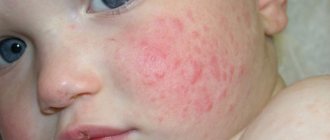
Keratoconjunctivitis and acute conjunctivitis are caused by inflammation of the conjunctiva of the eyes. First one eye is involved in the process, then the inflammation spreads to the second. The child is worried about lacrimation, pain, pain in the eyes, and a sensation of a foreign body. He begins to avoid bright light.
Upon examination, a pediatric ophthalmologist detects swelling and moderate redness of the eyelids, granularity and hyperemia of the conjunctiva, and sometimes the appearance of a whitish-grayish film on it. Conjunctivitis can be catarrhal, membranous or follicular. At the beginning of the second week of the disease, the development of keratitis, characterized by corneal syndrome, is possible.
Mesadenitis with inflammation of the mesenteric lymph nodes of a viral nature is manifested by paroxysmal pain in the navel area or in the right lower abdomen, reminiscent of attacks of acute appendicitis. Accompanied by fever and vomiting.
Diarrheal syndrome in children is an independent manifestation of adenoviral infection or one of the signs of mesenteric lymphadenitis or catarrh. It is more often observed in children under one year of age with an intestinal form of infection. At the height of the disease, the frequency of bowel movements reaches 7-8 times. Mucus without blood is found in the stool.
Forms of infection:
- light;
- moderate severity;
- heavy;
- complicated;
- not complicated.
In severe cases of the disease, parenchymal organs are affected, and severe adenoviral pneumonia often develops, complicated by severe respiratory failure.
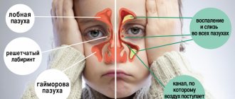
Other complications that develop with the addition of a secondary bacterial infection include the development of focal serous-desquamative pneumonia, otitis media, and sinusitis.
Etiology and pathogenesis
Symptoms of adenovirus infection in children arise due to infection with the pathogen of the same name. It is worth noting that adenoviruses are resistant to exposure to external conditions due to the fact that their DNA has something like a capsule. Thanks to it, they can exist for about two weeks at room temperature and are not afraid of treatment with ether and solutions with an acidic pH. However, they can be destroyed by treatment with ultraviolet light or chlorine. They are also afraid of boiling, but at low temperatures down to −40 °C they can live for more than two months.
The source is an infected child, who remains dangerous to others for 3 to 4 weeks from the moment of infection. Virus transmission occurs:
- by airborne droplets - during communication, kissing, sneezing;
- by nutritional means - in case of non-compliance with personal hygiene, through hands, objects, toys;
- water - when swimming in a pool with a carrier of the infection.
The incubation period can last from 2 to 12 days. Isolation of the pathogen often occurs even before the symptoms of adenoviral infection appear.
Diagnostics
Diagnostic criteria that serve as a basis for suspecting adenoviral infection:
- fever;
- polyadenitis;
- catarrh of the respiratory tract;
- signs of conjunctivitis;
- hyperplasia of pharyngeal lymphoid tissue;
- sequence of symptoms.
Positive laboratory results confirm that a disease caused by an adenovirus is developing in the body:
- immune electron microscopy (IEM);
- immunofluorescence reactions (RIF);
- enzyme immunoassay (ELISA);
- analysis for complement fixation reaction (CFR);
- determination of hemagglutination inhibition reaction (HAI);
- bacteriological culture of a scraping or smear from the conjunctiva;
- examination of a swab from the nose and throat for microflora.
Differential diagnosis of various forms of this disease is carried out with infectious mononucleosis, influenza, other types of acute respiratory viral infections, yersiniosis, mycoplasma respiratory infection, diphtheria of the eyes and pharynx.
Treatment of adenovirus in adults
Most adults are not at risk of contracting this virus. However, elderly patients and people with reduced immunity, as well as pregnant women, are quite susceptible to this disease. Since the virus persists for quite a long time on various surfaces indoors, the infection is often localized in nursing homes, boarding schools, military schools, barracks, correctional institutions, and sometimes in schools and kindergartens.
Treatment of adult patients is also comprehensive, that is, it includes both symptomatic and antiviral therapy. Treatment is always prescribed by a doctor, taking into account concomitant and chronic diseases.
According to studies, the results of which were published in the article by Yu.A. Klimova and co-authors “Immunotherapy of patients with adenovirus infection”, the inclusion of the drug VIFERON in complex therapy for this disease contributes4:
- reducing the duration of cough by 6.6 days (for sore throat);
- reduction in the number of days of hospitalization by 6.4 days (for pneumonia);
- reducing the duration of cough by 4.2 days (for bronchitis).
Treatment of adenovirus infection in children
Hospitalization is required only for children with a severe course of the disease and serious complications. More often, treatment is carried out at home. A pediatrician called to your home helps you choose a therapy. Sometimes consultation with an infectious disease specialist, otolaryngologist or ophthalmologist is required.
Recovery is facilitated by bed rest, which is mandatory throughout the entire period of fever, plus the next 2-3 days after the temperature has returned to normal. The child is recommended to have a fortified diet with sufficient amounts of protein, as well as frequent drinking. Liquids in the form of jelly, fruit drink, dried fruit compotes, hot milk, herbal decoctions or infusions help remove toxins from the body - waste products of adenoviruses.
General etiotropic treatment is carried out with antiviral drugs (children's anaferon, arbidol, kagocel, ribavirin), taken according to the regimen. It is possible that the doctor may prescribe desensitizing agents (fenkarol, suprastin, tavegil) and one of the vitamin-mineral complexes (alphabet, multi-tabs, etc.) in an age-specific dosage.
Syndromic therapy consists of antipyretic (children's Panadol), mucolytic (lazolvan, broncholitin or ACC) drugs. Aggravation of adenoviral infection by bacterial complications requires the prescription of antibiotics.
Local treatment consists of intranasal application of oxaline ointment or cycloferon liniment, instillation of interferon into the nasal passages, inhalation with herbal infusions or soda solution after a decrease in temperature.
If the conjunctiva is affected, apply antiviral eye ointment, for example, acyclovir, behind the eyelid; instillation of eye drops, sodium sulfacyl solution or deoxyribonuclease, for example, into the conjunctival sac.
To reduce febrile temperature, wiping with an alcohol or vinegar solution is effective. The elbow and knee bends, the inner surfaces of the arms and thighs, as well as the sides of the neck are treated. This helps to avoid the use of antipyretic drugs or significantly reduce their frequency of administration and dose.
The prognosis for a disease that proceeds without complications is favorable. The child recovers in 10–14 days, with a mild form sooner.
Mortality among young children is observed in severe cases of the disease, aggravated by serious complications of a bacterial nature.
Complications that adenovirus can cause in children
At risk, the most vulnerable to the disease are children aged 6 months to 3 years, when their immune system is still in its formation stage. If the baby is bottle-fed and not breastfed, then he can also be infected with this infection, since he does not receive immune protection from his mother’s milk. Like other respiratory diseases, adenovirus infection is seasonal. The greatest number of diseases occurs in the autumn-winter period.
It is believed that the average duration of the disease in children is one week, provided that the course is uncomplicated. With a prolonged course, symptoms may appear over 2-3 weeks. It must be said that the symptoms of conjunctival irritation disappear earlier than inflammation in the nasopharynx and upper respiratory tract, which can last up to three weeks. Complications2 that develop against the background of this disease are usually caused by the active proliferation of pathogenic microflora, since the patient’s immunity is weakened. The bacterial infection primarily affects the respiratory system, which often results in bronchitis and pneumonia (adenoviral pneumonia).
If an adenovirus in children infects the lymph nodes of the peritoneum (mesentery) located in the abdominal cavity, the development of appendicitis is possible3.
Other possible complications include tonsillitis, sinusitis and exacerbation of chronic diseases.
Infants have a high risk of middle ear inflammation (otitis media).
Prevention
There is no specific prevention, just like vaccination. The remaining measures are suitable for preventing any type of viral infection:
- hardening of the body;
- seasonal course intake of vitamin and mineral complexes;
- during epidemics, taking immunomodulators, such as Immunal.
- isolation of sick children from healthy ones;
- in case of contact with a sick child, preventive use of children's anaferon or other antiviral agent.
- during illness, regular (2 times a day) wet cleaning of the room and daily ventilation; in the hospital, quartzing the room.
Laboratory diagnostics
One of the research methods is laboratory diagnosis of adenoviral infection. For this purpose, blood and urine tests are taken from the patient. There are no significant changes in the hemogram; the number of leukocytes remains within normal limits. In the initial period of the disease, lymphopenia can be observed, as well as slight leukocytosis with neutrophilia. However, the blood picture does not play a significant role in diagnosis, since it is not specific.
A urine test can show the presence of an inflammatory process in the body, but in most cases it remains within normal limits. Just like a blood test, it is not specific.