Age-related features of fractures
The physiological state of bone tissue and periosteum significantly affects the nature of possible damage. In young people, the bone tissue is elastic, the periosteum is dense; with age, the bone tissue of our body gradually loses these qualities.
Features of fractures in young children
The most common occurrences are depressed or. So-called “greenstick” fractures.
Depressed fractures are characterized by arc-shaped smooth depressions in the bone relative to the place of application of force, which is caused by compression of the “strength beams” of bone tissue.
A subperiosteal greenstick occurs in long bones due to the dense and elastic periosteum.
Osteoepiphysiolysis and osteoapothesiolysis are also common types of fractures that can only be found in children. They arise at the border of the bone growth zone. Damage to the growth zone itself in the case of epiphysiolysis further leads to disruption of its function with characteristic shortening of the bone. Further deformation of the bone, due to damage to the growth zone, can be as significant as the growth zone was damaged at an early age and how active it is.
Elderly age
Elderly people especially often suffer from bone fractures due to the loss of their strength due to age-related changes. Due to the loss of organic substances, the elasticity of the bones is disrupted and significantly weakened. They become brittle and grow together rather poorly. Fractures in older people most often result in a large number of sharp individual bone fragments. Weakened function of the periosteum and damage to it by sharp edges of fragments have a bad effect on further fusion.
Prevention of recurrent fractures in children and adolescents with low bone mineral density
Currently, the whole world is wary of the manifold increase in the incidence of skeletal fractures; thus, by 2050, the number of femoral fractures is expected to increase 4 times. There is a significant increase in the frequency of recurrent fractures, which are now healing worse, especially in children. Therefore, urgent attempts to reduce the predicted “epidemic of fractures” should begin now and in childhood [1]. In modern domestic and foreign literature on this problem, we have come across a few publications that indicate the lack of effective methods for treating and preventing fractures [2, 3]. Russia does not have its own osteoprotectors; even strategic vitamin D is bought abroad. Billions of dollars are being spent on this.
Currently, all over the world they are trying to solve the problem of osteoporosis and bone fractures by prescribing various osteoprotectors in order to increase bone mineral density. We have established that this is the wrong way. It is impossible to increase bone mineral density and reduce the incidence of bone fractures in this way, especially with calcium supplements, since children and adolescents have extremely diverse pathogenetic disorders of bone-mineral metabolism. As a result of an imbalance in the system that regulates calcium metabolism, an imbalance of osteoclasts and osteoblasts, the calcium that we prescribed to the patient is deposited not in the bones, but in other organs and tissues with the risk of calcification of soft tissues, blood vessels with the risk of hypermineralization, and in the future (in adults ) increasing the risk of strokes and heart attacks. Thus, the prescription of most modern osteoprotectors fails to solve the problem of osteoporosis and bone fractures, especially in the long term [4, 5].
Our work is aimed at solving the above problems. Its novelty is that by changing the diagnostic approach with an emphasis on identifying the main pathogenetic causes of osteoporosis (fracture), the therapeutic approach to osteoporosis is changing. We have proposed a new method for determining the therapeutic effectiveness of an osteoprotector, a new method for the treatment of osteoporosis, including the closure of cavity formations (patent N 2466732). New means have been created to treat fractures and reduce consolidation time; one of them will be discussed in this article.
The purpose of this work was to study the etiopathogenetic structure of bone fractures in children and adolescents and, based on this, to develop a personalized approach to the treatment and prevention of bone fractures. To study the effectiveness of the treatment of osteoporosis in children with the new drug Osteo-vit D3.
Materials and research methods
From 2007 to May 2013, at the Penza CSCH, 37 children and adolescents with low mineral density of bone tissue (25 boys, 12 girls), with fractures of the long tubular bones of the distal extremities, who made up the main contingent, were examined. Fractures of the upper extremities predominated - 28 cases, lower extremities - 9. The study included only children and adolescents with bone fractures aged 10 to 18 years. With bone mineral density (BMD) less than –2.0 standard deviations (SD), with bone fractures, and the presence of cavity formations in the trabecular bones. The diagnosis of “osteoporosis” was made on a Z-scale below –2 SD, taking into account the content of bone minerals (BMC), bone mineral density (BMD), the presence of the main criterion - fracture(s), height and weight indicators using the tables of L. A. Sheplyagina ( 2013). The content of calcium, inorganic phosphorus, 25(OH)-vitamin D, parathyroid hormone, thyroid-stimulating hormone, and thyroid hormones T3 and T4 in the blood serum was determined.
Children with secondary osteoporosis due to various systemic diseases, osteogenesis imperfecta, tubulopathies, hyperparathyroidism, and treatment with glucocorticoids were excluded from the study.
The examination of patients included anamnesis, examination, general clinical, laboratory and biochemical studies: calcium, inorganic phosphorus, alkaline phosphatase, 25(OH)-vitamin D in blood serum, and x-ray methods. Diagnosis of vitamin D deficiency was carried out by the content of 25(OH)-vitamin D in the blood less than 20 ng/ml. BMD was determined using the X-ray absorption method before and after treatment in 9–10 months using the Osteometer DTX-100 apparatus. In the process of an in-depth examination of the main contingent of children, three groups of causes of repeated fractures were identified: 11 children with vitamin D deficiency (group 1), 7 children with the presence of bone cavities in the trabecular sections (group 2), the rest - 19 children with low bone mineral density (group 3).
A new approach to solving the problem was the use of drone solution (drone jelly) as a fortifier (amplifier) of osteoprotector (vitamin D). Drone milk contains a high content of protein (up to 41%), amino acids (up to 37%), including essential (about 38% of the total amino acids), nucleic acids (1.1–1.3%), enzymes (lipases , proteases, phosphatases, urease, dehydrogenase, amylase, etc.), phospholipids (1.1–1.5%), carbohydrates (fructose, glucose, etc.), a complex of lipid fraction substances, micro- and macroelements, vitamins A, D, E, group B and other biologically active components, so it is not for nothing that drone brood is called a “bank of biologically active substances”. This composition of drone brood (jelly) has therapeutic and prophylactic effects: antioxidant, immunomodulatory, antitumor, osteoprotective [6–9]. Beekeepers themselves do not have severe forms of osteoporosis. Based on this, we have developed Osteo-Vit D3 (a dietary supplement), containing 500 IU of vitamin D3 and 100 mg of drone brood in 1 tablet as a corrector of metabolic processes.
The effectiveness of Osteovit D3 in the treatment of osteoporosis was determined by the dynamics of the clinical picture of the disease, BMD and closure of cavity formations in the bones. Children of the 1st and 2nd groups received Osteo-vit D3 in three-month courses three times a year, 1 tablet 2 times a day with monthly breaks. Children of group 3, depending on the method of treatment, were divided into two comparable subgroups according to age and severity of the disease: subgroup A - 10 patients received Osteo-vit D3 orally, 1 tablet 2 times a day (morning and at night, i.e. 200 mg brood/day, 1000 IU vitamin D3/day) in three-month courses three times a year. Subgroup B (control) - 9 patients received Calcium-D3 Nycomed, containing an adequate amount of vitamin D and 1.0 g of calcium carbonate per day. All children underwent osteometry before and after treatment 9–10 months later.
Research results
Children of group 1 (with vitamin D deficiency) treated with Osteovit D3 had positive dynamics in the main clinical, instrumental and biochemical parameters. The frequency of pain and its severity decreased by 3 times after 1–2 months. There was a faster formation of callus, by 7–10 days, compared to children who did not receive Osteo-vit D3. The level of 25(OH)-vitamin D after 6 months of treatment increased from the “deficiency level” to normal.
In the 2nd group of patients receiving Osteo-vit D3, positive dynamics were also noted in the form of disappearance and reduction of pain, accelerated formation of callus, increased mineral density, closure of cavities or reduction in their size in 2/3 of patients (Fig. 1).
A third of patients in group 2 did not obtain a positive result. These children had pronounced disturbances in hormonal status (deficiency of sex hormones, dysfunction of the thyroid and parathyroid glands).
A comparative analysis of the effectiveness of therapy with Osteovita D3 and Calcium-D3 Nycomed in group 3 showed that the domestic drug was not inferior to the recognized foreign drug and even surpassed it in increasing BMD and closing cavitary formations.
The discussion of the results
The structure of reduced bone mineral density and causes of bone fractures in children referred for examination to the Osteoporosis Center turned out to be heterogeneous and pediatric determined. It has been established that, first of all, in children with repeated fractures, if such children are diagnosed with osteoporosis, it is necessary to examine them for vitamin D deficiency - the main cause of reduced mineral density in children. The findings are consistent with the existing literature on the widespread prevalence of vitamin D deficiency (MF Holick, 2007).
If there are cavity formations in the trabecular bones in children with fractures, it is necessary to examine the hormonal status and exclude osteopathies of endocrine origin (dysfunction of the thyroid gland is well compensated by the use of thyroid-vit (cinquefoil white), the deficiency of sex hormones is compensated by andro-vit (drone brood homogenate). This is will increase the effectiveness of treatment and is important for the prevention of recurrent fractures. Diseases of the gastrointestinal tract (peptic ulcer of the stomach and duodenum, chronic gastroduodenitis, malabsorption) can cause reduced mineral density and bone fractures. With these diseases, calcium absorption in these parts of the intestine is reduced (R. T. Galeeva, 2009) Thus, taking into account the data obtained, we can with good reason believe that bone fractures in children against the background of low mineralization are caused by various pediatric diseases and without taking these factors into account, it is impossible to prevent recurrent fractures, especially in follow-up.
In addition, other important risk factors for bone fractures in children and adolescents were identified: 75% of children (28 out of 37) received little or no milk. 25% (9 children), although they received enough dairy products, did not receive vitamin D supplements. Therefore, milk, although an excellent source of calcium, lacks protective effects on the bone skeleton (due to ineffective absorption of Ca without vitamin D). In this regard, it is important to establish the production of D-vitaminized milk.
Osteovit D3 was used in the treatment of osteoporosis in all studied groups of patients with positive results. The drug significantly improves the absorption of calcium from dairy products and thereby increases bone mineral density. In the follow-up study of children who received three courses of treatment with Osteo-vit D3, not a single case of recurrent fractures was noted. This can be explained by the fact that it reduces the frequency of falls by improving bone mineralization and strengthening the ligamentous and muscular apparatus. Further follow-up studies are needed.
conclusions
- The etiopathogenetic structure of low bone mineral density in children with fractures of long tubular bones is heterogeneous and pediatrically determined by various factors: D-deficiency, diseases of the gastrointestinal tract, insufficient consumption of dairy products without vitamin D, dysfunctions in the endocrine system and other factors.
- The introduction of Osteovit D3 for the prevention and treatment of osteoporosis in children and adolescents will significantly reduce the incidence of bone fractures, the incidence of vitamin D deficiency, and reduce dependence on the import of expensive foreign osteoprotectors, which will provide a significant economic and social effect.
- When studying the comparative effectiveness of Osteo-vit D3 with Calcium-D3 Nycomed, the domestic drug was not inferior to the foreign drug and even surpassed it in closing and reducing cavity formations, and the rate of consolidation of fractured bones for 7–9 days.
Literature
- Shilin D. E. Modern strategy for overcoming calcium and vitamin D deficiency in children and adolescents from the standpoint of preventing osteopenia and fractures // Questions of practical pediatrics. 2006. T. 1. No. 2. P. 50–56.
- Holick MF Vitamin D deficiency // N Engl J Med. 2007, 357: 266–281.
- Cooper S., Dennison E. M., Leufkens H. G. M., Bishop N., van Staa T. P. Epidemiology of Childhood Fractures in Britain: A Study Using the General Practice Research Database // J. Bone Miner. Res. 2004. Vol. 19. No. 12. P. 1976–1981.
- Strukov V.I., Galeeva R.T. et al. Current problems of osteoporosis. Rostra, 2009. P. 341.
- Strukov V.I., Elistratov D.G. et al. Known and new technologies in the treatment and prevention of osteoporosis. Penza, 2012.
- Belyaev V. A, Safonovskaya E. V. Adaptogenic properties of a drug based on drone brood // Beekeeping. 2009. No. 6. P. 52–53.
- Budnikova N.V. Biologically active compounds in drone brood // Beekeeping. 2009. No. 6. P. 52.
- Dubtsova E. A. Clinical and experimental substantiation of the use of beekeeping products in the complex therapy of certain diseases of the digestive system. Abstract of dissertation. Doctor of Medical Sciences M.: Central Research Institute of Gastr., 2009. P. 38.
- Krivtsov N.I. et al. Theory and means of apitherapy. M., 2007.
V. I. Strukov*, 1, Doctor of Medical Sciences, Professor D. G. Elistratov** Yu. G. Shcherbakova* A. T. Kuptsova* R. T. Galeeva*, Candidate of Medical Sciences L. G. Radchenko*, Candidate of Medical Sciences L. A. Burmistrova**, Candidate of Biological Sciences
* GOU DPO PIUV, Penza ** Federal State Budgetary Institution Research Institute of Beekeeping, Rybnoye
1 Contact information
Local features of traumatic bone injuries
Today we will look at the most common injuries to the skeleton of the chest, shoulder girdle and limbs.
Rib fractures
Occurs as a result of chest injury due to direct or indirect exposure to excessive force. The middle, most convex and protruding ribs (from the 5th to the 8th) are most often damaged. Paradoxically, multiple injuries are more common than isolated ones.
Fractures of the bony parts of the ribs cannot always be immediately identified using classical radiography; in this case, the most informative would be the use of spiral computed tomography.
Rib fractures can also cause additional damage to the surrounding soft tissue: bone fragments can injure: pleura, lung tissue, diaphragm, and even the liver and spleen. In turn, damage to the pleura can lead to an additional complication: the development of pneumo or hydrothorax, possibly both.
Also, rib fractures can be combined with fractures of the clavicle, scapula, and humerus.
Sternum
It breaks extremely rarely, most often due to direct force. Usually breaks in the body area (slightly below the upper section).
In the next article we will look in detail at the most common fractures of the clavicle, scapula, humerus, and limbs in general.
OSTEO-VIT. Vitamins for bone health
Osteoporosis (Greek osteon - bone and poros - time) is a multifactorial metabolic disease of the skeletal system, characterized by a decrease in bone mass per unit volume, a violation of bone microarchitecture, leading to excessive fragility of bones and a high risk of fractures.
Relevance. Osteoporosis (OP) is a pressing problem, since the damage from it is measured in billions of dollars, hundreds of thousands of sick and disabled people. In terms of prevalence, AP ranks fourth after cardiovascular pathology, diabetes mellitus and cancer. The number of osteoporotic fractures in the world is progressively increasing. The relevance of this problem is due not only to the increase in fractures, but also to the social and economic consequences. Thus, the cost of treating osteoporosis, excluding the cost of long-term home care in Europe in 2000 amounted to 31.7 billion euros, and by 2050 it will increase to 76.7 billion euros. The progressive increase in the number of patients with AP is currently due not only to the aging of the population, but also to the rejuvenation of the disease. Thus, osteoporosis has already acquired the character of a silent epidemic [1].
For a long time, AP was considered only a disease of older people, which was associated with bone loss. Now the view on this disease is changing. It has been proven that the origins of OP lie in childhood, which is characterized by an intense increase in bone mass. Interest in the problem of osteoporosis in children is associated with the work of Dent CE, after he reported in 1973 that senile osteoporosis is a pediatric disease [2]. Later, a large number of works were devoted to the problem of osteoporosis in children [1, 3, 4, 5, 6, 7], which showed that this disease in children occurs much more often than it is diagnosed. Thus, according to the Children's Health Center of the Russian Academy of Medical Sciences, when densitometry was carried out in 1000 children aged 7-15 years, a decrease in bone mineral density (BMD) was found in 40% of those examined. In severe osteoporosis, bone mass can decrease by up to 50% of the age norm. As a result, fractures easily occur without trauma or after minor trauma.
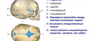
As is known, the formation of peak bone mass in children is a genetically programmed process. The final growth program consists of achieving programmed not only linear body dimensions, but also BMD, morphofunctional, anthropometric and psychometric indicators. Their close relationship ensures the harmonious development of the growing organism. However, there are many endogenous and exogenous factors that can cause deviations from the genetic program of bone tissue development in children, both before and after birth. Under these conditions, various osteopathies can form. Table 1, based on an analysis of literature data and our own research, presents the main reasons contributing to the development of osteoporosis in children and adults.
Table 1
The main reasons contributing to the development of osteoporosis (osteopenia) in children
(V.I. Strukov, 2009):
Intrauterine
- Hypoxic - ischemic lesions of the fetus.
- Disorders of calcium transport in the “mother-placenta-fetus” system.
- Pregnancy against the background of chronic diseases of the mother (pathology of the kidneys, endocrine system, gastrointestinal tract, etc.).
- Pregnancy due to maternal osteopenia.
- Bad habits of the mother (smoking, alcohol, drugs).
- Social factor (poor housing conditions, low income, etc.).
- Occupational hazards in the mother.
After birth (early age)
- Artificial feeding.
- Premature babies.
- Children from multiple pregnancies.
- Hypothyroidism (even transient).
- Poor absorption of fat-soluble vitamins in the intestines (premature babies).
- Insufficient bile production.
- Hypovitaminosis D.
- UFO shortage.
- Polyhypovitaminosis.
In older children and adolescents:
- Lack of health protection for healthy people;
- Poor nutrition, deficiency of protein, dairy products;
- Deficiency of ultraviolet radiation, vitamin D;
- Low calcium in the diet;
- Low physical activity;
- Diseases of the gastrointestinal tract, kidneys, liver, rheumatic, etc.
- Endocrine diseases, amenorrhea, etc.;
- Bad habits;
- Industrial toxicants, radionucleins.
Iatrogenic osteopenias:
- Steroid hormones for systemic use;
- Thyroid hormones;
- Anticonvulsants, phenobarbital, etc.;
- Heparin (long-term therapy for 3 months or more);
- Chemotherapy drugs;
- Antacids for long-term use (especially AL-containing drugs);
- Radiation therapy;
- Tetracycline;
- Cyclosporine;
- Gonadotropin, etc.
All of these factors have a negative impact on the formation of the bone peak; contribute to various functional abnormalities, which can develop into pathological osteomalacia with osteoporosis, the risk of deformities and bone fractures, both in young and old age. Thus, there is a certain dependence of osteoporosis in adults on the state of mineralization and accumulation of bone mass in childhood and adolescence. Therefore, constant monitoring of BMD is necessary throughout a person’s life from birth to old age. This must be done in the same way as we monitor the condition of our teeth.
Osteoporosis 15 years ago was considered a non-childhood disease and, if encountered, was secondary, as a rule, after the use of a number of medications, mainly corticosteroids. Currently, a decrease in BMD is observed in every third child with reduced muscle mass, accelerated growth, and frequent recurrent fractures [7]. When we carried out densitometry in 900 children aged 10-18 years, we found that in 38% of them BMD did not meet age standards.
OP in children has not been sufficiently studied in comparison with age, therefore, in the literature, many issues are discussed at the level of discussions, in particular, about the possibility of diagnosing osteoporosis only by reducing BMD, when there are no bone fractures yet. What is the main symptom of AP: a fracture or a decrease in BMD?
Currently, there is no acceptable classification of AP in children and adolescents. Below is a draft of our classification with division of OP:
- according to etiology:
congenital, acquired;
— by age:
OP pre-pubertal age (up to 12 years);
AP in puberty (12-14 years); OP in postpuberty; juvenile OP. — by distribution:
system, local;
— by the nature of morphometric changes in trabecular bone:
with cavity formations, without cavity formations, with the presence of microfractures (according to the TBS scale);
- BMD:
deficiency, normal, hypermineralization;
- according to the location and nature of the fractures:
typical (vertebral body, distal radius, proximal humerus, femoral neck and trochanteric region of the femur) and
not
a typical location (atypical).
The classification allows doctors (pediatricians, traumatologists, etc.) to navigate this problem and carry out timely treatment and prevention of AP in children. The classification was developed taking into account the recommendations of the World Congress on Osteoporosis in Spain (April 2-5, 2014, Seville, 2014) on the need to revise the WHO classification of OP and take into account not only BMD, but also the microarchitecture of trabecular bones. The justification for revising the WHO classification is the fact that now fractures often occur with a slight decrease in BMD. Thus, in St. Petersburg, fractures of long tubular bones in children and adolescents only in 50% of cases occur against the background of BMD deficiency [1]. Therefore, to assess the quality of trabecular bone tissue, a TBS scale for vertebral bodies (Trabecular Bone Score, Med-Lmaps patent France, 2006; https://www.med-imaps.com) has been proposed to identify microfractures. We have developed our own TBS scale for the bones of the forearms (11). This program can be used on simple osteometers, which is cheaper and more accessible.
According to the literature, in all countries of the world the frequency of skeletal fractures is increasing many times over, and the frequency of repeated fractures, which heal worse, has increased significantly, especially in children. Therefore, efforts to reduce the predicted epidemic of fractures must begin in childhood. Modern literature on this problem states the lack of effective methods for treating and preventing fractures [6, 7, 8, 9]. Now they are trying to solve the problem of bone fractures by prescribing osteoprotectors. This is the wrong way, since it is impossible to increase BMD and reduce the incidence of bone fractures with calcium supplements. Children have extremely diverse pathogenetic disorders of bone and mineral metabolism. As a result of an imbalance of the calcium regulatory system, an imbalance of osteoclasts and osteoblasts, prescribed calcium is deposited more not in the bones, but in other organs and tissues with the risk of hypermineralization [6].
This paper presents the results of a study of the etiopathogenetic structure of bone fractures, the main symptom of AP in children and adolescents, and based on this, a personalized approach to treatment and prevention is proposed. The effectiveness of the treatment of osteoporosis in children with the new generation osteoprotector “Osteo-Vit” (vitamin D3) was studied (patent No. 2498811).
Material and research methods.
From 2007 to 2014 on the basis of the Penza Regional Children's Clinical Hospital named after N.F. Filatova examined 39 children and adolescents (26 boys, 13 girls) with low BMD and fractures of the long tubular bones of the distal extremities. Fractures of the upper extremities predominated (30 cases); there were 9 fractures of the lower extremities. The study included only children and adolescents with bone fractures aged 10 to 18 years, with BMD less than 2.0 SD, with bone fractures, and the presence of cavity formations in the trabecular parts of the bones. The diagnosis of osteoporosis was made using a Z-score below -2 SD, taking into account IUD, AMD, and the presence of fracture(s); height and weight parameters were assessed according to the tables of L.A. Sheplyagina (2013). Patients with osteogenesis imperfecta, tubulopathies, and steroid-induced AP were excluded from the study.
All patients underwent medical history, examination, general clinical, laboratory and biochemical examinations. The levels of Ca, inorganic phosphorus, alkaline phosphatase, 25(OH)D, parathyroid hormone, TSH, T3, T4 were determined in the blood serum. Vitamin D deficiency was diagnosed when the level of 25(OH)D in the blood was less than 20 ng/ml. BMD was assessed using the X-ray absorption method before treatment and 9–10 months after it using the Osteometer DTX-100 device. Depending on the causes of repeated fractures, children were divided into 3 groups: 12 children had vitamin D deficiency (group 1); 8 – presence of bone cavities in the trabecular sections (group 2); the remaining 19 children had low BMD (group 3).
To solve these problems, we used the new domestic drug “Osteo-Vit” (vitamin D3) developed by us, based on drone brood as a fortifier (amplifier) of vitamin D. Drone jelly has a high protein content (up to 41%), amino acids (up to 37% ), including essential amino acids (about 38% of the total), nucleic acids (1.1–1.3%), enzymes (lipase, protease, phosphatase, dehydrogenase, amylase, etc.), phospholipids (1.1 –1.5%), carbohydrates (fructose, glucose), a complex of lipid fraction substances, micro- and macroelements, vitamins A, D, E, group B and other biologically active components. Thanks to this composition, drone brood (jelly) has a therapeutic and prophylactic effect: antioxidant, immunomodulatory, osteoprotective. "Osteo-Vit" (vitamin D3) in 1 tablet contains 500 IU of vitamin D3 and 100 mg of drone brood.
The effectiveness of the drug "Osteo-Vit" (vitamin D3) in the treatment of osteoporosis and bone fractures was determined by the dynamics of the clinical picture of the disease, BMD and closure of cavity formations in the bones.
Children of groups 1 and 2 received the drug in 3-month courses 3 times a year, 1 tablet 2 times a day with monthly breaks. Children of group 3, depending on the treatment method, were divided into 2 subgroups comparable in age and severity of the disease. Subgroup A – 10 patients – received the drug “Osteo-Vit” (vitamin D3) orally, 1 tablet 2 times a day (morning and at night, i.e. 200 mg of brood per day, 1000 IU vitamin D3 per day) 3- monthly courses 3 times a year, subgroup B (control) – 9 patients received “Calcium D3 Nycomed”, containing an adequate amount of vitamin D and 1.0 g of calcium carbonate per day, in the same courses. All children before and after 9–10 months. after treatment, osteometry was performed.
Discussion of results and conclusions
In children of group 1 (with vitamin D deficiency), when treated with the drug “Osteo-Vit” (vitamin D3), there was a positive trend in the main clinical, instrumental, and biochemical parameters; they developed callus 7–10 days faster than children who did not receive Osteo-Vit (vitamin D3). The level of 25(OH)D after 6 months of treatment increased from the level of deficiency to normal.
In group 2 (patients with cavities in the bones who received Osteo-Vit (vitamin D3)), positive dynamics were also noted in the form of disappearance and reduction of pain, acceleration of callus formation, increased BMD, closure of cavities or a decrease in their size in 2/3 of patients (Fig. 1). In 1/3 of patients of group 2, a positive result was not obtained. These children had severe hormonal imbalances (deficiency of sex hormones, dysfunction of the thyroid and parathyroid glands).
A comparative analysis of the effectiveness of therapy with the drugs “Osteo-Vit” (vitamin D3) and “Calcium D3 Nycomed” in the 3rd group showed that the domestic drug was not inferior to the foreign one and even surpassed it in increasing BMD and closing cavitary formations. When studying the timing of callus formation in fractures in children, depending on therapy, it was noted that in patients taking Calcium D3 Nycomed, callus formation was observed only at the 4th–5th week of immobilization.
Rice. 1. Patient A.; a – cavities before treatment; b – after treatment the cavities closed
If children received the drug "Osteo-Vit" (vitamin D3), bone callus appeared on radiographs at 3-4 weeks, therefore, this drug helps reduce the time of immobilization, which allows for earlier rehabilitation.
The structure of the causes of decreased BMD and bone fractures in children referred for examination to the Osteoporosis Center turned out to be heterogeneous and pediatric determined. According to the analysis of the survey results, it was revealed that, first of all, children with repeated fractures should be examined for vitamin D deficiency (the main reason for the decrease in BMD in children), which corresponds to literature data [8].
Children with cavity formations in the trabecular parts of the bones should be examined for hormonal status, osteopathies of endocrine origin should be excluded (thyroid dysfunction, deficiency of sex hormones, etc.) and adequate therapy should be prescribed, which is important for the prevention of recurrent fractures. The data obtained suggest that bone fractures in children against the background of low mineralization are caused by osteoporosis due to vitamin D deficiency and hormonal imbalances. Without taking these conditions into account, it is impossible to prevent recurrent fractures. 70% of children had dietary errors, insufficient consumption of dairy products, or consumption of milk not fortified with vitamin D, which resulted in ineffective absorption of calcium. All patients examined showed a positive result when treated with Osteo-Vit (vitamin D3). This can be explained by the fact that the drug contains drone brood, a vitamin D fortifier that significantly improves its genomic effects on the absorption of calcium from foods and increasing BMD. In the follow-up study, no recurrent fractures were noted in children who received 3 courses of treatment with Osteo-Vit (vitamin D3) (observations are ongoing).
Conclusions:
- The main cause of repeated fractures in children is osteoporosis, which should be included in the diagnosis. The etiopathogenetic structure of osteoporosis and bone fractures is heterogeneous and pediatrically determined by various factors: D-deficiency, diseases of the gastrointestinal tract, insufficient consumption of dairy products, including those without vitamin D, dysfunction of the endocrine system, etc.;
- the drug "Osteo-Vit" (vitamin D3) has a pronounced therapeutic effect in the treatment of osteoporosis in children, increases the concentration of 25(OH)D in the blood serum and significantly reduces the incidence of recurrent bone fractures;
- the domestic drug "Osteo-Vit" (vitamin D3) is not inferior to the foreign drug "Calcium D3 Nycomed" in the treatment of AP and bone fractures, improving BMD and is superior to it in closing cavity formations and the rate of bone consolidation in fractures;
- "Osteo-Vit" (vitamin D3) meets all the requirements for import-substituting drugs. Its introduction into practical healthcare reduces dependence on the import of a number of foreign osteoprotectors. This will provide significant economic and social benefits.
- Apparently, in the coming years, under the current socio-economic conditions, it will be quite difficult to solve the problem of preventing adolescent osteoporosis with the help of a nutritious diet rich in protein, calcium and vitamins. Therefore, you should pay attention to mineral-containing food supplements, which, in combination with the principles of a healthy lifestyle, have a holistic normalizing effect on physiological processes in a growing body.
Authors: V.I. Strukov1, D.G. Elistratov2, A.I. Kislov1, O.V. Strukova-Jones3, M.S. Bazhenov4, M.N. Maksimova1, D.V. Agafonov4, G.V. Dolgushkina1, T.A. Kuptsova1, Yu.G. Shcherbakova1 1Penza Institute for Advanced Medical Studies, 2Parapharm LLC, 3Mansfield Medical Center, Fort Worth, Texas, USA, 4Penza Regional Children's Clinical Hospital named after. N.F. Filatova
Literature
- D. A. Tyrtova, M. V. Erman, L. V. Tyrtova, T. M. Ivashikina. Osteoporosis in childhood and adolescence. State of the problem // Bulletin of St. Petersburg University. Ser. 11. 2009. Issue. 2
- Dent CE.Keynote address: problems in vetabolic bone disease. Clinical aspects of Bone Disease. Excerpta Medica, Amsterdam 1973, p.6.
- Chesnut S.H. Is osteoporosis a pediatric disease?//Sandoz revue 1/91. P.24-26. 27.
- Korovina N.A., Zakharova I.N. Modern approaches to the prevention and treatment of phosphorus-calcium metabolism disorders in children: A manual for doctors. - M., 2000. - 24 p.
- Frolova T.V., O.V. Okhapkina O.V., Berus A.V. Osteoporosis in children and adolescents: a modern view of the problem. Journal “Child's Health” (Kharkov).-2006: 2(2):13-17.
- Strukov V.I. and others. Current problems of osteoporosis. – Rostra, 2009. – P. 341.
- Sheplyagina L.A., Petrova I.N., Moiseeva T.Yu. The origins of osteoporosis in adults lie in childhood. Treatment and practice. – 2013; 1 (5): 5–12.
- Cooper S., Dennison E., Leufkens H. et al. Epidemiology of Childhood Fractures in Britain: A Study Using the General Practice Research Database // J. Bone Miner. Res. – 2004; 19 (12): 1976–1981.
- Shilin D.E. Modern strategy for overcoming calcium and vitamin D deficiency in children and adolescents from the standpoint of preventing osteopenia and fractures // Questions of practical pediatrics. – 2006; 1 (2): 50–56.
- Strukov V.I., Krutyakov E.N., Elistratov G.K. A method for the prevention and treatment of osteoporosis and bone fractures and a drug for the prevention and treatment of osteoporosis and bone fractures. Patent for invention No. 2498811. Priority of the invention dated 04/19/12.
- Boykov I.V., Strukov V.I., Semerich Yu.S. State registration certificate N2013660284. A program for determining the extent of a patient's osteoporosis. "PROST v.1" dated 10/30/2013.
← Arthrosis of the shoulder joint. Symptoms, causes and treatment.
Osteomyelitis. Causes, symptoms and treatment. →
The disease of our time. Why is osteoporosis being diagnosed more and more often in children?
The genetic program of each person initially determines what the maximum volume and strength of his bones will be. But due to various external and internal factors, failures in the implementation of this program are possible. And not only after a person is born, but also while he is in his mother’s womb.
The Scientific Center for Children's Health of the Russian Academy of Medical Sciences examined the condition of the skeletal system of 1000 young patients (7–15 years old). 400 of them (40%) had bone density below normal. Similar results were obtained by the Russian professor, founder of the Volga Region Osteoporosis Center V.I. Strukov. Of the 900 children and adolescents aged 10–18 years he observed, bone density was reduced in 342 people (38%).
“If the amount of bone tissue in the skeleton (bone mass) decreases to 50% of the age norm, fractures will occur after minor injuries and even in the absence of them,” the professor warns. The scientist names the following as the main causes of bone fractures in childhood.
- Before birth: oxygen deficiency, exposure to toxins, disturbances in the delivery of calcium from mother to child, chronic diseases of the mother (including bone and hormonal) and bad habits, poor nutrition and living conditions of the mother.
- Early age: artificial feeding, premature birth, deficiency of thyroid hormones, insufficient formation of bile, lack of a number of vitamins at the same time (including vitamin D3), poor absorption in the intestines.
- Older childhood and adolescence: diet low in protein, calcium, vitamin D3, rare walks in the sun, sedentary lifestyle, bad habits, poor environment, some diseases (digestive tract, excretory and reproductive systems, joints, hormonal imbalances).
- Regardless of age: taking medications and using therapeutic methods that affect bone health. Here V.I. Strukov included steroid hormones and thyroid hormones, antiepileptic drugs, blood thinners and immune suppressants, antibiotics, heartburn drugs, as well as radiation therapy.
In the vast majority of cases, parents are unable to influence these factors. But they are quite capable of stronger