Localization of symptoms
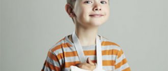
To suspect a broken leg in a child, it is not necessary to see an open wound and bone fragments. Sometimes external signs cannot be determined by eye due to compression or incomplete fracture of the “green branch”. Any area of the leg is susceptible to injury, the symptoms will vary:
- Sharp unbearable pain in the hip joint, shortening, pathological mobility of the limb, the leg is turned outward, bruises, swelling in the groin - these symptoms indicate a displaced femoral neck fracture. Cervical injuries without displacement have mild symptoms. The child can even walk.
- Knee pain, swelling, and swelling associated with bleeding into the joint characterize a fracture of the kneecap. The child cannot even bend his leg. If the fragments have separated by more than 0.5 cm, the supporting function suffers.
- Deformation, pathological mobility of the leg, severe pain, hematoma, swelling indicate a double fracture of the tibia and fibula. If only one bone was damaged, the child will still be able to lift the leg, and the deformation of the lower leg will be minimal.
- Severe pain in the leg, worsening if the child tries to move the leg, hemorrhage, swelling, partially impaired support function - symptoms of a fracture of the foot bones.
- The heel is turned outward, sharply thickened, the motor functions of the ankle joint are impaired - these are signs of a fracture of the heel bones.
- Pathological mobility, unnatural position of the toe, traces of a hematoma under the nail, on the skin, swelling, acute intense pain that intensifies when the baby tries to lean on the foot, indicate a fracture.
Source of troubles
There are many reasons why this type of injury increasingly affects people who are not yet very old. A diet that lacks calcium-rich foods, vegetables and fruits, and is dominated by fast food, a sedentary lifestyle, and no adequate physical activity - all this undermines our frame. As a result, bone and muscle tissue, as well as joint tissue, suffers. And there it’s not far from the turning point.
The mechanism of injury to the hip joint is usually simple: you fell, hit your side, and could no longer stand up on your own. In older, sedentary people, a fracture can occur even without a fall. The injury usually occurs while walking, when the patient suddenly feels severe pain in the joint.
Unlike other types of fractures in the hip area, which usually heal, the femoral neck recovers very poorly, since it has a different blood supply anatomy. After an injury, practically no blood flows into the fracture area. In addition, the nutrition of the femoral head is disrupted, and aseptic necrosis or coxarthrosis develops. In these cases, the patient's life is at risk. Severe complications occur especially often in old age, when the process of tissue restoration suffers.
How to help a child?
The quality of treatment for any fracture depends on timely first aid. Therefore, while waiting for the ambulance to arrive or before independently transporting the victim to the emergency room, you must take the following actions:
Calm the child down and give him pain relief. Prolonged stress combined with painful shock has a bad effect on the nervous system and can even cause fainting.
Find the fixing material. Traumatologists use special Kramer and Dichters splints and plaster splints, but improvised means are also suitable. You can use any suitable object: board, umbrella, stick.
Ask your child to relax his leg as much as possible, place it in the most comfortable position, and wrap it in a soft cloth so as not to pinch the joints. Tie your leg to the splint with a bandage, pieces of fabric, or belts.
- Interesting information: how to help a child with a broken nose
If the child has an open fracture accompanied by heavy blood loss, apply a tourniquet to the palm above the wound. Sprinkle streptocide on the wound.
Remember: without medical skills, do not try to set broken bones yourself, as you may cause additional damage or cause infection.
Treatment of femoral neck fracture
If the femoral neck is fractured, the child must be hospitalized in the traumatology department. If the fracture occurs without displacement, the leg fusion occurs in traction. An adhesive plaster and a small weight are used. The leg is retracted outward, then a splint is applied, in which the child will spend the next 2–2.5 months.
A displaced femoral neck fracture in a child requires treatment with skeletal traction, also with leg abduction. Epiphysiolysis of the femoral neck requires 2 months of traction. The transcervical and trochanteric fracture heals in 3–4 weeks with traction. Afterwards, a plaster cast will be applied to the entire hip area for 1.5 months.
Rehabilitation of the femoral neck begins from the first days of treatment. The child is forced to lie in traction for a very long time, so that bedsores do not form, he is prescribed a course of exercise therapy. Therapeutic exercise is the basis for restoring the functions of the femoral neck. The exercises are developed by the attending traumatologist, who will take into account the characteristics of the injury, the condition and age of the child.
Also, the femoral neck rehabilitation program includes a set of therapy that restores the anatomical structure of the joint and accelerates bone fusion in the form of UHF, massage and physiotherapy.
- Information for inquisitive parents: pelvic fractures in children
Principles of treatment of fractures in childhood
Damaged bone tissue is restored in several stages.
First aid
Proper and timely emergency care contributes to the successful treatment of fractures of the bone elements of the musculoskeletal system. Initially, the child should be given painkillers, since under the influence of pain shock and stress he may lose consciousness.
The following medications are suitable for children:
- Ibuprofen – 10 mg per day per 1 kg of body weight, should be taken in 3-4 doses;
- Paracetamol – 15 units per 1 kg of weight;
- Nimesulide - take 3 mg per 1 kg of body weight after meals, this amount of the drug should be divided into 2-3 times;
- Analgin – 10 mg per 1 kg, the dosage is divided into 3-4 doses.
Fixation of the bone diaphysis
This is the most severe damage to the femur, according to statistics, accounting for 60% of all fractures. Usually the child is prescribed conservative treatment. One of the methods is used:
- The child lies in traction until the fracture heals completely;
- Traction is combined with immobilization. A plaster, plastic bandage is used. For about three weeks, the child lies in traction, when a callus forms, it is replaced with a plaster cast. The baby will stay in it until complete recovery;
- The victim is immobilized using a coxite bandage.
The traumatologist decides which method to use, taking into account the characteristics of the injuries. The first two dressing options are most often used. Plaster application without traction is allowed only for impacted and subperiosteal fractures. Patients under five years of age are recommended to undergo adhesive traction, and for older children - skeletal traction. Complete bone fusion occurs on average in 4–8 weeks.
Surgical intervention is prescribed to a child only if it is impossible to provide him with high-quality traction, if there was a comminuted fracture with subsequent tissue interposition. In such cases, hip osteosynthesis is performed.
The operation uses locking screws. Doctors try not to use metal structures; they cause active growth of periosteal tissue in children. The procedure takes place under anesthesia. Afterwards, the child will be put in a cast, in which he will stay for 6–8 weeks.
Treatment of a tibia fracture
Treatment of this type of lower leg injury is often conservative. Surgery is performed if:
- successful reduction of the leg bones cannot be performed;
- with skeletal traction it is difficult to achieve the standing of fragments;
- there was an open fracture of the tibia;
- skin, blood vessels, nerves can be damaged by bone fragments;
- there is tissue interposition.
During surgery on the lower leg, intramedullary structures are used. Preference is always given to fixation with pins, as well as osteosynthesis with locking rods. Bone plates are very rarely used in pediatric traumatology, since they cause active growth of the periosteum.
When choosing a metal structure, the doctor is guided by the rule of minimal trauma. Only a design is used that will provide convenience when performing rehabilitation measures and will provide the opportunity to move and load the limb immediately after surgery or at least after a short period of time.
Treatment of a foot fracture
The foot consists of the tarsus, metatarsal bones and phalanges. Depending on the location of the injury, the following types of foot fractures are distinguished:
- Tarsal injury . When exposed to external forces, the heel bone is damaged, and less commonly, the talus bone. Other types of fractures practically never occur in children;
- Metatarsus injury . Fractures can be very severe, multiple, accompanied by displacement of fragments and damage to surrounding tissues;
- Finger injury . Injuries to the first and third fingers in children are common and may have signs of open and closed fractures.
Heel injury
A non-displaced heel fracture is treated in a trauma center. The doctor places a special splint on the foot, in which the arch of the foot is carefully modeled. After 3-4 days the bandage is circulated. Children aged 8–10 years spend about 3 weeks in a cast, older ones – 4–5. After removing the bandage, the patient uses orthopedic instep supports for 6 months.
If displacement of the fragments is detected, the child is hospitalized in the department. Here the traumatologist will perform immediate reduction under general anesthesia. After which a plaster cast will be applied to the entire leg, including the middle third of the thigh. The leg is in the optimal position - the knee is bent at a right angle, the foot is also bent. After 2 weeks of fusion, the plaster is removed, the foot is given a natural position, and the bandage is replaced with a special plaster boot. The heel will remain in a fixed position for about seven more weeks.
Metatarsus fracture
If the damage occurs without displacement, or the displacement is no more than ½ of the normal diameter, children are prescribed outpatient treatment. A splint is applied to the injured foot, and after a week the bandage is circulated. The length of time spent in a cast varies depending on age. Children under 10 years old wear the bandage for about 3 weeks. Older children – a week longer. You can lean on the sore leg only 10–12 days after the fracture.
The presence of greater or angular displacement of the metatarsal bones indicates the need for hospitalization. In the trauma department, reduction will be done under general anesthesia. Then a similar treatment is carried out - a week in a cast. Fusion takes place in 5–6 weeks, but from 15–18 days you can lean on your leg. After complete recovery, you should use an arch support for six months.
Surgery for this type of fracture is rarely performed. Only if there are open wounds, soft tissues are pinched by fragments, or bone fragments cannot be properly fixed. The operation is also performed under anesthesia, but implants are not used. The doctor stitches the fragments together or fixes them with a knitting needle.
Toe injury
A fracture of the toe does not require hospitalization of the child, so treatment will take place in a trauma center. If no displacement is detected, the foot is cast for 7–10 days.
Despite the good prognosis, traumatologists recommend not using an adhesive bandage. It compresses the small blood vessels of the finger, provokes swelling and is practically useless for an active child.
For displaced finger fractures, reduction is performed under local anesthesia. Fragments that cannot be securely fixed with plaster are fastened through the skin with a knitting needle or an injection needle.
To fix the fragments of the main phalanx of the finger, it is bent and until the knitting needle is removed, it remains in this position. Similar manipulations on the middle and nail phalanx are carried out in an extended position of the finger.
Next, a plaster cast is applied, the wire is covered with sterile material, and dressings are performed every 1–2 days. After 12–15 days, the child undergoes an X-ray control. If signs of a callus are visible, the needle can be removed.
Dislocations and fractures in children: what parents should do
Fractures and dislocations in children differ from injuries in adults, which is due to the structural features of children's bones and joints. Some types of injuries occur exclusively in childhood.
What are dislocations and fractures?
Bone fracture is a violation of the integrity of the bone (complete or partial), resulting from exposure to a load that exceeds the strength of the injured area.
Joint dislocation is a disruption of the shape of the articular surfaces of bones as a result of mechanical stress. Dislocation may be accompanied by a violation of the joint capsule or may not affect its integrity.
Most often in childhood, fractures of the arms occur, in particular the elbow joint, radius, humerus and forearm bones. Among the dislocations, the most common are dislocations of the bones of the forearm, head of the radius, and elbow joint. In childhood, fractures and dislocations can occur, combining two types of injury.
Features of bone fractures in childhood.
Young children fall frequently, but they are rarely seriously injured. This pattern is due to the child’s lower body weight and the structural features of his bones:
- thinness, strength and elasticity of bones, associated with a smaller amount of mineral salts in their composition;
- the periosteum is thick and abundantly supplied with blood vessels, which serves as a case for the bone and gives it flexibility.
These features, on the one hand, minimize the possibility of injury, but on the other hand, they are associated with a number of injuries that are characteristic only of childhood.
Types of dislocations and fractures in children
The most common types of fractures in children are:
Complete (bone broken on both sides):
Compression - occurs as a result of compression of the tubular bone along.
Like a green branch - when the bending of the bone exceeds the strength of its plasticity, but there is no complete fracture. The injured area is supported by the periosteum.
Bending (plastic deformation) is most typical for the elbow and knee joints and is caused by insufficient pressure to cause a complete fracture.
Depending on the integrity of the skin, the following types of fractures are differentiated:
- closed – bone fragments are isolated, the skin is not damaged;
- open – the integrity of the skin at the site of injury is compromised.
According to the degree of separation of bone fragments, fractures are:
- without displacement - bone fragments are not separated;
- with displacement - bone fragments are far from each other.
Causes of damage
In most cases, fractures and dislocations are the result of mechanical stress on the bone, but in some cases their appearance may be a consequence of the pathology of pregnancy and childbirth.
Main causes of damage
Falls of children at different ages lead to various injuries.
Infants with a relatively large head weight often suffer fractures of the parietal or occipital bone when falling.
Children learning to walk more often suffer from dislocations and fractures of their legs, in particular the phalanges of the fingers, metatarsals, and ankles.
Active games often cause dislocation of the cervical vertebra in children 3–5 years old, as well as dislocations of the elbow and knee joints.
Child abuse leads to a range of injuries from minor dislocations to fractures of the tibia, which may go unnoticed in childhood.
Carelessness of parents. For example, uncalculated force when pulling the arm can lead to sprained ligaments and dislocation of the shoulder and elbow joint.
Lifting heavy weights as a child can lead to dislocations of the elbow and shoulder joints.
Incorrect methods of massage or exercise therapy can cause dislocations of any joint.
The main signs by which parents may suspect a fracture:
- sharp pain when moving;
- broken limb deformity;
- limited movement in the injured limb;
- hematoma at the fracture site, which quickly grows under the skin;
- in the case of an open fracture, there is a wound through which the broken bone can be seen;
- Often children's body temperature rises to subfebrile levels.
Main symptoms of dislocation:
- pain in the injured joint;
- swelling or hematoma in the joint area;
- joint deformity;
- lengthening or shortening of the affected limb;
- movement in the joint is absent or difficult.
Diagnosis of injuries
Diagnosis of fractures in children is difficult. If you suspect damage, you should contact a pediatric traumatologist. The doctor examines and palpates the injured limb.
To determine a fracture or dislocation, the following is carried out:
X-ray (usually an X-ray is taken of the damaged and healthy limb to compare images);
MRI can detect greenstick fractures, closed non-displaced fractures, and bone fractures.
How to cure a child
If your baby has a dislocation or fracture, you should never adjust it yourself. Parents should adhere to the following procedure when providing first aid:
- provide the child with peace;
- immobilize (immobilize) the injured limb;
- fix broken bones or dislocated joints (apply a splint or bandage);
- if there is an open wound, it must be disinfected;
- in case of bleeding, apply a tourniquet above the damaged area;
- in case of severe pain, the child can be given an analgesic before the ambulance arrives.
Doctor pays attention !!!
You cannot independently choose a set of rehabilitation exercises and perform massage, since sometimes it is completely contraindicated. For example, with intra-articular or periarticular injuries, massage can provoke the formation of callus.
To fix a broken limb, you can use any hard object (board, book, rolling pin, etc.). Remember that your task is to secure the broken bones and prevent them from dislodging or moving until the ambulance arrives.
If a child is injured, it is important for parents to orient themselves correctly, provide emergency assistance and create favorable conditions for recovery.
If you have a fracture, you can go to a trauma center, which is open 24 hours a day, 7 days a week, or call an ambulance.
The material was prepared by doctor Konon T.A.