During the second trimester of pregnancy, a woman's body continues to change. This is the period from 13 to 26 weeks. Every day the baby in the womb grows and develops, which leads to an enlarged belly.
The second trimester is considered the best time to prepare for the arrival of your baby. Most women who did not feel very well in the first trimester, starting from the 13th week, notice a significant improvement in their condition. Every day, nausea and vomiting in the morning occur less and less often, although there are exceptions when unpleasant symptoms remain. The expectant mother has more energy and strength, and the incidence of mood swings decreases as hormone levels become more balanced.
In the second trimester, a woman’s body weight grows even faster. There is a need for special clothing for pregnant women.
Will my breasts continue to grow?
Yes, breasts grow at the same intensity in the second trimester due to an increase in the volume of the mammary glands and fat deposition. It is these changes that prepare the expectant mother for feeding her baby.
The skin on and around the nipples will become darker. A woman may notice the appearance of peculiar tubercles around her nipples. These are glands that secrete a special substance that prevents the skin of the nipples from drying out.
A yellowish liquid may also begin to be released - an intermediate form of milk (colostrum), which plays an important role in the first days of the baby’s life. Having a laxative property, colostrum helps the newborn get rid of meconium (original feces), which consists of epithelial cells, prenatal hair, mucus, amniotic fluid, bile and water digested in the prenatal period.
Where does it start to grow from?
Those who become pregnant for the first time do not always know where the belly begins to grow during pregnancy. The most reliable answers to all your questions can be obtained from a doctor who monitors the condition of the expectant mother and baby.
The gynecologist should tell you that first the volume of the bottom part of the uterus increases. It is located above the pubis, so the first bulge can be noticed there.
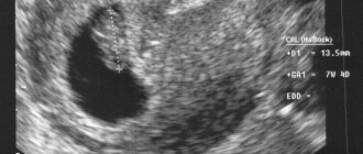
During the first examination of a pregnant woman, the doctor determines where the abdomen begins to grow, while palpating the enlarged organ. Its size helps the doctor understand how long ago the conception occurred. A more accurate period can be determined by ultrasound examination.
During each examination, the doctor measures the longitudinal and transverse dimensions of the abdomen and compares the data obtained with the result of the previous measurement. If all is well, you should see an increase each time.
What are stretch marks and can they be prevented?
Some areas of a pregnant woman's body naturally grow and enlarge. In such places, the skin becomes tense and the elastic fibers underneath are torn. This is how stretch marks are formed - stripes with indentations on the skin. Stretch marks usually appear on the abdomen and breasts, the areas most susceptible to growth during pregnancy.
This is a fairly common problem, although not every pregnant woman develops stretch marks. It is impossible to completely prevent the appearance of unpleasant stripes on the skin.
The main thing you can do is control your weight. Do not gain more weight than what your doctor recommends. There are many lotions and oils available in the cosmetic industry to prevent stretch marks, but the effect of such products has not been proven. However, it is worth noting that keeping the skin well hydrated will reduce itching.
Typically, after childbirth, stretch marks become less noticeable. In some cases they disappear completely.
What other changes can you expect?
Skin problems do not bother all pregnant women. Typically, such changes completely disappear or decrease in intensity after the birth of the child. Common skin problems during pregnancy include:
- dry skin on the abdomen;
- acne;
- increased sensitivity to the sun. Because of this, the skin burns much faster in the sun. When spending a long time outdoors, you should always take sunscreen with you;
- a dark line (“linea nigra”) from the navel to the pubis right in the middle of the abdomen and dark spots on the face (sometimes called the “mask of pregnancy”). This is melasma. It occurs due to the action of estrogen (the hormone responsible for preparing a woman’s body for childbirth and then for lactation).
Often dry skin on the abdomen is accompanied by itching. However, this may be caused by cholestasis (reduced bile flow from the liver) and gestational diabetes (a carbohydrate metabolism disorder that occurs during pregnancy). You should be careful with these symptoms and consult a doctor if you suspect any complications.
When does it stop increasing?
While in a state of anticipation, a woman usually strictly monitors changes regarding her well-being and appearance. Accurately notices when the belly begins to grow during pregnancy. Knows how quickly it is increasing.
Any incomprehensible nuances when expecting a child make you worry. One of these is when the belly stops growing during pregnancy. The reasons for this can be harmless (increase is not noticeable on a full figure) or dangerous:
- slow development of organs, low body weight of the baby;
- transverse position;
- intrauterine death;
- small volume of liquid in which the child remains.
There is no need to guess what is the reason for the growth stop. You won’t be able to identify it on your own; you need to contact a specialist.
What other changes can you expect in the second trimester?
Pregnant women in the second trimester are often bothered by leg pain and cramps during sleep. Such phenomena are associated with the pressure exerted by the child in the womb. It compresses the nerve endings and vessels that supply blood to the lower extremities. To avoid pain and cramps, it is recommended to sleep on your side rather than your back.
A more dangerous symptom is deep vein thrombosis. A thrombus is a blood clot in a vessel that occurs due to disturbances in the coagulation system. The main signs of the disease are pain and swelling of one lower limb. If you notice such symptoms, you should immediately consult your doctor.
Swelling is one of the most common problems in pregnant women. Typically the ankles, arms and face become swollen. During pregnancy, the volume of fluid in the mother's body doubles. This includes the fetal circulatory system and amniotic fluid. In addition, due to hormonal changes, sodium is retained in the tissues, which tends to retain water. Swelling can appear at any stage of pregnancy, but most often occurs at the end of the second trimester.
Back pain occurs due to the growing belly of a pregnant woman. It takes extra effort to maintain it. And this is against the backdrop of relaxation of the ligaments holding the bones of the pelvis and hips. This happens due to specially released hormones that prepare a woman’s body for childbirth, thus making the birth canal more mobile.
A pregnant woman is worried about abdominal pain. The muscles and ligaments that support the uterus stretch as it grows. This causes mild pain or cramping.
Sometimes pregnant women notice tooth decay. To build a child's skeleton, a lot of calcium is consumed. When there is not enough calcium, the mother's special hormones are activated, and calcium is washed out of the woman's bones and teeth. In addition, the acidity in the oral cavity changes, which increases the risk of infection and the development of caries. To prevent this, it is necessary to visit the dentist regularly throughout your pregnancy.
The occurrence of nasal congestion, nosebleeds and bleeding gums is associated with increased blood flow in the mucous membranes of the nasal passages and oral cavity.
Heartburn usually begins if it was not there, or gets worse in the second trimester of pregnancy. Some may never encounter it. The uterus quickly increases in volume, thereby putting pressure on the stomach, which is why food and stomach acid enter the esophagus, which provokes discomfort.
In the second trimester of pregnancy, there is a risk of contracting urinary tract infections. The outflow of urine slows down due to hormonal changes, while the bladder is not able to empty completely because the enlarged uterus puts pressure on it. Untreated urinary tract diseases can cause premature birth. If there are any signs of infection, you should make an appointment with your doctor: the urge to urinate frequently, a burning sensation, the presence of blood, or an unpleasant, persistent smell of urine.
Braxton-Hicks contractions (“false contractions”) are contractions of the uterine muscles. In this way, the uterus prepares for childbirth. These contractions make the abdomen tense and rigid, causing discomfort. Contractions are irregular in timing and should subside within a few minutes. You should call your doctor if contractions become regular, painful, and do not go away when you change position. This condition may indicate that premature labor has begun.
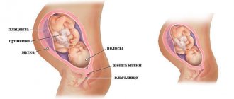
How the uterus grows during pregnancy
The uterus increases in size throughout pregnancy. For the first few weeks, the uterus is pear-shaped. At the end of the 2nd month of pregnancy, its size approximately triples and it has a rounded shape. During the second half of pregnancy, the uterus retains its round shape, and at the beginning of the third trimester it acquires an ovoid shape. Before pregnancy, the weight of the uterus averages 50-100 g, and at the end of pregnancy - 1000 g. The volume of the uterine cavity at the end of pregnancy increases by more than 500 times. During pregnancy, each muscle fiber lengthens 10 times and thickens approximately 5 times. The vascular network of the uterus increases significantly; in terms of its oxygen regime, the pregnant uterus approaches such vital organs as the heart, liver and brain.
These parameters can be determined by measuring the uterus using external obstetric examination techniques. To do this, the so-called value of the uterine fundus is measured, but at the beginning of pregnancy, until the uterus extends beyond the pelvic bones, an increase in the size of the uterus can be determined using a vaginal examination (it is carried out during a gynecological examination) or ultrasound.
The doctor determines the height of the fundus of the uterus at each regular examination using a centimeter tape: this helps to navigate the rate of growth of the abdomen. The doctor measures the distance from the upper edge of the symphysis pubis to the upper part of the uterus - its bottom. Approximately, the height of the uterine fundus in centimeters corresponds to the gestational age in weeks. For example, if the height of the uterine fundus is 22 cm, the gestational age is 22 weeks.
When will I feel my baby move?
Just in the middle of the second trimester, a pregnant woman begins to feel her baby. The first tremors are more like trembling movements deep in the stomach. Over time, the sensations intensify.
Of course, the child had moved before, but it was very weak, almost imperceptible. Usually, women who give birth for the second or third time can feel the first tremors much earlier, because they are already familiar with this sensation. The day of the first tremors should be marked on the calendar and the doctor should be told about this at the next consultation so that he can transfer the data to the pregnancy chart.
Will my interest in sex return?
Due to unpleasant symptoms such as morning sickness and vomiting, frequent mood swings and lack of energy, a woman may experience decreased sexual activity. She simply does not want sex due to emotional or physical condition and fatigue. However, many women note a return to the desire to have sex in the second trimester.
Doctors say that sex is not contraindicated regardless of the stage of pregnancy. But only the attending physician can say for sure. There are individual situations when sex is prohibited: threat of miscarriage, multiple pregnancies, bleeding.
What does the volume and shape of the abdomen indicate during pregnancy?
During pregnancy, the belly grows due to the development of the child, the growth of the uterus and an increase in the volume of amniotic fluid. At the very beginning of pregnancy (up to about the 12th week), the size of the abdomen of expectant mothers does not change, since the growing uterus does not yet reach the edge of the pubis. But starting from the 2nd trimester, the uterus gradually rises to the navel, the woman’s belly begins to round, and those around her notice changes in her condition.
Of course, all this is very individual, and a pregnant woman’s belly may grow earlier or later, and may also be slightly behind or ahead of the norm, depending on the following factors:
- fetal size and position;
- anatomical features and build of a woman;
- weight gain of the expectant mother;
- number of embryos;
- heredity;
- volume of amniotic fluid.
How does a pregnant belly grow?
Throughout pregnancy, the circumference and shape of a woman’s abdomen changes in accordance with changes in the size of the baby, uterus and amniotic fluid volume approximately as follows:
- At a period of 4 weeks, the uterus is similar in size to a chicken egg, and after another four weeks - to a goose egg.
- At the 12th week, its upper edge reaches the edge of the womb, and its bottom can be felt through the wall of the peritoneum.
- 16 weeks - the bottom of the organ is located approximately in the middle between the pubic symphysis and the navel.
- 20 weeks - the uterus continues to actively grow, and its edge almost reaches the navel (located somewhere two centimeters below). During this period, the pregnant woman's belly begins to noticeably increase and become rounded.
- 24 weeks – the fundus of the uterus can be felt in the navel area.
- 28 weeks - the bottom of the organ is located approximately two centimeters above the navel.
- 32 weeks - the uterus is located at the midpoint of the distance between the sternum and the navel, and the navel itself literally “disappears” from the surface of the abdomen.
- 38 weeks - The upper edge of the uterus reaches its highest level between the ribs and the lower part of the sternum.
- 40 weeks – a woman’s navel protrudes strongly forward, and the uterus drops slightly downwards.
Belly Measurement Options
The main parameters that the gynecologist will measure at each preventive examination from about the fifteenth week are the circumference of the woman’s abdomen and the height of the uterine fundus. It is these indicators that are most informative; that is, based on them, it is possible to monitor the dynamics of the child’s growth and its disturbances. The general measurement standards are that before the procedure you need to empty your bladder, then lie down on a hard surface, straighten your knees and completely relax.
Abdominal circumference during pregnancy
The abdominal circumference should be measured so that in front the meter tape passes along the line of the navel, and in the back along the line of the lumbar deflection. Abdominal circumference (AC) norms during pregnancy are determined using a special table, but it should be noted that it allows an error of approximately two to three weeks - this is about 10 cm.
Week of pregnancy OJ, cm
- 20 weeks — 70–75
- 22 weeks — 72–78
- 24 weeks — 75–80
- 26 weeks — 77–82
- 28 weeks — 80–85
- 30 weeks — 82–87
- 32 weeks — 85–90
- 34 weeks — 87–92
- 36 weeks — 90–95
- 38 weeks — 92–98
- 40 weeks — 95–100
If your abdominal circumference is greater than normal
If, during the next gynecological examination, it turns out that the pregnant woman’s life expectancy is much higher than normal, she is recommended to undergo additional examination to exclude the following pathologies:
- polyhydramnios pregnancy (when the volume of amniotic fluid is about 2–5 liters, and in some cases reaches 10–12 liters);
- hydatidiform mole;
- chorionepithelioma.
In addition, if a woman’s pregnancy is more than 38 weeks, an overly large belly may indicate a transverse position of the fetus, which is a rather serious pathology. Typically, in such cases, the patient is recommended to have a cesarean section.
If your abdominal circumference is less than normal
Too small an abdominal circumference, which is significantly “lag behind” the norm, may indicate:
- delay in the development of the baby due to placental insufficiency;
- oligohydramnios, which usually accompanies severe forms of gestosis;
- post-maturity.
Fundal height of the uterus during pregnancy
The height of the uterine fundus (UFH) is very important for diagnosing any abnormalities in the condition of the expectant mother, as well as for calculating the gestational age by week. On average, in women of reproductive age, the length of the uterus is approximately 8 cm, and during the period of bearing a child it gradually increases, and by the 40th week it reaches approximately the same length - 40 cm.
VMR is measured using a centimeter tape, the zero end of which must be applied to the beginning of the pubic bone, then move up and stop at the point where the more elastic part of the abdomen turns into a softer one. The resulting distance will be the indicator that gynecologists call the height of the uterine fundus. Normally, it should correspond to the intrauterine age of the child (gestational age by week) plus or minus 1–3 cm.
If the height of the uterine fundus is higher than normal
The fundus of the uterus in a pregnant woman is above normal in the following cases:
- child is too large;
- multiple pregnancy;
- incorrect presentation of the baby;
- polyhydramnios;
- narrow pelvis in a pregnant woman.
If the height of the uterine fundus is less than normal
This situation may indicate that the pregnant woman:
- the term is incorrectly defined;
- oligohydramnios;
- delayed fetal development;
- very wide pelvis.
By the way, sometimes these numbers can be used to approximately determine the weight of the fetus: for this, the height of the uterine fundus must be multiplied by the circumference of the abdomen. Obstetricians say that the error of this method is about 150 g, but pregnant women note that it is quite inaccurate, and sometimes the error can reach a kilogram.
It should be noted that it is not recommended to independently measure BMR and WC during pregnancy and make diagnoses based on the obtained values - the likelihood of an error is too high, fraught with unnecessary stress.
Belly shape
The shape of the expectant mother's tummy is another indicator that gynecologists and obstetricians often pay attention to. It is influenced by many factors, including the size of the child, the pace of its development and location, the weight of the expectant mother, the shape of the pelvis, the thickness of the abdominal fat layer, the number of embryos and the condition of the abdominal muscles.
Of course, it is quite difficult to talk about any standards in this case, because even one woman’s tummy can look completely different during several different pregnancies, but experts still note some connections between the characteristics of the expectant mother’s body and the characteristics of the course of pregnancy:
- Ideally, during a normal pregnancy, the abdomen has a regular, ovoid, slightly pointed shape, and in first-time mothers it is directed with the sharp end upward, and when carrying a second, third, etc. child - point down. If the mother has a narrow pelvis, then the pointed shape of her abdomen will be especially pronounced in the second trimester.
- A round or even square belly can be due to weak abdominal muscles, carrying a large child, or a large volume of amniotic fluid. Additionally, if a small, frail woman is carrying a baby that is slightly larger than normal, her belly may also appear large.
- A large, spherical belly also often indicates a polyhydramnios pregnancy, and therefore requires additional examination.
- A pear-shaped belly is also usually characteristic of pregnant women with too weak abs, the muscles of which are unable to support the baby properly, so the belly sag slightly downward.
- A large, pointed tummy occurs with twins, triplets, etc., as well as in mothers with characteristic anatomical features, that is, with a narrow pelvis.
- A small, neat, almost invisible tummy is sometimes a sign of oligohydramnios, but it can also occur in fragile women carrying an equally fragile, small-sized child.
- An uneven, asymmetrical shape indicates an incorrect (transverse or oblique) presentation of the fetus. For example, with a transverse presentation, a woman’s tummy will look like a horizontally elongated oval.
In what cases should a change in the shape of the abdomen bother a woman? You need to urgently inform your doctor about this fact when the stomach takes on an asymmetrical shape, and the pregnant woman feels discomfort or pain. This situation may indicate some disorders that require correction and even treatment.
Source