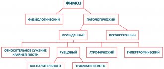
Indications
Synechiae in boys is formed as a result of phimosis, a disease characterized by narrowing of the folds of skin covering the outer end (head) of the penis and the inability to fully or partially expose it. Synechiae are areas of fusion of the foreskin and the glans itself. There is a natural and pathological form of adhesions. Depending on the form, course of the disease and complications, the doctor will have to choose which method will be used to separate the synechiae of the foreskin.
Phimosis is divided into:
- physiological;
- pathological.
Physiological manifests itself in most newborn boys and is characterized by incomplete dilation of the head of the penis and the presence of loose adhesions - synechiae. Starting from the age of five, synechiae tend to resolve under the influence of hormones and enzymes produced by the body itself.
This process can last until puberty and gradually disappear with the appearance of the first erection. It is a natural protection in the development of the child’s genitourinary system and prevents the development of bacterial, viral, parasitic and other diseases. Physiological phimosis does not require treatment.
If, over time, the synechiae do not loosen and begin to cause inconvenience, then they resort to conservative methods of treatment: physiotherapeutic procedures, warm enveloping ointments and proper care of the external genitalia. If in the future these methods do not provide the desired result, then the synechiae are separated surgically.
Pathological phimosis is an indication for surgical or medical intervention. Physiological phimosis, although it is a natural process, can also turn into pathology. This happens for some reasons:
- genetic predisposition;
- violation of metabolic processes;
- endocrine pathologies;
- inflammatory processes;
- mechanical damage.
An independent attempt to fully open the head of the penis causes ruptures of skin folds and the formation of bleeding wounds. This method provides fertile ground for infection and the development of inflammatory processes.
Very often, during masturbation or self-opening of the head of the penis, paraphimosis develops - a shift of the narrowed foreskin behind the head. Blood supply and urination are disrupted. The penis itself becomes cyanotic and swells, increasing in size. The danger of this complication lies in necrosis of the head of the genital organ. Urgent assistance is required.
Balanoposthitis often develops during the pathological process. The disease manifests itself when a large accumulation of smegma stagnates, which is normally produced by the sebaceous glands of the penis and serves as a lubricant, and also promotes the natural resorption of synechiae. The development of balanoposthitis is provoked by a lack of penile hygiene and thickening of smegma in the preputial sac, as a result of which infection occurs.
Synechia: symptoms
Most often, synechiae can be detected with the naked eye, primarily by parents, of course. Fusion usually occurs from the coronal groove of the penis to the urethra. In addition, since the consequence of synechia is often inflammation, additional symptoms of this disease can be considered:
- swelling of the glans penis and the foreskin itself;
- severe itching in the intimate area;
- redness of tissues, the appearance of small wounds (most often a consequence of itching and attempts to get rid of it);
- discharge from the urethra;
- pain when urinating or difficulty urinating.
The appearance of the described manifestations may indicate that, against the background of synechiae, the spread of infection has begun, which requires timely intervention by a specialist.
If parents notice that at the age of 3 years or more a boy has problems with exposure of the head of the penis (at first partial, and by 6 years full), it is necessary to make an appointment with a pediatrician or pediatric urologist to avoid the development of complications.
Contraindications
When the first symptoms of phimosis or synechiae appear, doctors recommend going to the clinic to diagnose the stage of the disease and determine the processes that are developing in the body. It is contraindicated to perform surgery if phimosis is physiological, when the child does not experience pain when urinating, and the head of the penis is mostly exposed for the purpose of superficial hygiene.
Contraindications for separating synechiae of the foreskin of the penis are:
- infectious diseases;
- inflammatory foci in the body, including acute inflammation in the head area;
- exacerbation of chronic diseases;
- purulent inflammation or balanoposthitis;
- narrowing of the foreskin, which makes it difficult to expose the head even in the absence of adhesions;
- rough cicatricial adhesions.
In case of inflammatory processes and exacerbation of chronic diseases, antibacterial therapy is prescribed. First, it is necessary to extinguish the source of inflammation, and then proceed to surgery.
The foreskin is endless
16.07.2019
10203
2
| Tarusin Dmitry Igorevich Doctor of Medical Sciences, Professor, City Center for Reproductive Health of Children and Adolescents, Morozov Children's City Clinical Hospital, Moscow |
During the urological Internet conference No. 3 “Diseases of the penis,” Doctor of Medical Sciences, Professor Dmitry Igorevich Tarusin described a wide range of diseases of the foreskin in children and adolescents, requiring a thoughtful, differentiated approach to treatment.
Foreskin and decency
At the beginning of his speech, Dmitry Igorevich recalled that between the diagnosis of “phimosis” and the state of the open glans penis, there are many intermediate conditions about which specialists in both urology and pediatric surgery are, unfortunately, little aware.
The history of the perception of human foreskin goes back thousands of years. In particular, in the ancient world (Ancient Greece and Rome) a long foreskin, which covered the head even during an erection, was considered ideal. The presence of a long foreskin was one of the criteria for exemplary male beauty, an indicator of health and respectability. Sometimes the foreskin was specially lengthened using a thin leather loop - kinodesma, which was tied to the foreskin with tension upward and worn around the waist. Wearing it was a sign of modesty and decency, as it prevented exposure of the head, which, in turn, was considered extremely indecent and offensive, since it was reminiscent of circumcision.
Since at least the 19th century, the foreskin has been actively studied by specialists in various fields, in particular, morphologists and embryologists [1,2,3].
The structure of the foreskin
This is one of the few human organs located outside, in the formation of which during embryogenesis the ectodermal, mesenchyal and neuroectodermal layers take part, ultimately forming a penthalaminar structure called the single structure of the cytokeratin complex. It consists of a mobile dermis, devoid of hair follicles, a muscular layer located inside, which is, in fact, a continuation of the shell of the testicle, its own epithelial plate, and then there is mucosa, which lines the epithelial plate from the inside. The organ contains a large number of nerve structures. You can imagine it in more detail by layers as follows:
- Stratified squamous cells undergoing keratinization. The structure is abundantly supplied with Langerhans cells, Merkel cells are modified, have a neuroendocrine apparatus, and have unmyelinated dendrites. Enolase, chromogranin A and cytokeratin 20 were determined.
- The dermis is abundantly supplied with receptors - there are 12 types of receptors, the meaning of 4 of which has not been reliably established today. Sparse sebaceous glands are present. Connective tissue of two types - pliable fibers and elastic - plays the role of a spring for the return of the foreskin after retraction.
- The muscular layer of the foreskin is able to tense and relax. It forms a complex with the fleshy membrane of the scrotum. The structure of the muscle is smooth, has powerful and long processes, and is temperature dependent. The distal part of the fibers is located subcircularly and causes the skin to wrinkle, forming a one-way valve. Steroid hormones can cause irreversible reorientation of fibers, promoting increased retractility of the foreskin. The nerve fibers of the foreskin, passing intramuscularly, can change their length (the only dendrocytes of their kind in humans).
- The most richly vascularized tissue, loose collagen with elongated trabeculae. Deprived of vellus hair follicles, sweat and sebaceous glands. The presence of Tyson's glands (smegma producers) remains questionable. Pheromone producers have been accurately identified in great apes.
- From the 11th week of intrauterine development, the structure contains intraepithelial nerves, the maturation of which occurs up to 2.5 years. Contains Langerhans cells, but does not contain melanocytes at all.
Preputial sac microbiome
In the preputial sac, the moist environment is provided by transudate from the rich choroid plexus of the dermis and the meatus, constantly secreted by the secretion of the Cooper and Littre glands, as well as by the waste products of saprophytic bacteria of the preputial sac. Bacterial colonies here include Cornybacterium, Bacteroidum melanmogemcus, Enterococcus, Coagulase-positive Staphylococcum and Mycobacteria smegmatis. Smegma contains squalene, beta-cholestanol, sterols, long-chain fatty acids, as well as 9,10-methyleneoctadecanoic acid, the amount of which is higher in aging men.
Options for the development of the foreskin
A cross-species phenomenon that occurs not only in humans is preputial adhesion (synechia of the foreskin). This is a physiological process that allows the proliferation system of the exfoliated (squamous) part of the epithelial lining to achieve a state of mature, regulated activity and proper apoptosis. The process of mobilization of the foreskin begins with the appearance of secretion activity of “small” androgens during the adrenarche period. The process is controlled by inhibition of type II COX [4]. With some violations in its course, variants of the development of the foreskin may form:
- proboscis (completely prevents the entry of exogenous pathogenic flora, provides additional stimulation for introitus);
- ocellar (by default, urination occurs correctly: through the slightly open head, there is no retention of urine in the preputial sac);
- slightly open;
- open (as in the case above, urination occurs correctly by default, the physiological “rolling” of the foreskin back when an erection occurs);
- absence of foreskin (deprives a man of 85–90% of adequate innervation, creates disorgastic sexual intercourse, reduces the volume of afferentation).
Russian classification of diseases of the foreskin in children
In 2004, Sergei Sergeevich Sadchikov defended his dissertation “Diagnostics and differentiated tactics for treating diseases of the foreskin in children,” which presented Russia’s first classification of diseases of the foreskin of the penis in children [5]. According to it, diseases are divided into inflammatory (dissanitary, chronic and acute balanitis) and degenerative (synechia, functionally narrow foreskin, hypertrophic and atrophic phimosis, as well as specific lesions of the foreskin).
Since then, a number of changes have been made to the classification. In 2015, according to a new version, diseases of the foreskin in children were divided into functional, atopic, inflammatory (acute and chronic), degenerative, reactive and cicatricial changes, cancer predictors, congenital malformations, as well as trauma and iatrogenicity [6].
Often signs of a malformation of the foreskin are “hump”, “hood” and “concord syndrome”. Such phenomena require therapeutic measures. In some cases, the condition of the foreskin with a “hidden” penis is accompanied by phimosis. In such a situation, performing circumcision is a mistake.
Functionally, narrow foreskin is a condition characterized by the possibility of carrying out hygienic measures during detumescence, but at the same time pain, discomfort, cracks in the foreskin (inner, outer or both layers) during an attempt to expose the head in a state of tumescence of the cavernous bodies. Causes discomfort, pain, chronic recurrent fasting, ignoring or avoiding hygiene measures, and subsequently avoiding sexual activity. In some cases, with this condition, there is a moderately present noose in the area of the coronary sulcus, which does not cause discomfort and pain, cracks and ruptures, and is capable of enlarging the glans penis due to “edema” - increased venous stasis in the glans area, which provides a set of specific sensations when sexual contact. With age and a decrease in the plasticity of the foreskin, this, however, becomes a problem.
An excessively pronounced contractile groove may also be observed, which interferes with masturbatory and sexual activity, causes chronic inflammation and trauma, thus requiring specific treatment. It often causes paraphimosis during initial sexual contact. Sometimes this configuration causes lifelong sex with the head closed.
In other cases, “dry foreskin” - desquamative marginal posthitis. This situation does not require surgical intervention and is often associated with a lack of vitamins, primarily B6.
The skin pattern of the foreskin is one of the criteria by which reactive (atrophic) changes can be assessed. Often, reactive changes in the tissue of the foreskin occur in response to the presence or exacerbation of a systemic disease that weakens the immune defense mechanisms. Such diseases include vitamin deficiencies, hypovitaminosis and diabetes mellitus. Changes are always secondary to the system-forming problem.
As Dmitry Igorevich emphasized, reactive changes in the tissue of the foreskin can occur in response to global changes in hormonal homeostasis, atrophic, as a rule, correspond to a decrease in the hormonal protection of the tissue of the foreskin, and hyperplastic or hypertrophic changes occur against the background of incrisis changes in homeostasis.
The lecturer also reminded of the need to examine not only the penis, but also the adjacent skin. In this way, for example, atopic allergic reactions can be detected.
Quite rarely, a picture of abscess formation of the foreskin and skin of the penis can be observed. Most often it occurs as a result of an ingrown hair or careless shaving of the skin.
If the patient is not trained in the rules of personal hygiene, the clinical picture of smegmal accumulations with the formation of plaque may develop. This phenomenon is conventionally designated as dissanitary balanoposthitis and belongs to the normal category.
An extremely unpleasant situation is purulent-necrotic posthitis , which often occurs in children with immune deficiency. Like ulcerative balanoposthitis, this case requires careful identification of the causes and competent development of therapy.
Keir's erythroplasia , an autoimmune disease of unknown etiology, leads to degenerative changes in the glans penis and foreskin. It is a predictor of the development of cancer of the glans penis, and therefore requires close attention. Vitiligo-type depigmentation is also a predictor of various neoplasms, so in this case, preserving the foreskin seems inappropriate.
Dermatological diseases include degenerative keratofolliculitis, in which preservation of the patient's foreskin often becomes impossible. Another reason for excision of the skin layer can be cicatricial changes in the foreskin - both with and without a keloid component. If only the outer leaf is affected, you can limit yourself to organ-preserving plastic surgery.
As Dmitry Igorevich noted, of the total number of patients with a non-opening head, real cicatricial phimosis is confirmed in only 2–3%. However, a picture of “fake” cicatricial phimosis with fistulas of the foreskin is possible.
Lichen sclerosus is a disease characterized by flat, confluent white nodules that form unfiltered plaques. Occasionally, vesicles develop on plaques due to the accumulation of fluid under the epidermis. The disease is determined by degeneration of the spinous layer of the dermis and always affects the glans penis. In such situations, conservative treatment is carried out after the procedure for removing the lesion.
Finally, traumatic injuries to the foreskin can be distinguished. Sometimes they can be healed in such a way that the head opens normally, but in some cases restorative surgical procedures are required to ensure full functioning of the organ.
Separately, the lecturer emphasized the fact that the patient’s young age does not guarantee that he does not have cancer, so neoplasms of the glans penis always require high alertness.
Material prepared by Yu.G. Boldyreva, special. corr. Urology Digest The full version of the report can be viewed on Uro.TV
Sources:
- Schweigger-Seidel, 1866
- Retterer, 1885–1915
- Glenister, 1921
- Cold CJ, Taylor JR, 1999
- Sadchikov S.S., 2004
- Tarusin D. I., 2015
Topics and tags
Urology Digest
Comments
Annenkov Andrey Viktorovich - 07.19.2019 - 13:41:13
Thanks for the interesting message
Asylov Almat Zeinetollaevich - 07/17/2019 - 10:28:27
very interesting!!!
To post comments you must log in or register
Carrying out separation
Separation of synechiae is a relatively simple operation that does not require special training or high-tech medical instruments. Initially, the patient is premedicated with local anesthesia and anesthesia of the nerve endings of the head and foreskin. The genitals contain a large number of nerve endings and are among the most sensitive in the body.
Next, under the influence of anesthetics, doctors carefully separate the adhesions. Most often, a small probe is used. If the adhesions are well interlocked, the doctor resorts to using a scalpel. The adhesive septa are carefully cut off with a scalpel. After removing the synechiae, an anti-inflammatory ointment or powder is applied to the wound.
The price of such an operation will be small due to the simplicity and ease of its implementation.
Causes of synechiae
- Genitourinary infections - vulvovaginitis, vulvitis, etc. Infection is possible in the maternity hospital or at home, when swimming in stagnant natural bodies of water.
- Violation of intimate hygiene rules. Both a lack of hygiene and its excess are harmful.
- Allergy in a child. A reaction can occur to food (from the diet of a girl or a nursing mother), components of care products, washing powder, etc.
- Difficulties during pregnancy - intrauterine infections, severe toxicosis. If a mother had difficulties during pregnancy, her daughter increases the risk of developing synechiae.
- Genetics. The tendency to fusion of soft tissues can be transmitted from mother to daughters.
- Wearing tight synthetic underwear, which creates all the conditions for inflammation.
- Reduced estrogen levels. This is normal before puberty, but in some girls it causes synechiae.
- Dysbacteriosis. Pathogenic intestinal flora can provoke adhesions. Source: Z.K. Batyrova, E.V. Uvarova, L.S. Namazova-Baranova, N.Kh. Latypova, A.E. Donnikov Fusion of the labia minora in girls during early childhood: tactics of a pediatric gynecologist // Issues of modern pediatrics, 2012, vol. 11, no. 2, pp. 118-121
Symptoms and signs of fusion of the labia majora and minora in a child
With complete adhesion:
- There are difficulties with urination. The child becomes very nervous when the urge arises, strains hard on the potty, cries, and refuses to go to the toilet.
- Stagnation of urine causes infections in the urethra, which are manifested by: rash;
- redness and peeling of the skin in the intimate area;
- secretions.
The mother can examine the girl herself. To do this, you need to separate the labia majora. With synechia, the small ones do not “come apart” and they are connected by a whitish-gray film, similar to a membrane. When trying to separate the labia minora, the child begins to cry - it hurts.
What is phimosis and how does it happen?
Phimosis is the inability to open the head of the penis due to insufficient size and poor extensibility of the external opening of the foreskin. Phimosis can be physiological (normal) or pathological (abnormal), and it can also be congenital or acquired. Phimosis should not be confused with synechiae or adhesions of the foreskin , when the head of the penis does not open completely due to adhesion or fusion of the inner layer of the foreskin with the head of the penis. Synechiae are eliminated by simple division with or without local anesthesia, and in most cases do not require surgical treatment.
Prevention
Since almost all newborn boys experience synechiae and this is considered a normal condition, a preventive set of measures is aimed at preventing inflammatory processes. For this purpose, it is recommended that parents wash the baby’s external genitalia daily with running water; it is very important that during the hygienic procedure water does not get into the preputial sac.
Under no circumstances should parents try to expose the boy’s head of the genitals on their own. This can only lead to the opposite effect - pinching of the head of the genital organ, painful sensations.
Parents should also ensure that their boy does not wear an overfilled diaper. For this reason, doctors recommend changing your baby's diapers every six hours. Every half hour to an hour the boy needs to have air baths to prevent inflammation and irritation on the genitals. If it is very hot, it is better for the baby not to wear diapers at all to prevent overheating of the genitals.
Parents also need to be attentive to their child’s clothing. Linen should be used only from natural cotton. Underwear should not rub, chafe, pull or compress the genitals.