Urologist
Mkrtchyan
Karen Gagikovich
13 years of experience
Candidate of Medical Sciences, member of the European Association of Urology and the Russian Society of Oncourologists
Make an appointment
Among the congenital pathologies of the penis in men, phimosis is the most common. This is the name given to the narrowing of the opening of the epithelial tissue of the foreskin, which prevents the free exit of the head. The main cause of phimosis is an insufficient number of elastic cells that form the structure of the foreskin. Physiological phimosis is observed in more than 95% of newborn boys. There is no need for its treatment; the presence of signs of the disease until the end of puberty is considered normal. However, for adults, this problem can cause disruption of excretory and reproductive functions, so if a narrow opening persists in patients over 20 years of age, it is recommended to seek medical help.
Causes and types in adults
The disease is congenital, due to the action of a hereditary factor. Less common is phimosis, which has become a complication of diabetes mellitus, serious illness, injury or failure to comply with the requirements of personal hygiene of the genital organs. A small proportion of the total number of diseases is occupied by the consequences of balanoposthitis, when the foreskin suffers an inflammatory process, and rough scars form in place of the damaged areas.
In the vast majority of patients, phimosis develops in the following sequence:
- first degree - the head of the penis is exposed freely with little effort;
- second stage - the head is exposed only at rest, during an erection it is impossible to open it;
- third stage - the head is not exposed even in a calm state, there are no problems with urination;
- fourth stage - the head is not exposed, problems arise with urine output.
Anatomical and physiological aspects
The formation of the penis begins from the 7th week of pregnancy and ends at the 17th week. The skin of the penis folds in front to form the foreskin and preputial sac. It performs many functions, the main ones being protective, immunological and erogenous. The foreskin is represented by a two-layer skin fold that merges with the head of the penis. It is connected to the undersurface of the glans penis by a highly sensitive tissue called the frenulum. The prepuce is richly vascularized and innervated. Conventional circumcision removes most of these sensitive areas. The glands present on the foreskin and glans produce a secretion containing lysozyme, which has a protective function. At birth and in the first few years of life, the inner part of the foreskin is intimately fused to the head of the penis and, therefore, the latter is not removed.
Physiological phimosis is observed in the first years of life and is associated with the presence of synechiae between the foreskin and the head. The foreskin gradually becomes more mobile with age and allows more and more exposure of the head of the penis. This is facilitated by erection, retraction and keratinization of the epithelium of the inner layer of the foreskin. Thus, the possibility of exposing the head of the penis increases.
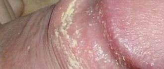
With pathological phimosis, the altered foreskin looks like a cone-shaped “cover” with a distal narrow part, which can be fibrous. It is important to distinguish pathological phimosis from physiological one, as this completely changes the tactics of observation and treatment. Physiological phimosis requires only conservative treatment, pathological phimosis can only be corrected through surgery.
Poor hygiene and repeated episodes of balanitis or balanoposthitis lead to the formation of scars on the inner layer of the foreskin, which leads to pathological phimosis. Strong retraction leads to microcracks in the opening of the preputial sac, which also leads to the formation of scars and phimosis. Older people are at risk for phimosis secondary to loss of skin elasticity and infrequent erections.
Symptoms of pathological phimosis in men may include painful erection, hematuria, recurrent urinary tract infection, pain in the foreskin, weakness, straining of the urine stream, and a feeling of incomplete emptying of the bladder.
Up to 10% of men have physiological phimosis before the age of 3 years, and a larger percentage of children will have only partially retractable foreskin. 1-5% of men have phimosis by age 16.
Symptoms
The main symptom of phimosis, which allows us to speak confidently about the existing disease, is the inability to remove the head from the foreskin at rest or erection. The phenomenon is accompanied by pain, urinary disturbances, and inflammation due to stagnant processes in the cells. In complicated cases, purulent contents are released from the opening of the fold, the groin lymph nodes become inflamed, and the body temperature rises.
Are you experiencing symptoms of phimosis?
Only a doctor can accurately diagnose the disease. Don't delay your consultation - call
Tracing the head of a child with phimosis
This method of eliminating phimosis in children is considered a surgical procedure, but it is a bloodless method of expanding the foreskin. The procedure involves inserting a special probe into the preputial cavity, after which the instrument is drawn around the head of the penis. This manipulation leads to the separation of synergies and the release of the head. Next, the surgeon performs mechanical stretching of the foreskin. Uncomplicated hypertrophic phimosis can be corrected, therefore, after 2-3 manipulations.
Common Complications
In the absence of medical control and unskilled attempts at self-medication, phimosis can cause the following complications:
- paraphimosis - compression of the head of the penis by a fold of skin when trying to expose it, this can cause circulatory problems and even partial tissue necrosis;
- impaired urination, development of inflammation in the urinary tract;
- intimate life disorders;
- gluing of the foreskin and glans, which can only be corrected surgically.
Phimosis in a child: when to go to the surgeon?
Almost everyone who has a boy experiences the phenomenon of phimosis. Some say that this condition is quite normal, while others believe that you need to urgently go to the surgeon. How to distinguish a physiological norm from a pathology, and what parents should do, told Yuri Yuryevich Glubokiy , pediatric surgeon, endoscopist.
Phimosis translated from Greek means “narrowing.” This is a condition in which the removal of the head of the penis is either difficult or completely impossible. In this case, there may not be any special complaints. But parents notice that before urination, a “bubble” first appears (the space between the foreskin and the head of the penis is inflated), and then the urine comes out in a thin stream or drops. At the same time, the child strains, which causes discomfort and can provoke psychological problems in the future.
Physiological phimosis
Phimosis is physiological until two to three years of age. According to statistics, in 50% - 90% of boys at birth it is impossible to remove the head of the penis.
At the same time, there is no need to sound the alarm, in half of the boys the head of the penis opens by the first year of life, by two years this figure increases to 80%, and by three years the head of the penis opens in 90%-95% of boys.
Depending on the type of narrowing of the foreskin, parents are usually advised to remove the glans penis themselves after a shower or bath. But in some cases, forced removal of the head is not recommended until at least 1.5-2 years of age are reached.
Signs of pathology
If by the age of 3 years it is still not possible to remove the head of the penis, then the boy is diagnosed with: “Congenital phimosis or hypertrophic phimosis . In this case, the foreskin most often has the shape of a “proboscis”. If you see such signs in your child, then you need to consult a pediatric surgeon. He will tell you about the best treatment methods. It is also recommended to show your baby to a pediatric surgeon or pediatric urologist in the first year of life to rule out possible congenital pathology and receive recommendations on care and hygiene.
Reasons for the formation of phimosis
- Inflammation of the foreskin and glans penis - balanoposthitis.
- Direct blow to the genital area.
- Attempts to roughly remove the head of the penis.
- Adhesions that prevent the head of the penis from being completely removed.
- Discrepancy between the development of the penis and foreskin.
Prevention
The main prevention is aimed at preventing complications such as balanoposthitis and paraphimosis (pinching of the glans penis by a ring of narrowed or swollen foreskin), and consists of a daily hygienic shower and following the recommendations of a pediatric surgeon or pediatric urologist. Recommendations for removing the head of the penis can only be given by a specialist, depending on the individual characteristics of the child. Under no circumstances try to remove the head yourself, this can lead to inflammation. Act only in accordance with
recommendations of the attending physician.
Treatment
If you are experiencing phimosis that needs treatment, there are two main treatments for phimosis, surgical and non-surgical.
The surgical method is an operation in which a circular excision of the foreskin is performed with further stitching of the inner and outer layers of the foreskin (most know it as “circumcision”). In CNMT it is carried out from the age of three, but depending on the presence of complications, earlier surgical treatment is possible. The operation is performed under general anesthesia.
Advantages of surgical treatment at CNMT:
- minimal preoperative examination;
- parents being in the room with the child, regardless of the child’s age;
- anesthesia is provided with the latest generation of drugs that are as safe as possible for the child;
- discharge is carried out on the day of surgery.
Indications for surgical treatment are:
- cicatricial phimosis;
- congenital phimosis;
- recurrent balanoposthitis.
A non-surgical treatment method includes mechanical removal of the glans penis, which is performed under the supervision of a physician. Special ointments are often used that help soften and stretch the foreskin. There is also an intermediate treatment method for separating synechiae of the foreskin under local anesthesia with ointment anesthetics.
Parents
Don't worry, phimosis is a fairly common condition and can be cured. Most importantly, even after reading this article, do not self-medicate.
If the first signs of concern appear, contact a pediatric surgeon.
Treatment in children
Under the age of 3 years, the disease does not pose any danger, but requires observation by a specialist and examination by a surgeon. If there is a threat of fusion of the foreskin with the head, manipulation of their separation is performed with local anesthesia. If, as the patient grows older, the problem does not go away and phimosis becomes pathological, surgical intervention to excise a narrow part of the foreskin is indicated. The decision is made by the urologist together with the surgeon if there is a threat of disruption of the urination process and possible complications in the intimate area.
When does phimosis require treatment?
Phimosis in children can be both physiological and pathological. The latter is accompanied by the manifestation of various complications, such as paraphimosis (infringement of the head by the foreskin, which develops impaired blood circulation in the tissues until they die)), balanopastitis (an acute inflammatory condition of the head of the penis caused by bacterial factors), as well as urinary retention in the preputial space. Attentive attention to the development and health of the child will allow parents to easily identify signs of a pathological process that require medical intervention, which include:
- Swelling of the foreskin;
- Redness of the tissues of the penis;
- Pain and discomfort during urination;
- Itching;
- Purulent or serous discharge from the preputial space;
- Difficulty urinating;
- Persistence of signs of phimosis over the age of six years.
The presence of at least one of the above manifestations is an indication for a medical examination and development of a therapeutic strategy.
Treatment methods for patients of different ages
Depending on the stage of phimosis, the patient may be prescribed:
- drug treatment - applying ointments to the foreskin to increase its elasticity. At the same time, it is possible to cope with swelling, cracks in the skin and the inflammatory process;
- non-drug treatment – most often used for children under 2 years of age. After a hot bath, the foreskin is gently pulled back to stretch it. The procedure is only permissible in consultation with a doctor;
- surgical intervention - partial circumcision of the narrowed skin fold. The cuts are made in the form of zigzags, the edges are carefully sewn together. Good results are obtained by using a laser, which ensures rapid healing of wounds.
Features of treatment in childhood
The option of therapeutic intervention depends on the age of the child, the type of phimosis, the severity of its course, the reasons that led to it and the presence of pathological conditions resulting from phimosis [1,3].
If you are sure that the boy’s phimosis is physiological, the attending physician explains to the parents that this condition is the norm for the child, typical for most male children.
Also, the attending physician should familiarize parents with the necessary techniques for preventing infections in this area.
For pathological phimosis, the following types of treatment are used:
- Local application of ointments and gels based on corticosteroids. The exact mechanism underlying the therapeutic effect of these dosage forms has not been established.
Perhaps the therapeutic effect is due to the local anti-inflammatory and immunosuppressive effects of the drugs. A positive effect from the use of steroid ointments/gels is observed in 6-9 cases out of 10.
Only a small part of the drug is resorbed and reaches the bloodstream, so taking these drugs is rarely accompanied by systemic side effects.
More often, local adverse reactions are recorded in the form of swelling of the foreskin, its redness, and local pain. Local use of steroids is the basis of the first line of treatment for pathological phimosis in children.
Despite the high effectiveness of local use of steroids, a few months after the end of the course (the course lasts 4-6 weeks), a relapse of phimosis is possible. In this case, a second course of steroids may be prescribed.
- Surgical treatment is divided into minimally invasive methods of foreskin plastic surgery and circumcision (circumcision).
With plastic surgery of the foreskin, the effect is achieved by eliminating the narrowing of the foreskin without completely removing it.
With plastic surgery in the postoperative period, pain and bleeding are less pronounced, and the risk of infectious complications is lower. In addition, normal sensitivity of this area remains, but there is a possibility of relapse of the disease.
Answers to frequently asked questions
Do they take you into the army with phimosis?
Phimosis is not an indication for medical exemption from military service. Especially if the disease proceeds without complications and does not cause inflammation of the urinary tract and penile tissue. If the patient’s pathology has signs of a complex fourth stage, preliminary surgery is possible. Based on its results, a decision is made on conscription or sending the conscript to alternative service.
How to treat phimosis?
Treatment of phimosis at home is strictly not recommended, because there is a possibility of skin injury, impaired blood flow, swelling and the development of an inflammatory process. Before using pharmaceutical drugs or traditional medicine, you should obtain a doctor’s advice and undergo an examination for possible inflammation of the tissues and urinary tract.
Is it possible to live with phimosis?
Phimosis is considered safe only for young and adolescent patients. In adults, it can cause the development of side effects, including tissue infection and inflammation in the area of the head and foreskin. Therefore, even with mild symptoms, it is worth consulting with a specialist and taking measures to prevent the progression of the disease.


