A clavicle fracture is a change in the physiological structure of the bone. The patient, during or after a clavicle injury, experiences pain in the supraclavicular and infraclavicular areas, with disruption of the usual previously available active movements in the upper limb on the injured side. In addition, the person notices swelling and changes in the shape of the collarbone.
When examining a bone injury, a traumatologist or surgeon relies on the patient’s complaints, medical history, general examination, and the results of instrumental studies. X-ray examination has high diagnostic value. Depending on the type of fracture, the doctor will determine treatment tactics. For example, in case of a “green stick” type injury (in this case, the integrity of the periosteum is preserved), a soft fixing bandage must be applied to the child. If a clavicle fracture is accompanied by displacement of fragments, then the tactics change: reposition is used with further fixation using a plaster cast. Finally, if there is a possibility of damage to nearby nerve trunks or vascular structures, the patient needs immediate surgical treatment.
Therefore, if your collarbone hurts , you should definitely contact a traumatologist! The doctor will conduct a qualitative examination and determine the causes of pain.
Causes of clavicle fractures
Often, a clavicle fracture occurs at home, and if symptoms of a clavicle fracture , you should immediately consult a traumatologist. Clavicle injury can occur for the following reasons:
- Direct mechanical impact on the bone. Due to direct trauma (for example, a sharp or strong blow) to the collarbone area, sharp pain in the collarbone may occur - a characteristic symptom of its damage. Direct trauma causes oblique, transverse and comminuted fractures of the clavicle. Situations in which a direct injury to the collarbone occurs are completely varied: a fall of a heavy object, a collision with a protruding part of a structure at work, a fight with the use of bats or large metal objects.
- Indirect bone injury. A broken collarbone can result from a person falling on an outstretched arm, shoulder or elbow. This mechanism causes the formation of oblique and oblique-transverse fractures. Often, indirect injuries occur during difficult weather conditions (ice), as well as when a person is under the influence of drugs or alcohol.
In addition, sudden muscle contraction can be a rare cause of a clavicle fracture. Such situations are observed in patients with epilepsy: after a generalized convulsive attack, the patient notes pain under or above the collarbone, as well as symptoms of limited motor functions of the arm.
causes and symptoms of birth trauma
Birth trauma occurs during the passage of a child through the birth canal, if any unsynchronized action occurs in the mother’s body at the time of birth.
The process of childbirth is greatly influenced by the anatomical features of the mother and child and their state of health. If any abnormalities occur during childbirth, failure to provide obstetric care can lead to injury. On the other hand, any medical impact also poses a risk of developing injury to the baby: drugs that stimulate labor, pressure on the abdomen during pushing, the application of obstetric forceps, a Caesarean section.
Even during normal physiological childbirth, the baby, passing through the birth canal, experiences enormous stress. In a newborn, the muscles and ligaments of the neck are very weak, so it is very easy to injure them during stress during childbirth. The use of obstetric aid increases the incidence of injuries to the cervical spine, but even without medical intervention, the risk of birth injury remains.
Regardless of the severity of the birth injury, the existing damage will certainly have consequences. The worst option is death. In this case, a diagnosis of sudden death syndrome is made. The cause of death is the progression of scarring of the injury site in the soft tissues of the neck, which affects the vital centers of breathing and heartbeat, which is incompatible with life.
Quite severe consequences of birth trauma can be the development of cerebral palsy (CP), paresis or paralysis of the limbs.
Sometimes there are no obvious consequences of the injury, and it does not manifest itself for several months or years. This does not mean that the child is absolutely healthy. When the cervical spine is damaged, vertebral displacement often occurs and protective muscle spasm develops. As a result of birth trauma, cerebral blood flow begins to suffer, and at a certain time the baby begins to show the consequences of the trauma suffered from obstructed cerebral blood flow.
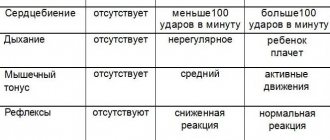
In the first days and months of life, a violation of cerebral blood flow manifests itself in the form of sudden short-term stops in breathing (from the first day), in the form of trembling of the chin, arms or legs, and irregular heart rhythm. The child is restless and often cries a lot, sleeps poorly, has high or low muscle tone in the arms or legs, squint, and a clearly noticeable short neck. Some children have delays in motor development.
After a year, the following problems may arise: delayed motor and emotional development, frequent hysterics, crying, hyperactivity or lethargy, attention deficit, poor memory. Such children may have an increased head size, deformations of the skull and thoracic region, mental abnormalities, convulsions (episyndrome), and paresis of the limbs.
Some consequences appear only in preschool and school age. At that time, few people associate them with the trauma received at birth. These consequences include: dysarthria, impaired fine motor skills, enuresis, headaches, vegetative-vascular dystonia (VSD), increased intracranial pressure, neuroses. At this age, as a result of a birth injury, problems with the spine (scoliosis, poor posture), clubfoot, flat feet, and different leg lengths appear. Consequences also include problems with the immune system, persistent allergic reactions, and diseases of the ENT organs.
Symptoms of a clavicle fracture
A closed clavicle fracture is accompanied by the following characteristic symptoms:
- intense pain in the bone, while the intensity of pain in the collarbone area does not decrease when taking any position;
- limitation of available movements on the side of the bone injury, severe pain when performing previously habitual actions;
- the occurrence of swelling in the area of the shoulder girdle, its shortening;
- changing the standard shape of the collarbone;
- drooping of the shoulder, its displacement inward and slightly anteriorly.
Often, a doctor may suspect a clavicle fracture based on the patient’s characteristic posture. So, a person often holds a limb that has been injured with his healthy hand. The patient may also press the injured limb against the body, explaining this by reducing pain when using this position.
As a rule, in the first seconds of a collarbone injury, a person feels a sharp and severe pain in the supraclavicular or infraclavicular area. The pain syndrome intensifies when the limb hangs freely, which explains the forced position - pressing the injured limb to the body. The free position of the injured side leads to the fragments of the collarbone shifting in different directions, changing their position in the wound, causing the person additional discomfort. Thus, a person needs to make a minimum number of movements so as not to provoke displacement of bone fragments when the clavicle is fractured.
Over time, the body's physiological response to a clavicle fracture is observed. Edema, hyperemia (redness of the damaged area), hyperthermia (increased temperature in the damaged area) is formed. This is a typical inflammatory reaction of the body to trauma to the bone base and soft tissues. Then the pain syndrome increases and the dysfunction of the hand progresses. Above the area of the broken clavicle, a significant swelling is visually visible, corresponding to the area of displacement of the clavicle under the tension force of the sternocleidomastoid muscle.
During an objective examination, the doctor pays attention to the following symptoms of a clavicle fracture:
- the presence of areas of hemorrhage in the damaged area;
- the formation of pathological mobility in the supraclavicular or subclavian region, with any movement causing pain to the patient;
- crepitation (crunching) of bone fragments.
In addition, open fractures are accompanied by the formation of a wound from which some portion of the clavicle protrudes, depending on the area of the fracture. If a collarbone injury has led to a violation of the anatomical integrity of any neurovascular bundle located in the neck, then the patient complains of severe limb weakness, numbness, and crawling.
Often, dizziness and general weakness are added to the list of symptoms of a clavicle fracture, which can be explained by internal bleeding. In severe situations, shortness of breath and lack of air are added to the typical signs of a collarbone injury, which are caused by damage to the pleura covering the lungs by fragments of the collarbone. This leads to the formation of pneumothorax in a person, and is accompanied by symptoms characteristic of such a complication.
How to treat
In your opinion, breaking a child’s collarbone is serious damage to bone tissue. But don’t despair, a child will lose a fracture much faster than it would happen to an adult. This injury is not associated with casting and does not require surgical intervention.
The applied bandage consists of swaddling a part of a limb with a soft roller inserted into the armpit, which fixes the bone in its proper place and prevents it from moving or compromising its integrity during unconscious movements of the newborn.
It is important to ensure that the bandage is not tight. This can lead to poor circulation, a visible sign of which will be the baby's blue fingers. The bandage is applied for a week. It is better to entrust the first dressing to a doctor who will show you how to do it correctly and prescribe medications that promote healing, pain relief and relieve swelling.
The elimination of hematomas is facilitated by Traumeel C ointment, which, due to its properties, has a fairly effective effect on the damage and contains natural ingredients and useful minerals. According to the manufacturers' recommendations, the drug can be used from the moment of birth and supplemented with intramuscular injections of vitamin K. This complex will not only numb the area, but also relieve inflammation.
During health restoration procedures, the baby should sleep and feed only on the healthy side.
Complications
Like any disease, a bone injury can be complicated by many pathological conditions if treatment for a clavicle fracture is not started in time. Fortunately, complications from injury are quite rare because patients often seek medical attention immediately. However, possible complications due to poor-quality or untimely medical care cannot be excluded:
- Injury to a vascular or nerve formation. A displaced clavicle fracture can cause damage to a large vascular trunk or nerve formation, which leads to the formation of characteristic symptoms. In the case of damage to the nerve fiber, disturbances in the motor or sensory sphere in the injured area are observed as long-term consequences of the damage. The severity of neurological symptoms depends on the nature and extent of nerve damage, however, one should not forget about the likelihood of injury to nerve fibers. Damage to the vessel often leads to serious bleeding, especially if a sharp bone fragment has injured a large major vessel, resulting in serious blood loss.
- Damage to the pleura . A life-threatening condition for a person, pneumothorax, is formed when displacement during a clavicle fracture causes damage to the parietal pleura. Air enters the pleural cavity, which is accompanied by a characteristic clinical picture (shortness of breath, lack of air, lag of one of the halves of the chest when breathing). Pneumothorax requires prompt action by medical professionals and immediate treatment.
In most cases, fracture complications can be prevented thanks to competent and proper care for a clavicle fracture.
Causes of birth injuries to the collarbone in infants
Even after completing all the preparatory measures, unforeseen circumstances cannot be avoided. During childbirth, the body of a woman and baby experiences enormous stress - the nervous, cardiovascular and musculoskeletal systems take the brunt. The baby's bone tissue is not yet fully formed, which is why the child's bones and joints are quite flexible and fragile. There is no need to worry prematurely, since bone damage during childbirth goes away without a trace in the vast majority of cases.
Why does dislocation happen?
The shoulders are the widest part of the baby's body, and, for anatomical reasons, are inactive, which significantly increases the likelihood of displacement or fracture in this area during birth. Clavicle injuries (especially the right one due to fetal position) are the most common injuries.
Dislocation is the release of the head of the bone from the articular cavity under the influence of mechanical factors (pressure, stretching, sudden displacement). When the baby comes out, the obstetrician can help him by pulling his arm. Among the reasons, one can note congenital underdevelopment of the joints, which can also cause the clavicle bone to come out of the joint.
For what reasons is a fracture possible?
A fracture is primarily a mechanical damage to the collarbone that occurs due to a load that is not typical for this part of the body.
The causes and factors that increase the risk of a fracture are in many ways similar to the reasons for joint displacement in an infant:
- the impact of medical personnel on the position of the child (when presentation changes), the use of forceps, vacuum extraction,
- atypical presentation of the fetus (transverse, breech), in such positions children are often born with injuries,
- rapid labor process - the child does not have time to take the correct position, which leads to a fracture or other injuries,
- narrow and unprepared birth canal.
Diagnosis of a clavicle fracture
Diagnosis of a clavicle fracture with or without displacement is carried out by a traumatologist. As a rule, the doctor performs the entire list of diagnostic measures in the emergency department of a hospital or in a trauma center. The main methods that the doctor uses are as follows:
- Objective research data . The traumatologist conducts a thorough examination of the injury site. A clavicle injury is characterized by severe deformation of the area, severe swelling of the damaged area, and in some cases, especially with a displaced fracture of the clavicle, crepitus (crunching) of bone fragments is observed. During an objective examination, the doctor necessarily checks for the presence of neurological damage: determines the sensitivity of the hand on the side of the injury, the presence or absence of active movements. In addition, the doctor needs to evaluate possible complications. Thus, during the examination, the traumatologist determines the presence of symptoms of blood loss due to damage to large vascular bundles or difficulty breathing, shortness of breath as symptoms of the formation of pneumothorax.
- X-ray diagnostics. X-ray is the standard examination method for suspected clavicle injury. With its help, the doctor determines the site of the fracture and the displacement of the collarbone. A clavicle fracture in a child is often accompanied by an angular injury. In this case, displacement of bone fragments occurs, however, the integrity of the periosteum is preserved. X-rays are traditionally performed in a direct projection, but in difficult cases, if the traumatologist is unable to determine the location of all fragments of the clavicle, the diagnosis is additionally carried out in a lateral projection.
- An additional research method, if X-ray diagnostics is insufficiently informative, is computed tomography . Three-dimensional reconstruction allows you to fully see the picture of the clavicle injury and determine the degree of displacement of bone fragments when the clavicle is fractured.
Finally, if the traumatologist suspects complications in the form of trauma to large neurovascular bundles, he refers the patient for consultation with a neurologist and vascular surgeon.
Fracture of the collarbone in a newborn during childbirth
A clavicle fracture in a newborn, sustained during childbirth, is a partial or complete violation of the physiological integrity of the clavicular bone. This type of birth injury is one of the most common. This usually happens at the final stage of the birth process, when the baby is helped to be born using various physical influences (pulling the handle, using forceps or a vacuum). Causes of occurrence The shoulders of a newborn are the widest part of his body, therefore it is the clavicle bone that is subject to the greatest load, especially at the final stage of labor. A clavicle fracture can occur:
- if the size of the mother’s pelvic bones does not correspond to the size of the child;
- due to the use of auxiliary methods during childbirth (instrumental application of forceps or vacuum);
- due to the rapid progress of labor (the birth canal and pelvic bones do not have time to open sufficiently);
- if the baby was in a breech or transverse presentation until birth.
A fracture can also occur if the doctor was not very careful in his actions during obstetrics. Symptoms The clinical picture of such an injury directly depends on the type of fracture. So, if a partial fracture occurred (the upper part of the periosteum was not affected, but only the inner part was damaged), then the symptoms will not be noticeable immediately. Doctors may suspect such a fracture even a few days after birth. In this case, the child experiences:
- absence or only partial impairment in the motor activity of the affected arm;
- on days 2-3 a slight swelling may appear at the site of injury;
- the area of soft tissue and skin above the fracture becomes bluish, and a hematoma occurs.
If the collarbone is broken, but both ends of the bone remain in their normal physiological position, then they speak of a fracture without displacement. The symptoms in this case will be more pronounced:
- on the first day after birth, the child develops swelling and hematoma at the site of injury;
- on palpation you can hear crackling and clicking sounds;
- the child is restless, appetite becomes noticeably less;
- Swaddling or other pressure on the injured arm causes sudden crying.
A displaced fracture is characterized by a condition where parts of the broken bone are in different planes from their normal location. This type of fracture is the most serious and causes the greatest concern on the part of the child:
- the newborn refuses to breastfeed;
- a violation of the motor activity of one of the hands is clearly recorded;
- the soft tissues over the damaged area swell greatly;
- the hematoma is pronounced;
- any physical action with a sore arm is accompanied by sharp crying.
Treatment First, the newborn will have an x-ray to determine the presence and type of fracture. If the diagnosis is confirmed, then the baby’s sore arm is fixed to the body using a soft elastic bandage. In this case, a roll of cotton wool and a bandage is placed in the armpit. Wearing this bandage for a non-displaced fracture takes 7-10 days, and after surgery the wearing time increases to 25-30 days. In addition to fixing the limb to relieve symptoms of hematoma and tissue swelling, vitamin K is administered intramuscularly for 3 days. To relieve pain, it is possible to use local agents (ointments) with an analgesic and regenerating effect. Throughout the entire treatment period, the baby should not be turned over or placed on the side of the body on which the fracture is located. Usually the damaged bone heals after 20-25 days. And only with complex displaced fractures may surgical intervention be necessary. The recovery time increases to 1.5-2 months. Rehabilitation
- At the final stage of treatment and upon its completion, additional procedures are recommended:
- electrophoresis (by applying medications and exposure to low power current, regeneration and healing processes are accelerated);
- massage (performed only after completion of treatment);
- magnetic therapy;
- therapeutic exercises (after consultation with a specialist, the mother herself is able to carry out physical therapy exercises).
- The use of any additional methods of treatment and recovery is carried out only in consultation with a doctor.
Consequences The baby’s body is fragile, but accelerated metabolism and an active period of growth contribute to the rapid healing of the damaged bone. After a collarbone fracture, with adequate treatment, the baby will not have any cosmetic or physical defects.
Treatment
The therapeutic tactics chosen by the doctor depend on many parameters. the degree of clavicle fracture matters , whether the clavicle fracture occurred in a child or an adult, and whether there is displacement of bone fragments. In addition, when deciding on treatment tactics, the doctor also evaluates the presence of developed complications. If there is no threat of damage to large nerve or vascular formations, and a person has a well-reduced clavicle fracture, then the patient does not require long-term hospitalization.
If there is a high risk of complications or exacerbation of the clinical situation, then treatment of the collarbone must be carried out in the trauma department. According to statistics, in a significant number of cases, patients do not require surgical intervention; only ordinary therapeutic treatment is sufficient.
Providing first aid for a fracture
At the first stage, you must definitely call an ambulance . Within a few minutes after a clavicle injury, active swelling begins to form in the damaged area, which subsequently creates difficulties in performing closed reposition of the fragments. The more time passes, the more likely it becomes that the patient will undergo open reduction. Treatment of a broken collarbone by a traumatologist consists of a surgical operation, during which special access to the site of injury is performed, and then comparison of bone fragments. Of course, if closed reduction is not possible, then the open method is used to treat the clavicle. However, when performing an open surgical operation, the risk of infectious agents, pathogenic microorganisms entering the wound, as well as the occurrence of purulent complications (abscess, phlegmon, secondary osteomyelitis) naturally increases. The ambulance transports the victim to the emergency department of the trauma hospital, and during the provision of first aid, medical workers will provide high-quality pain relief, as well as immobilization of the fracture site. Thus, the ambulance carries out primary actions aimed at reducing the likelihood of complications developing in the long term, as well as transporting the patient as carefully as possible. It is important to remember that a certain amount of time will pass before the ambulance arrives, which can be properly used to alleviate the patient’s condition.
The main rule that you need to remember when providing assistance with a fracture of the collarbone is that you cannot independently try to carry out medical procedures to eliminate the displacement of the collarbone by performing sudden movements or jerks. It is imperative to limit the mobility of the injured limb : due to sudden movements, secondary displacement of fragments or complications may occur. Competent first aid consists of immobilizing a limb (reducing its mobility by fixing it to the body) using a scarf. In this case, the arm must be fixed in a special way: press the humerus to the body, and only then bandage it using a scarf. However, there is a rule: if a person reports increased pain during shoulder adduction, then it is necessary to leave the injured limb in a position that provides minimal pain.
Immobilization is considered an important aspect when providing first aid. Primary immobilization before the arrival of the ambulance team will reduce the severity of pain in the patient, because the injured area will be firmly fixed and immobilized with a bandage. Secondly, immobilization will help to avoid the development of complications in the form of traumatization by bone fragments of vascular or nerve formations during transportation of the injured person to the emergency department of a trauma hospital. For immobilization, a scarf bandage is actively used on the arm, during which the limb is bent at the elbow joint and pressed against the body. It should be remembered that during long-term transportation of the patient to the hospital, the supporting function of the bandage weakens over time. Therefore, rewinding of the bandage may be necessary to maintain sufficient fixation force. Often, for immobilization, in addition to the scarf dressing, the following types of dressings are used:
- Velpo;
- Deso;
- 8-shaped bandage;
- Delbe rings.
We must not forget about painkillers . Pain from a broken collarbone is a strong irritant to which a person may have an unexpected reaction. This, in turn, can complicate the provision of first aid to the victim: he may resist and refuse any action. Besides. intense pain during the arrival of an ambulance will complicate the diagnosis of the condition and the collection of anamnesis, because the person will not be able to correctly and clearly answer the doctor’s questions. Non-steroidal anti-inflammatory drugs are actively used as painkillers at home. Ketanov, Ibuprofen, Spazmalgon, Paracetamol are actively used. However, before using these medications, you need to be sure that the person is not allergic to one or more components of the drug. In addition, non-steroidal anti-inflammatory drugs should be used very carefully in patients with gastric or duodenal ulcers. The drug will begin to act within 15-30 minutes when taken orally, 5-10 minutes after intramuscular administration. If the effect of the drug is not observed, you cannot increase the dosage or introduce a new dose of the drug, because there is a possibility of an overdose of the drug, which requires special assistance for intoxication.
If you do not have the opportunity to use painkillers to relieve pain, then you can use a proven and effective method - applying cold to the injured area. Cold has a local effect without affecting other tissues and systems of the human body. Additionally, applying cold is the only option if you don't know if the person has an allergic reaction to the pain medications available to you. The mechanism of action of cold is very simple: in the area of the collarbone fracture, a cold object reduces the temperature of the soft tissues, where many nerve endings are located. When the tissue reaches a temperature of 4-5 degrees, the intensity of the transmission of pain impulses in it decreases, which means that irritation does not enter the brain. Thus, there is a temporary reduction in the severity of pain in a person. However, a broken collarbone requires a special approach in applying cold. You should not apply a whole piece of ice to the damaged area, but crushed ice. The specificity is explained by the anatomical structure of the area: in the clavicle area there is a supraclavicular and subclavian fossa, which forms an uneven relief. Crushed ice will work much better on the damaged area than a single piece of ice. In addition, you need to apply ice exclusively over the fracture site: if you move the ice pack to the neck area, the body may react in the form of a sharp drop in blood pressure and heart rate. The fact is that in the neck area there is a sinocarotid nerve plexus, and careless influence on this formation can lead to serious consequences.
about our treatment of birth trauma
As an alternative to standard treatment of birth injury, we offer a rehabilitation complex aimed at eliminating the consequences of birth injury to the neck and restoring cerebral blood flow.
It is important to restore cerebral blood flow for the proper functioning of the central nervous system. When disturbances in the functioning of the brain are eliminated, the child begins to develop correctly and the manifestations of birth trauma disappear. With proper trauma treatment, not only the injury itself is eliminated, but also its neurological consequences, in the form of specific disorders in the child.
The essence of treating any injury is traction and fixation. To treat birth trauma in children, we use an orthopedic splint of our own design, which provides measured traction and fixation of the cervical spine. This product is called a “traction collar”, patented (Patent No. 2587960) and has been successfully used for more than 25 years.
At the first stage, damaged anatomical structures of the cervical spine are treated. Thanks to traction and therapeutic exercises, in a short time the displaced vertebra is realigned, muscles are strengthened, neck ligaments are fused, and hematomas are resolved. The collar has a beneficial effect on the state of blood supply to the brain by releasing blood vessels from compression. Without this stage, the effectiveness of treatment of birth trauma is reduced several times.
At the next stages, therapy is carried out aimed at improving the functioning of blood vessels and brain cells (including amino acids), resolving deep hematomas and scars near the vertebrae, restoring motor functions of the cervical spine and neck muscle tone.
Treatment is carried out on an outpatient basis (outside the hospital) and involves the child wearing a traction collar of an original design (
) in combination with taking natural medications to normalize brain activity.
Our treatment quickly and effectively solves the problem of birth trauma in a child, while it is safe and reliable.
The first effects are felt within a few days from the start of treatment: the child’s well-being improves significantly
Maximum effectiveness is achieved thanks to a fundamentally different approach to solving the problem of birth trauma - our treatment is aimed at eliminating the cause, rather than suppressing the symptoms of the disease
The treatment we offer is completely safe, since the traction collar does not have a negative effect on the child’s body, and the prescribed drugs do not cause complications or side effects
After the course of treatment, seizures no longer occur, since during treatment the main cause of the disease is eliminated and the natural functions of the brain are restored
Thus, the traction therapy we offer, due to its obvious advantages, is an excellent and often the only alternative not only to taking anticonvulsants, but also to other methods of treating birth trauma.
Who should I contact?
Letay.org is the best assistant in finding a doctor!
The organization is engaged in providing treatment for complex orthopedic injuries in clinics in Spain, selecting a attending physician for each patient in accordance with the established diagnosis. Letai will organize a preliminary consultation with a doctor for you, find a hotel and arrange a flight. Letai guarantees examination and treatment in the best Spanish clinics!
Why will you definitely choose Letai? Due to the large number of advantages of the organization:
- Modern equipment and use of innovations. Letai uses only the latest techniques for treating and rehabilitating patients. This allows you to shorten the period of hospital stay or treatment. In addition, innovative approaches ensure maximum restoration of the functions of the human body in a fairly quick time.
- Fast reaction. Letai is not affiliated with any one clinic. This is a significant advantage, because mobility allows you to organize an operation for the patient in the shortest possible time!
- Doctors are professionals. Letai provides patients with the opportunity to be treated by the best Spanish doctors who have significant clinical experience. Specialists from the best clinics in Spain, recognized in European countries, and some throughout the world, guarantee patients high-quality treatment and a responsible approach to therapy!
- Price policy. It is worth noting that the cost of all services provided by Letai is within acceptable limits for each person! Thus, anyone in need of quality treatment can take advantage of Letai’s help in the treatment and rehabilitation of injuries.
Letai's activities cover three main areas:
- Joint diseases. An experienced orthopedic doctor will diagnose and treat diseases of the human musculoskeletal system. Why endure discomfort if you can turn to a competent specialist who will ensure timely diagnosis and correct treatment?
- Treatment of injuries. Often, patients come to Letai because of an injury that causes significant pain and significant discomfort. The doctors of our clinics guarantee treatment of any injuries according to the latest protocols used in the world!
- Rehabilitation after injuries. The recovery period after injury is no less significant. Doctors at Spanish clinics will write you an individual rehabilitation program, and then monitor the dynamics and adjust the plan depending on the activity of restoring the functions of the musculoskeletal system.
Letay.org will find the best doctor for your speedy recovery and quick rehabilitation!
Procedures and care for a child during the rehabilitation period
During the recovery period, it is necessary to strictly follow the recommendations of specialists. In the first week after the injury, decongestants may be prescribed for the hematoma (administration of vitamins K), if there is one. The consequences of the bone coming out of the joint pass faster than with a fracture, however, even such an injury in newborns heals within 7-10 days, if there are no complications. The rehabilitation period may take about 30 days.
READ ALSO: natal birth injury of the cervical spine in a newborn
After the bone has healed, procedures may be prescribed to restore mobility, such as massage and exercise therapy. In some cases, electrophoresis or magnetic therapy is used. Sometimes the attending physician may prescribe vitamins (calcium, magnesium, phosphorus) or recommend adding foods rich in these elements to the mother’s diet (for breastfeeding).
To read: Shaping underwear for women after cesarean section and childbirth for the abdomen, hips and waist: can you wear it, how to choose?
Treatment in children
The main method of therapy is strong fixation, that is, immobilization of the limb. In children, the immobilization time is 2-3 weeks. To firmly hold broken bone fragments, special Delbe rings are often used. Pediatric clavicle fractures often occur as a “greenstick” type, that is, with preservation of the integrity of the periosteum. In this case, a fixing bandage on the collarbone is used. A fixing bandage is sufficient even if the clavicle injury is not accompanied by displacement.
In cases where the fracture is characterized by displacement of fragments, reposition is performed.
Is it possible to foresee?
There are risk factors that every doctor knows about. Among them:
- fetoplacental insufficiency (disturbances in the “mother-placenta-fetus” system), various types of hypoxia and asphyxia;
- discrepancy between the size of the child’s head and the mother’s pelvis (the baby is too large, and the woman’s pelvis is too narrow), including against the background of diabetes mellitus in the mother;
- incorrect fetal position (for example, breech or transverse presentation);
- premature or delayed birth;
- rapid or prolonged labor.
In these cases, competent management of childbirth is very important. The doctor must assess the risks and, if necessary (for example, a woman is giving birth to a “hero” or the green water has broken, which means the child is experiencing hypoxia), perform a caesarean section on time.
Treatment in adults
Immobilization of the limb is carried out for 1 month, using a Chizhin frame or other bandages. If displacement of the fragments occurs, then reposition is required (returning the fragments to their normal physiological position) with local anesthesia. Then fixation occurs with a soft or plaster cast.
The reposition is followed by a recovery period, and after some time a control photograph is taken. It is actively used during the rehabilitation period of UHF; in the presence of severe pain, the doctor prescribes painkillers. As soon as the need for immobilization disappears, the doctor refers the patient to massage and physical therapy.
Conservative treatment consists of using soft or plaster casts, as well as fixing the limb. This type of therapy is used for uncomplicated fractures in both adults and children. Immobilization is carried out by a traumatologist who actively monitors the restoration of the integrity of the patient’s bone.
The main goal of conservative therapy is to ensure the correct position of the bone and rigid fixation of the clavicle in this position for a certain time. If displacement of the fragments has not occurred, then immobilization of the shoulder girdle is sufficient for high-quality healing to occur. In cases where displacement has occurred due to a clavicle fracture, preliminary reposition is required; at the next stage, immobilization occurs. In traumatology, there are two types of reposition - closed and open techniques.
Closed reduction is non-invasive, that is, the doctor does not perform an open surgical approach. Using non-invasive medical techniques, the traumatologist performs temporary immobilization, and then x-rays are taken. If the bone fragments have been juxtaposed correctly, then temporary immobilization is changed to permanent plaster fixation. In the case where the bone fragments could not be compared as accurately as possible, a repeated closed reduction or open reduction is performed according to the decision of the traumatologist.
Open reduction has a number of strict indications, because during its use open surgical access to the fracture area is performed. Then the doctor compares the bone fragments in the wound and fixes them, which is called intraosseous osteosynthesis and refers to the surgical treatment of a clavicle fracture.
debunking myths about birth trauma
Today, there are a number of misconceptions about the problem of birth trauma in children, generated mainly by the lack of objective information among parents who are faced with this problem about the causes of birth trauma and the proposed treatment. We will try to clarify this issue and debunk the most popular myths.
Osteopathic sessions are the best method of treating birth trauma
Indeed, many deformities of the skull and spine can be corrected with the help of osteopathy, compression and twisting can be eliminated. But the spasm of the cervical muscles in the suboccipital region after a birth injury is so strong that it cannot be overcome by hand, and therefore the reduction of many subluxations of the atlas is problematic for an osteopath. Also, osteopathic techniques cannot dissolve microscars that form during childbirth due to hemorrhages in the soft tissues of the neck. If the child continues to have increased muscle tone in the neck, pain in the suboccipital region and asymmetrical turns of the head, or NSC in the SHOP and PPCNS, then it is necessary to undergo complex treatment by a vertebroneurologist.
Osteopathy is enough to treat birth trauma
An osteopath does not restore damaged brain tissue in severe trauma (PPCS). An osteopath eliminates various birth deformities of the skull and spine as one aspect of a birth injury, but does not treat the injury as a whole. Treatment of birth trauma should occur in stages, affecting all damage and disorders: elimination of pathological spasm of the cervical muscles, fusion of vertebral ligaments, realignment of displaced vertebrae, resorption of scar tissue and restoration of nerve cells in the brain.
Chiropractors adjust vertebrae following a birth injury
Manual therapy is extremely dangerous for birth injuries of the cervical spine, especially in infants. A chiropractor, seeing external signs of a neck injury, conducts manual sessions using rough jerking techniques. There are cases when, after manual sessions, a child’s neurological symptoms intensify, since during the sessions, vertebral ligaments and neck vessels can be additionally damaged, internal hematomas can form, followed by the formation of scars.
Birth trauma treated with massage
Medical neck massage is often prescribed for birth trauma to relieve pathological spasms of the neck muscles. The cervical spine can be massaged at the final stage of treatment of a birth injury, as well as any injury in general, after realignment of the vertebra and pain relief. At the beginning of the course of treatment, traction and rest are required. Massaging an injured neck is painful for a child, so he cries loudly during it. Even after several courses of massage, spasm of the neck muscles cannot be eliminated, since the spasm occurs reflexively in the presence of displaced vertebrae.
An osteopath and a chiropractor can diagnose a birth injury with their hands, and it is not necessary to take an x-ray
Using your hands you can diagnose spasms of the neck muscles, pain in the vertebral joints, ligaments, and incorrect position of the vertebra. To confirm the diagnosis of subluxation and to monitor the position of the vertebra after treatment, we recommend taking x-rays in three projections. An x-ray gives a complete and objective picture of the condition of the cervical spine BEFORE and AFTER treatment. This is how injuries and ruptures of the ligaments of the cervical vertebrae are detected, which cannot be diagnosed manually. The x-ray also reveals abnormalities in the development of the spine, in which manual therapy is contraindicated, and treatment by an osteopath is ineffective.
Birth trauma does not need to be treated; it goes away with age
Birth trauma as an acute condition passes, but its unresolved consequences appear throughout life, replacing each other. In case of birth trauma, the existing high tone of the cervical muscles, displaced vertebrae, compressed vessels and nerve endings lead to disruption of blood flow at the level of the cervical spine (NSC in the CS) and the brain (PCNS), which determines the presence of neurological complaints. For example, in childhood – poor sleep, hyperactivity, speech disorders, enuresis, etc.; in adults – headaches, increased fatigue, heart rhythm disturbances, etc. Therefore, birth trauma must be treated.
Birth trauma is always manifested by an inclined position of the head (torticollis)
Indeed, the cause of an inclined head position (i.e. torticollis) in 95% of cases is a birth injury. But birth trauma of the cervical spine occurs without torticollis or other visible deformities of the neck or head. Signs indicating a birth injury to the neck include vascular spots on the forehead or back of the head, as well as neurological symptoms that are difficult to treat. To identify a birth injury to the neck without external signs, for children under 3 years of age, we prescribe an ultrasound scan of the neck, an ultrasound scan of the vessels of the neck and brain, and after 3 years, an X-ray of the cervical spine in three projections and an ultrasound scan of the vessels of the neck and brain.
When treating a birth injury in the Shants collar, the neck muscles atrophy
This is indeed a widespread myth, as muscle atrophy is often observed after treatment of arm or leg injuries in a cast. The neck muscles belong to the axial group of muscles and are constantly toned to maintain a vertical position of the head. Atrophy of these muscles can only develop after damage to the nerves through which the muscles receive impulses. Treatment in our traction collar is safe for the muscles, as the neck muscles receive nerve impulses and maintain their tone. In addition, the neck muscles can be trained with special exercises even in a collar, which will ensure the creation of additional good tone of the neck muscles, including in infants.
Foam collar treats birth injury to neck
The foam collar acts as a temporary fixation bandage to stabilize the cervical spine. Treatment of birth trauma in a foam collar is impossible, since it does not provide uniform long-term neck traction, without which the spasmodic neck muscles cannot be relaxed and the displaced vertebra cannot be realigned. To do this, we use a traction (pulling) collar of our own original design, which provides conditions for the successful treatment of birth trauma, namely: elimination of muscle spasm and displacement of the vertebrae, fusion of damaged ligaments.
As can be seen from the examples described above, an objective look at the problem of birth trauma from the point of view of the causes of its occurrence and methods of solution helps the parents of a child who has been diagnosed with this disease to promptly and thoughtfully approach the choice of an effective and safe treatment method.
Surgery
surgical treatment is necessary if the patient has an open fracture of the clavicle or a closed injury, however, complicated by acute conditions. For example, a bone fragment damaged a large nerve trunk, which caused a disruption of the innervation on the side of the injury or a major vessel, which caused bleeding. In some cases, bone fragments damage the parietal layer of the pleura, which threatens the development of an emergency condition - pneumothorax.
Surgical interventions are routinely performed for a clavicle fracture if the patient has a significant displacement of bone fragments or the shoulder girdle area is externally significantly deformed. Surgical treatment consists of performing osteosynthesis of the clavicle using one of the following techniques:
- Intraosseous method. Used for comminuted fractures. During the operation, a special pin or Bogdanov nail is used.
- Bony technique. During the operation, a curved plate is used. This method is used for injuries with many fragments in the wound.
- Spoke method . Rigid fixation is performed using wires that are literally passed through fragments of the bone base. The ends of the knitting needles are fastened, and they are first brought outside the collarbone.
In the postoperative period, antibiotics are used to prevent the development of infectious complications, and painkillers are used to relieve pain. Physiotherapeutic procedures provide faster and more complete restoration of the functions of the injured limb. Discharge from the hospital becomes possible after the stitches are removed, which occurs on the 8-10th day of hospital stay.
We suggest adding your company to a thematic directory of organizations, for example in Sport365days - this is an excellent example of a thematic aggregator tied to topics such as sports, health, sporting goods and beauty. If you are the owner of a business focused specifically on these areas, you can create your own website absolutely free, find business partners with the ability to post information on your page or a partner’s page. (Example: Medart).
Recovery
Physiotherapy is widely used in medicine for more active recovery.
Physiotherapeutic methods of rehabilitation after a clavicle fracture are actively used in medicine to accelerate the healing of the injury, as well as reduce pain and discomfort for the patient. In addition, physiotherapy has an anti-inflammatory, anti-edematous effect, stimulates metabolism in the area of damage and tissue regeneration.
The classification of physiotherapeutic procedures is based on the time of their implementation. Thus, there are methods of physiotherapy that are used only during the period of plaster immobilization, after removal of the plaster, and also regardless of whether the patient has a plaster cast.
During plaster immobilization, SUV irradiation in erythemal doses . At the local level, the procedure provides relaxation of muscle tissue in the damaged area, expansion of the capillary network, which stimulates blood flow in the irradiated tissue. Finally, tissue swelling is reduced, as well as the severity of pain due to a decrease in the sensitivity of pain receptors. The general effect of SUV irradiation in erythemal doses is manifested in stimulating the formation of vitamin D, which accelerates the process of callus formation. The second procedure, electrophoresis of painkillers, ensures the accumulation of painkillers in the subcutaneous fat and muscle tissue, which long-term reduce the intensity of the patient’s pain syndrome.
After the moment when a person's plaster cast is removed, it is possible to use new physiotherapeutic procedures. If the traumatologist has removed the plaster, then the callus has already formed quite well, which means that it is possible to put small loads on the bone to increase its strength. For this purpose, therapeutic massage, high-frequency magnetic therapy, amplipulse therapy, UHF therapy, ultrasound therapy, and remote shock wave therapy are used.
Therapeutic massage provides active and persistent dilation of blood vessels in the injured area. Active blood flow accelerates the transformation of formed callus into healthy functioning bone tissue. Therapeutic massage has a significant overall effect through a reflex effect on the vasomotor center located in the brain. Thanks to therapeutic massage, blood pressure in patients is normalized.
High-frequency magnetic therapy provides an analgesic effect by influencing nerve tissue. In addition, the tension in the patient's skeletal muscles decreases.
Amplipulse therapy creates an analgesic effect and leads to relaxation of muscle fibers. The physiotherapeutic method actively influences blood vessels, dilating them, which enhances tissue trophism. Thus, amplipulse therapy stimulates the reduction of swelling, the resorption of infiltrates, and also activates recovery processes in the injured area.
UHF therapy creates an active warming effect, which directly affects cellular metabolism. Thus, microcirculation processes are activated, the muscle layer of blood vessels relaxes, and the swelling of the damaged area decreases. As a general effect, UHF therapy reduces the tone of the sympathetic nervous system, increasing the tone of its parasympathetic department.
Ultrasound therapy actively stimulates blood circulation processes, as well as lymph circulation. At the local level, capillary permeability increases, which affects the intensity of wound healing.
Remote shock wave therapy activates the synthesis of special biologically active components. They provide vasodilation in the area of injury, which stimulates the processes of proliferation of cells and tissues in the injured area. Cell division ensures the direct process of repair of the bone and cartilage base.
Regardless of immobilization, electrophoresis of vasodilators, low-frequency magnetic therapy and mineral water intake can be performed. Low-frequency magnetic therapy becomes an excellent stimulating factor for reparative processes. In addition, this method has a good anti-inflammatory effect. Mineral waters restore the balance of electrolytes in the human body and provide the necessary minerals for the active healing of damaged bone and cartilage tissue.
Question answer
What is the prognosis for clavicle fractures?
In the majority of cases, if a bone injury occurs without complications from other pathological conditions, doctors note a favorable prognosis for the patient. As a rule, bone fragments heal well, and even if there is some residual displacement, this does not affect the active movements of the arm on the injured side. In the case when a person develops complications (for example, damage to the pleura or a large nerve trunk, a great vessel), the outcome of the disease depends on the quality, time and literacy of first aid provided to the patient.
What is the patient's recovery time after surgery?
Absolute restoration of the structure of the clavicle and the function of the damaged limb occurs within 6 to 8 months. After surgery, removal of auxiliary structures (wires, screws or medical plates) is performed approximately 6-12 months after the intervention, depending on the intensity of the restoration processes in the bone.
Typical symptoms of birth trauma
A collarbone injury is diagnosed immediately after childbirth, but sometimes the initial examination reveals nothing. According to some signs, it is determined over the next few hours or days:
- swelling and/or redness, hematoma in the clavicular area (we recommend reading: how is a hematoma on a child’s head that appears during childbirth treated?),
- pain when touching the injured area,
- decreased appetite, anxiety,
- limited limb mobility,
- crunching sound when you move your hand,
- uncharacteristic position of the shoulders relative to each other,
- hand hanging,
- incorrect head position.
To read: How is an ultrasound of the abdominal organs performed on children: how to prepare for the examination and what does it show?