Definition of newborn asphyxia
Translated from Latin, asphyxia means suffocation, that is, lack of oxygen. Asphyxia of newborns is a pathological condition in which gas exchange in the newborn’s body is disrupted, which is accompanied by a lack of oxygen in the child’s tissues and blood and the accumulation of carbon dioxide.
As a result, a newborn who was born with signs of a live birth either cannot breathe independently in the first minute after birth, or he experiences isolated, superficial, convulsive and irregular respiratory movements against the background of an existing heartbeat. Such children are immediately given resuscitation measures, and the prognosis (possible consequences) for this pathology depends on the severity of asphyxia, the timeliness and quality of resuscitation.
Causes
There are a number of reasons why asphyxia may develop. Experts group them into groups:
- The cause may be a lack of oxygen in the mother's body or an excess of carbon dioxide. This happens if the mother has diseases of the cardiovascular or respiratory system, if the mother suffered severe shock during pregnancy, or if the mother’s body is intoxicated.
- The umbilical cord entwined around the fetal neck is a serious and one of the most common causes. If the attending physician was unable to prevent entanglement, then he will no longer be able to rid the fetus of it.
- In case of disturbance of placental circulation. It can be a consequence of post-term pregnancy, it can occur during late toxicosis, in the presence of pathologies of the umbilical cord or placenta. During childbirth, this pathology, as a rule, causes oxygen starvation of the fetus.
- The cause may be various diseases of the fetus associated with the cardiovascular or respiratory system, birth injuries and much more.
Every pregnant woman should be attentive to her health and the health of her unborn child. Asphyxia is a serious problem that, unfortunately, is becoming increasingly common among young mothers.
Factors provoking the development of asphyxia
This pathological condition is not an independent disease, but is only a manifestation of complications during pregnancy, diseases of the woman and the fetus. Causes of asphyxia include:
Fruit factors
- birth injury (traumatic brain injury) in a child;
- Rhesus conflict pregnancy;
- anomalies in the development of organs of the bronchopulmonary system;
- intrauterine infections;
- prematurity;
- intrauterine growth restriction;
- obstruction of the respiratory tract (mucus, amniotic fluid, meconium) or aspiration asphyxia;
- malformations of the heart and brain of the fetus.
Maternal factors
- severe gestosis occurring against the background of high blood pressure and severe edema;
- decompensated extragenital pathology (cardiovascular diseases, diseases of the pulmonary system);
- anemia of pregnant women;
- endocrine pathology (diabetes mellitus, thyroid disease, ovarian dysfunction);
- woman's shock during childbirth;
- disturbed ecology;
- bad habits (smoking, drinking alcohol, taking drugs);
- insufficient and malnutrition;
- taking medications contraindicated during gestation;
- infectious diseases.
Factors contributing to the development of disorders in the uteroplacental circle:
- post-term pregnancy;
- premature aging of the placenta;
- premature placental abruption;
- umbilical cord pathology (umbilical cord entanglement, true and false nodes);
- constant threat of interruption;
- placenta previa and bleeding associated with it;
- multiple pregnancy;
- excess or lack of amniotic fluid;
- anomalies of labor forces (weakness of labor and incoordination, rapid and rapid labor);
- drug administration less than 4 hours before completion of labor;
- general anesthesia for women;
- C-section;
- uterine rupture;
Secondary asphyxia is provoked by the following diseases and pathologies in the newborn:
- impaired cerebral circulation in a child due to residual effects of damage to the brain and lungs during childbirth;
- heart defects that were not identified and did not appear immediately at birth;
- aspiration of milk or formula after a feeding procedure or poor-quality sanitation of the stomach immediately after birth;
- respiratory distress syndrome caused by pneumopathy: the presence of hyaline membranes;
- edematous-hemorrhagic syndrome;
- pulmonary hemorrhages;
- atelectasis in the lungs.
Mechanical asphyxia
Asphyxia is suffocation caused by compression of the respiratory tract, closing of their lumen with mucus, food, compression of the neck, chest and abdomen and leading to oxygen starvation and excessive accumulation of carbon dioxide in the tissues and cells of the body, which threatens severe disorders of the nervous system, breathing and circulation , can lead to the death of a child.
In a newborn, this condition can be caused by the following reasons:
- the presence of an external obstacle to free breathing: a diaper or corner of a blanket may fall on the child’s face; the baby may bury himself or crawl under the blanket when he sleeps restlessly or wakes up, covering the child’s airways with a soft object (pillow, toy);
- frequent regurgitation or vomiting;
- foreign bodies entering the respiratory tract (small toys, buttons, rings, coins, etc.);
- milk entering not into the child’s esophagus, but into the larynx and bronchi;
- squeezing the neck area with chains, braid with crosses (talismans), or a pacifier;
- playing with plastic bags and packaging (children often put bags on their heads while playing, which tightly fit their face while breathing and prevent air from entering);
- sleeping together in the same bed between the baby and parents or with older children.
PREVENTION OF ASPHIXIA:
- do not leave the child unattended!
- After each feeding, let the baby burp air; to do this, hold the baby upright for a while, do not put him in the crib immediately after feeding; children (especially premature or weakened ones) may burp repeatedly. To prevent inhalation of contents, always turn the baby's head to one side, but if the baby regurgitates frequently and profusely, you should definitely consult a doctor;
- When choosing clothes and bedding for a child, give preference to products without ribbons, laces, laces, ties, etc.
- You should not leave older children near the newborn for a long time or put the baby to sleep with them in the same bed. It is optimal for the baby to sleep in the parent’s bedroom, but not in the parent’s bed, leave the baby crib close to yours, lower the side bars - this will ensure the safety of the baby and you will be in close proximity to your child, it will be convenient to feed him at night, and then, without getting up, transfer to the crib;
- a child's mattress should be semi-rigid, the child should not be placed on a pillow, instead of a baby blanket, you can use a special sleeping envelope, do not use blankets that are too large;
- You should not put ribbons with nipples or chains around the baby’s neck.
Signs indicating the need to urgently seek medical help:
- prolonged holding of breath (more than 20 seconds), accompanied by:
- cyanosis or pallor of the skin;
- rapid or difficult breathing;
- pronounced decrease in muscle tone (lethargy of the child).
First aid procedure:
- call an ambulance immediately;
- clear the child’s airways (remove the diaper, blanket from the face, remove objects squeezing the neck);
- open the window, provide oxygen access to the room;
- if a foreign object (part of a toy, wrapper) is visible in the child’s mouth, try to remove it; do not remove round objects or stuck food, as this can push them deeper into the respiratory tract;
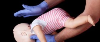
- place the child on your hand (forearm) so that the face rests on the palm, fix the child’s head with your fingers, slightly tilt the body down and make 5-6 tangential pats on the child’s back (between the shoulder blades);
- You can also take the child by the legs, turn him upside down and slap him on the back several times;
- Having wrapped your arms around the child, turn him over onto his back (the head should be lowered below the body) and with 2-3 fingers press 5 times on the central part of the sternum (pressure depth 1-2 cm).
Repeat these steps until the foreign body is cleared from the respiratory tract.
Be careful when removing a foreign body from an infant's mouth! Do this if you see a foreign object and are confident that you will not push it further down your child's throat.
Mechanism of development of asphyxia
It doesn’t matter what caused the lack of oxygen in the body of a newly born child, in any case, metabolic processes, hemodynamics and microcirculation are rebuilt.
The severity of the pathology depends on how long and intense the hypoxia was. As a result of metabolic and hemodynamic changes, acidosis develops, which is accompanied by a lack of glucose, azotemia and hyperkalemia (later hypokalemia).
In acute hypoxia, the volume of circulating blood increases, and in chronic and subsequent asphyxia, the blood volume decreases. As a result, the blood thickens, its viscosity increases, and the aggregation of platelets and red blood cells increases.
All these processes lead to microcirculation disorders in vital organs (brain, heart, kidneys and adrenal glands, liver). Disturbances in microcirculation cause swelling, hemorrhages and areas of ischemia, which leads to hemodynamic disturbances, disruption of the functioning of the cardiovascular system, and, as a consequence, all other systems and organs.
Asphyxia
Asphyxia (from the Greek a - negative particle and sphygmus - pulse) is suffocation and associated acute oxygen starvation with the accumulation of carbon dioxide, characterized by respiratory and circulatory disorders. The literal meaning of the word “asphyxia” does not correspond to its understanding. Asphyxia of the fetus and newborn is understood not as suffocation, but as anoxia, or more precisely, hypoxia, which occurs in the perinatal period and is caused by various reasons. Anoxia is a complete lack of oxygen in tissues. True anoxia almost never occurs. As a rule, hypoxia occurs - oxygen starvation, or oxygen deficiency in the tissues of the fetus. The term "asphyxia" for the state of oxygen deficiency in the fetus and newborn is imprecise. However, this term has become firmly established in medicine and is widely used both abroad and in our country.
Asphyxia occupies a major place in the pathology of the perinatal period and can occur in the ante- and intranatal periods and after childbirth. Ante- and intrapartum asphyxia is called intrauterine asphyxia of the fetus that occurs after childbirth - asphyxia of the newborn.
Antenatal fetal asphyxia. It is directly related to placental insufficiency.
Etiology and pathogenesis of intrapartum asphyxia. Intrapartum asphyxia, in addition to placental insufficiency, which plays a major role in disrupting the fetus’s adaptation to labor stress, can arise from complications of the act of childbirth itself: weakness of labor, breech presentation of the fetus, inconsistency of the fetal head with the mother’s birth canal (narrow pelvis, fetal macrosomia), rupture a short umbilical cord, when a long umbilical cord is entwined around the fetal neck, when the umbilical cord loop falls out and is subsequently pressed by the fetal head, etc.
Etiology and pathogenesis of newborn asphyxia. The basis of asphyxia in a newborn is always a violation of the act of spontaneous breathing. Extrauterine breathing is disrupted when the respiratory center is damaged due to severe intrauterine hypoxia, with massive intracranial hemorrhages that occur both during childbirth and in the newborn, as a result of various reasons that prevent the straightening of the lung tissue and filling it with air. A common reason that prevents the lungs from filling with air is aspiration of amniotic fluid and the contents of the mother’s birth canal when the carbon dioxide content in the fetal blood increases during the intrapartum period. Carbon dioxide irritates the respiratory center of the fetus, and it develops premature respiratory movements, leading to aspiration. With deep aspiration, the contents of the birth canal and amniotic fluid fill not only the bronchi, but also the respiratory sections of the lung and prevent gas exchange; aspiration is more often observed in full-term fetuses, since their respiratory center is more sensitive to hypoxia.
Thus, disruption of extrauterine breathing may be a consequence of intrauterine asphyxia or observed in a newborn born without intrauterine asphyxia, and then depends on the development of pulmonary complications - pneumopathy or aspiration pneumonia.
Pathological anatomy. Hypoxia is accompanied by atony of the walls of blood vessels, primarily the microvasculature, which leads to congestion of the internal organs. With the antenatal death of the fetus from acutely developed oxygen deficiency, only congestion of the internal organs is detected. It is difficult to establish a diagnosis of acutely developed intrauterine asphyxia without a detailed analysis of clinical data. Not only the paucity of morphological changes interferes, but also maceration of the skin and autolysis of internal organs. Chronic oxygen deficiency is accompanied by impaired permeability of vascular walls - damage to the endothelium and basal membrane, which leads to stasis, edema, hemorrhage and subsequent dystrophic and necrotic changes in organs. Hemorrhages are also caused by disorders of the coagulation and anticoagulation systems of the fetus (newborn), and the development of intravascular disseminated coagulation. Multiple fibrin thrombi developing in the microvasculature lead to consumption coagulopathy. The latter, together with impaired vascular permeability, causes hemorrhages.
Macroscopically , dark liquid blood is detected in the cavities of the heart, cyanosis of the skin and visible mucous membranes, dropsy of the cavities, congestion of the internal organs and brain, multiple petechial hemorrhages on the visceral and parietal pleura, under the epicardium around the coronary vessels, in the septa and cortex of the thymus. The lungs have a fleshy consistency, bluish-red color, do not fill the chest, and their airless pieces of tissue sink in water. When the fetus is revived and artificial respiration is performed, air pockets may appear in the lungs. In this case, ruptures of the alveoli are sometimes observed and interstitial emphysema develops - air bubbles in the interstitium of the lung and under the pleural layers.
Clinical picture
The main symptom of asphyxia in newborns is considered to be respiratory failure, which entails a malfunction of the cardiovascular system and hemodynamics, and also impairs neuromuscular conduction and the severity of reflexes.
To assess the severity of the pathology, neonatologists use the Apgar assessment of the newborn, which is carried out in the first and fifth minutes of the child’s life. Each sign is scored 0 – 1 – 2 points. A healthy newborn gains 8–10 Apgar points in the first minute.
Degrees of newborn asphyxia
Mild asphyxia
With mild asphyxia, the number of Apgar points in a newborn is 6 - 7. The child takes the first breath within the first minute, but there is a weakening of breathing, slight acrocyanosis (cyanosis in the area of the nose and lips) and a decrease in muscle tone.
Moderate asphyxia
The Apgar score is 4 – 5 points. There is a significant weakening of breathing, possible disturbances and irregularity. Heartbeats are rare, less than 100 per minute, cyanosis of the face, hands and feet is observed. Motor activity increases, muscular dystonia develops with a predominance of hypertonicity. Possible tremor of the chin, arms and legs. Reflexes can be either reduced or enhanced.
Severe asphyxia
The condition of the newborn is serious, the number of Apgar scores in the first minute does not exceed 1 - 3. The child does not make breathing movements or takes separate breaths. Heart beats are less than 100 per minute, severe bradycardia, heart sounds are muffled and arrhythmic. The newborn does not cry, muscle tone is significantly reduced or muscle atony is observed. The skin is very pale, the umbilical cord does not pulsate, reflexes are not detectable. Eye symptoms appear: nystagmus and floating eyeballs, possible development of seizures and cerebral edema, DIC syndrome (impaired blood viscosity and increased platelet aggregation). Hemorrhagic syndrome (numerous hemorrhages on the skin) intensifies.
Clinical death
A similar diagnosis is made when all Apgar indicators are assessed at zero points. The condition is extremely serious and requires immediate resuscitation measures.
Asphyxia in children of the first year and its prevention
Mechanical asphyxia (suffocation) - holding the breath for more than 20 seconds due to a mechanical obstruction (compression of the neck, chest, abdomen), closing the airway lumen with mucus, food, soft or hard objects.
Asphyxia due to a violation of the supply of oxygen to the respiratory tract is accompanied by a disturbance in well-being, a change in skin color (blueness or pallor), a pronounced sharp decrease in muscle tone, and lethargy. May cause the death of a child.
In infants, this condition can be caused by:
- covering the child’s respiratory tract with foreign objects: pillow, blanket, toy;
- covering the airways with the mother's breasts during feeding;
- entry of foreign bodies into the respiratory tract: small toys, buttons, rings, coins, etc.
- choking and the entry of milk or other food into the larynx, sometimes into the bronchi;
- frequent regurgitation;
- squeezing the neck area with chains or braid (when wearing crosses, talismans, pacifiers);
- sleeping in the same bed with parents and other children.
Signs of mechanical asphyxia are:
- lack of breathing for more than 20 seconds;
- lethargy, unusual muscle weakness;
- pale, bluish skin tone.
If you notice these signs, immediately call an ambulance by calling 103 and 112!
Don't lose your composure, the child needs your help!
1. Check the breathing rate by approaching the baby's mouth and nose, trying to catch the movement of the chest.
2. Make sure there are no foreign objects in the trachea by opening the mouth and tilting the child's head back slightly. If present, try to clear the airways.
3. Provide fresh air by opening the window slightly.
4. Assess skin color: respiratory arrest is indicated by pale skin and blue lips.
5. Before the ambulance arrives, if breathing stops and there is no heartbeat, begin performing cardiac massage and artificial respiration at the same time
Prevention of asphyxia
1. Never put your baby down immediately after feeding. Babies swallow air along with food; to get rid of it, you need to hold the child upright for a while.
2. Do not place the child on a soft feather bed or pillow. A children's mattress should be semi-rigid.
3. When choosing children's bedding and clothing, avoid all kinds of ties. Do not cover the baby with blankets, use an envelope for sleeping.
4. Do not swaddle your baby tightly. The baby should sleep on his back or side.
5. Do not use pillows or soft toys to support the child on his side.
6. Do not put chains, ribbons, laces with pacifiers, crosses or other objects around the child’s neck.
7. You should not leave older children alone near an infant for a long time, relying on their age and prudence.
8. Sleeping together in the same bed with parents (and with other children) is unsafe - it can cause the baby to suffocate.
9. Keep small objects out of the child’s reach; they can get into the respiratory tract or stomach.
Don't leave your children unattended!
Modified 01/20/2021 15:55::
Diagnostics
When making a diagnosis: “Asphyxia of a newborn,” data from the obstetric history, how the birth proceeded, the child’s Apgar assessment at the first and fifth minutes, and clinical and laboratory tests are taken into account.
Determination of laboratory parameters:
- pH level, pO2, pCO2 (test of blood obtained from the umbilical vein);
- definition of base deficiency;
- level of urea and creatinine, diuresis per minute and per day (function of the urinary system);
- level of electrolytes, acid-base status, blood glucose;
- level of ALT, AST, bilirubin and blood clotting factors (liver function).
Additional methods:
- assessment of the functioning of the cardiovascular system (ECG, blood pressure control, pulse, chest x-ray);
- assessment of neurological status and brain (neurosonography, encephalography, CT and NMR).
Treatment
All newborns born in a state of asphyxia are given immediate resuscitation measures. The further prognosis depends on the timeliness and adequacy of treatment of asphyxia. Resuscitation of newborns is carried out using the ABC system (developed in America).
Primary care for a newborn
Principle A
- ensure the correct position of the child (lower the head, placing a cushion under the shoulder girdle and tilt it back slightly);
- suck out mucus and amniotic fluid from the mouth and nose, sometimes from the trachea (with aspiration of amniotic fluid);
- intubate the trachea and examine the lower respiratory tract.
Principle B
- carry out tactile stimulation - a slap on the baby’s heels (if there is no cry within 10 - 15 seconds after birth, the newborn is placed on the resuscitation table);
- jet oxygen supply;
- implementation of auxiliary or artificial ventilation (Ambu bag, oxygen mask or endotracheal tube).
Principle C
- performing indirect cardiac massage;
- administration of drugs.
The decision to stop resuscitation measures is made after 15–20 minutes if the newborn does not respond to resuscitation measures (there is no breathing and persistent bradycardia persists). Termination of resuscitation is due to the high probability of brain damage.
Administration of drugs
Cocarboxylase diluted with 10 ml of 15% glucose is injected into the umbilical vein against the background of artificial ventilation (mask or endotracheal tube). Also, 5% sodium bicarbonate is administered intravenously to correct metabolic acidosis, 10% calcium gluconate and hydrocortisone to restore vascular tone. If bradycardia appears, 0.1% atropine sulfate is injected into the umbilical vein.
If the heart rate is less than 80 per minute, indirect cardiac massage is performed with the mandatory continuation of artificial ventilation. 0.01% adrenaline is injected through the endotracheal tube (can be into the umbilical vein). As soon as the heart rate reaches 80 beats, cardiac massage stops, mechanical ventilation is continued until the heart rate reaches 100 beats and spontaneous breathing appears.
Further treatment and observation
After providing primary resuscitation care and restoring cardiac and respiratory activity, the newborn is transferred to the intensive care unit (ICU). In the intensive care unit, further treatment of asphyxia of the acute period is carried out:
Special care and feeding
The child is placed in an incubator, where constant heating is provided. At the same time, craniocerebral hypothermia is performed - the newborn’s head is cooled, which prevents brain swelling. Feeding of children with mild and moderate asphyxia begins no earlier than 16 hours later, and after severe asphyxia, feeding is allowed after 24 hours. The baby is fed through a tube or bottle. Breastfeeding depends on the baby's condition.
Prevention of cerebral edema
Albumin, plasma and cryoplasma, and mannitol are administered intravenously through the umbilical catheter. Drugs are also prescribed to improve blood supply to the brain (Cavinton, cinnarizine, vinpocetine, sermion) and antihypoxants (vitamin E, ascorbic acid, cytochrome C, aevit). Diuretic and hemostatic drugs (dicinone, rutin, vikasol) are prescribed.
Carrying out oxygen therapy
The supply of humidified and warmed oxygen continues.
Symptomatic treatment
Therapy is carried out aimed at preventing seizures and hydrocephalic syndrome. Anticonvulsants are prescribed (GHB, phenobarbital, Relanium).
Correction of metabolic disorders
Intravenous sodium bicarbonate is continued. Infusion therapy with saline solutions (saline and 10% glucose) is carried out.
Newborn monitoring
The child is weighed twice a day, the neurological and somatic status and the presence of positive dynamics are assessed, and the incoming and excreted fluid (diuresis) is monitored. The devices record heart rate, blood pressure, respiratory rate, and central venous pressure. From laboratory tests, a complete blood count with hematocrit and platelets, acid-base status and electrolytes, blood biochemistry (glucose, bilirubin, AST, ALT, urea and creatinine) are determined daily. Blood clotting indicators and blood vessels are also assessed. cultures from the oropharynx and rectum. X-rays of the chest and abdomen, ultrasound of the brain, and ultrasound of the abdominal organs are indicated.
Antenatal fetal death (Intrauterine fetal death)
If the fetus dies in the first trimester, a miscarriage is possible. If a miscarriage does not occur, a medical abortion is performed. In case of death in the second trimester, the fetus is usually not expelled on its own; urgent delivery is necessary. After a comprehensive examination and study of the state of the blood coagulation system, the patient undergoes labor induction. First, estrogens, glucose, vitamins and calcium supplements are administered, then oxytocin and prostaglandins are prescribed. To speed up the first stage of labor, an amniotomy is performed.
In case of antenatal fetal death in the third trimester, spontaneous childbirth is possible. In the absence of labor, stimulation is prescribed. According to indications, fruit-destroying operations are performed. In case of hydrocephalus, frontal and pelvic presentation, threat of uterine rupture and serious condition of the patient, a craniotomy is performed. If the transverse presentation is advanced, decapitation or evisceration is performed; if the shoulders are retained in the birth canal, cleidotomy is performed.
Tactics for multiple pregnancies
In case of multiple pregnancy and suspected antenatal fetal death, immediate hospitalization is indicated for examination and decision on pregnancy management tactics. During the examination, the gestational age and the number of chorions are determined, the condition of the living fetus is assessed, and somatic diseases and diseases of the mother’s reproductive system are identified. In case of general chorion and antenatal fetal death, diagnosed at the beginning of the second trimester, parents are advised to consider terminating the pregnancy due to the high risk of developing intrauterine pathology in the second twin.
At 25-34 weeks of gestation, a thorough examination of the surviving fetus (ultrasound, MRI) is necessary. If the condition of the fetus is satisfactory, prolongation of pregnancy is indicated. The need for urgent delivery in case of antenatal fetal death is determined taking into account the condition of the mother and surviving child, the likelihood of developing intrauterine disorders and the risk of complications caused by prematurity.
Indications for delivery on the part of a pregnant woman include somatic diseases and diseases of the reproductive system that prevent the prolongation of pregnancy. Relative indications from the fetus are anemia, terminal blood flow and the threat of fetal death with arteriovenous anastomoses. Antenatal fetal death after 34 weeks of multiple pregnancy is considered as an absolute indication for delivery.
If there are two chorions, urgent delivery is usually not required. The patient is placed under constant monitoring, which includes daily monitoring of temperature, blood pressure, swelling and discharge, as well as regular tests to assess the condition of the blood coagulation system. The condition of the living fetus is assessed based on the results of Doppler measurements of uteroplacental blood flow, biometry and echography of the brain. After birth, an autopsy of the deceased twin is performed and the placenta is examined to identify the cause of antenatal fetal death.
Consequences
Asphyxia of newborns rarely goes away without consequences. To one degree or another, the lack of oxygen in a child during and after childbirth affects all vital organs and systems. Particularly dangerous is severe asphyxia, which always occurs with multiple organ failure. The baby's life prognosis depends on the Apgar score. If the score increases in the fifth minute of life, the prognosis for the child is favorable. In addition, the severity and frequency of consequences depend on the adequacy and timeliness of resuscitation measures and further therapy, as well as on the severity of asphyxia.
Frequency of complications after hypoxic encephalopathy:
- in case of I degree of encephalopathy after hypoxia/asphyxia of newborns - the child’s development does not differ from the development of a healthy newborn;
- with stage II hypoxic encephalopathy – 25–30% of children subsequently have neurological disorders;
- with stage III hypoxic encephalopathy, half of the children die during the first week of life, and the rest, 75–100%, develop severe neurological complications with convulsions and increased muscle tone (later mental retardation).
After suffering asphyxia during childbirth, the consequences can be early and late.
Early complications
Early complications are said to occur when they appear during the first 24 hours of the baby’s life and, in fact, are manifestations of a difficult course of labor:
- cerebral edema;
- cerebral hemorrhages;
- convulsions;
- increased intracranial pressure and hand tremors (first small, then large);
- attacks of apnea (stopping breathing);
- meconium aspiration syndrome and, as a result, the formation of atelectasis;
- transient pulmonary hypertension;
- due to the development of hypovolemic shock and blood thickening, the formation of polycythemic syndrome (a large number of red blood cells);
- thrombosis (blood clotting disorder, decreased vascular tone);
- hypoglycemia;
- heart rhythm disorders, development of posthypoxic cardiopathy;
- disorders of the urinary system (oliguria, renal vascular thrombosis, swelling of the renal interstitium);
- gastrointestinal disorders (enterocolitis and intestinal paresis, digestive tract dysfunction).
Late complications
Late complications are diagnosed after three days of the child’s life and later. Late complications can be of infectious and neurological origin. The neurological consequences that appeared as a result of cerebral hypoxia and posthypoxic encephalopathy include:
- Hyperexcitability syndrome
The child has signs of increased excitability, pronounced reflexes (hyperreflexia), dilated pupils, and tachycardia. There are no convulsions.
- Reduced excitability syndrome
Reflexes are poorly expressed, the child is lethargic and adynamic, muscle tone is reduced, dilated pupils, a tendency to lethargy, there is a symptom of “doll” eyes, breathing periodically slows down and stops (bradypnea, alternating with apnea), rare pulse, weak sucking reflex.
- Convulsive syndrome
Characterized by tonic (tension and rigidity of the muscles of the body and limbs) and clonic (rhythmic contractions in the form of twitching of individual muscles of the arms and legs, face and eyes) convulsions. Opercular paroxysms also appear in the form of grimaces, gaze spasms, attacks of unmotivated sucking, chewing and tongue protruding, and floating eyeballs. Possible attacks of cyanosis with apnea, rare pulse, increased salivation and sudden pallor.
- Hypertensive-hydrocephalic syndrome
The child throws back his head, the fontanelles bulge, the cranial sutures diverge, the head circumference increases, constant convulsive readiness, loss of function of the cranial nerves (strabismus and nystagmus are noted, smoothness of the nasolabial folds, etc.).
- Syndrome of vegetative-visceral disorders
Characterized by vomiting and constant regurgitation, disorders of intestinal motor function (constipation and diarrhea), marbling of the skin (spasm of blood vessels), bradycardia and rare breathing.
- Movement disorder syndrome
Residual neurological disorders (paresis and paralysis, muscle dystonia) are characteristic.
- Subarachnoid hemorrhage
- Intraventricular hemorrhages and hemorrhages around the ventricles.
Possible infectious complications (due to weakened immunity after multiple organ failure):
- development of pneumonia;
- damage to the dura mater (meningitis);
- development of sepsis;
- intestinal infection (necrotizing colitis).