Causes of urolithiasis in children
Crystallization of salts leads to the development of urolithiasis. Over time, the salts turn into sand, and then into stones. There are many reasons for the development of such a mechanism:
- Infections . If microbes penetrate the kidneys or urinary tract, an inflammatory process develops. At the same time, the composition of urine changes and stones form. Often we are talking about phosphate stones. When the surgeon removes them, he takes into account the possibility of infection. To avoid the spread of infection to surrounding organs, the stone is first hidden in a special “case”.
- Diseases of the urinary system . A number of pathologies provoke stagnation of urine, the formation of salt deposits, and the development of foci of infection. Such diseases include, for example, narrowing of the pelvis, prolapse of the kidneys, underdevelopment of the ureters, etc.
- Pathologies of the parathyroid and thyroid glands . If the glands do not work properly, hormonal imbalances occur. As a result, metabolic processes are disrupted and salts begin to be deposited.
- Unbalanced diet . Lack of fluids, clean water, frequent consumption of fast food and purine-rich dishes increase the risk of developing pathology.
- Genetics . Genetically, some children's intestines are characterized by increased absorption of calcium. As a result, the process of deposition of salt stones starts.
- Lack of inhibitors . Inhibitors are special substances that inhibit or stop the process of crystallization of salts.
- Injuries of the genitourinary system , including surgical interventionsSource: O. L. Chugunova, M. V. Shumikhina, L. V. Pavlushkina. Main risk factors for the development of urolithiasis in children // Pediatrics, 2021, No. 97 (5), p. 93-103..
Types of deposits
All nephropathies are divided into oxalate, phosphate and urate. There are primary (formed against the background of congenital diseases) and secondary (arise as a result of the negative influence of various factors on the body). The classification is based on the type of salt sediment. Remember: an increase in salt levels in a child’s urine may indicate not only the course of diseases of the excretory system and kidneys, but also other diseases not related to these organs.
Oxalates
Oxalate salts in urine are common; the causes of pathology are:
- eating vitamin C in unlimited quantities. This category includes sorrel, celery, spinach, radishes, and citrus fruits. The formation of oxalates is affected by the consumption of chocolate, ascorbic acid, cocoa, currants, sour apples in large quantities,
- problems in the metabolism of oxalic acid can be caused by congenital pathologies, which can lead to urolithiasis or pyelonephritis in a child,
- diabetes mellitus, ulcerative colitis,
- The cause of an increase in salts in a child’s urine is sometimes poisoning with chemical liquids, for example, antifreeze,
- the course of inflammatory processes in the child’s intestines.
Oxalates are active crystals, moving through the excretory system, they can damage the mucous membranes, leading to pain. Often this pathology develops against the background of a constant lack of fluid in the body. Symptoms of oxalate nephropathy are: changes in the color and odor of urine, the appearance of blood in urine. The child constantly complains of weakness or is capricious, and there is pain in the lumbar region.
Symptoms and diagnosis of urolithiasis in children
The disease may not show any signs for a long time. It is often discovered during a random examination and comes as a surprise. If symptoms do occur, they appear in the form of:
- renal colic, which is accompanied by severe pain in the abdomen, lower back, and genitals;
- elevated temperature;
- reducing urine output;
- blood in the urine;
- vomiting, nausea.
It is the severe symptoms that help to begin treatment of urolithiasis in children in a timely manner. If the pathology is not detected in a timely manner, dangerous complications may develop: inflammation or suppuration of the kidney, renal failure, urosepsis (spread of infection from the kidney through the blood to other organs), etc.
To make a diagnosis, the doctor prescribes a series of diagnostic laboratory and instrumental examinations. Source: T. V. Otpuschennikova, L. A. Deryugina Urolithiasis in children - causes, diagnosis, treatment // Attending physician, 2015, No. 4, p. 64-72.:
- ultrasound examination of the kidneys and urinary tract;
- X-ray of the kidneys and renal pelvis with contrast (urography and pyelography);
- computed tomography and magnetic resonance imaging – to exclude the presence of malignant tumors;
- urine analysis with bacteriological examination;
- biochemical and clinical blood tests, as well as testing for thyroid and parathyroid hormones;
- crystallography - analysis of the stone if it has already begun to recede.
Forms and degrees of VUR in children
The disease can be primary or secondary . The cause of primary VUR is a congenital defect of the ureteric orifice or bladder wall. A secondary disease is a continuation of diseases of the urinary system, for example recurrent cystitis.
Also, the disease can be permanent or transient. Permanent PMR is always present, while transient PMR appears in other diseases - acute prostatitis, cystitis.
PMR degrees
| Degree | Description |
| I | Reflux of small volume of urine is limited to the pelvic ureter, which is not dilated. There are no symptoms, the risk of complications is minimal. |
| II | Backflow of urine along the entire length of the ureter, but without its expansion. Urine does not reach the kidneys and pelvicalyceal system. There are no obvious symptoms, there is a small risk of infection, and reflux progresses quickly. |
| III | Urine enters the kidneys, but the pelvis does not dilate. Kidney function may be reduced by 20%. The ureter expands, urine stagnates in the excretory system, which increases the risk of infectious complications. Moderate symptoms appear. |
| IV | The ureter is significantly dilated, the pyelocaliceal region is deformed, kidney function is reduced by 50%, and less urine is produced. Symptoms are pronounced, body temperature rises, and swelling appears. If VUR is bilateral, life-threatening conditions may develop for the child. |
| V | All the signs of the previous stages are preserved, the kidneys are severely affected, their parenchyma is thinned. The ureter acquires knee-shaped bends. Symptoms of renal failure increase - nausea, vomiting, decreased urination, itching. |
Treatment
The therapeutic plan for combating urolithiasis for a child should be drawn up not only by a urologist, but also by a pediatrician, nephrologist, and also a surgeon if surgery is necessary. Therapy can be conservative and surgical.
Conservative treatment
Conservative therapy is used if the child has no indications for surgery. This is possible if the outflow of urine is not impaired, hydronephrosis and other complications have not developed. The main goal of such treatment is to remove the symptoms of the pathology and be sure to eliminate the factors that provoked the formation of stones.
Conservative therapy includes:
- Drug treatment . The child is prescribed medications that help dissolve stones, relieve spasms, and reduce inflammation. If there is an infection, antibiotics and uroseptics are needed. They also select medications that correct calcium metabolism in the body.
- Diet therapy . The doctor must prescribe a diet that includes dietary restrictions, which we will discuss below.
Conservative correction is a long process and is effective only with an integrated approach.
Surgery
If drug therapy is unsuccessful, surgery is considered. The intervention is performed endoscopically, under general anesthesia, and monitored using X-rays. To crush stones, one of the modern methods is used: remote shock wave, transurethral, percutaneous contact lithotripsy and lithoextraction. Open surgeries are rare. They are only needed if there are kidney abnormalities or if the child is diagnosed with staghorn stones.
After surgery, it is important to prevent relapse of the disease. Source: M. F. Trapeznikova, V. V. Dutov, A. A. Rumyantsev. Modern aspects of diagnosis and treatment of urolithiasis in children // Medical class, 2004. No. 3, p. 8-12.. For this purpose, anti-infective drug therapy is prescribed, as well as preventive measures.
Methods for diagnosing salt deposits in the kidneys
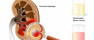
It is possible to determine the presence of phosphate or urate salt in general tests completely by accident during a preventive examination or medical examination. A low concentration of minerals may occur, but provided that the tests are normalized after proper nutrition, which is individually prescribed by the doctor if there are salts in the kidneys.
Red blood cells and white blood cells may appear in the urine - these are symptoms of the presence of an inflammatory process. An infection quickly attaches to the painful tissue - this is indicated by detecting the presence of bacteria in urine tests.
It is important! In appearance, urine tests may be cloudy or have sediment. Diagnostic methods such as ultrasound examination do not make it possible to determine the presence of sand in the urinary system. But ultrasound is excellent for diagnosing complications of pathology. Finding stones and measuring their sizes.
Diet for urolithiasis in children
A therapeutic diet is an obligatory component of the treatment plan, not only as part of the conservative fight against the disease, but also as an important element of postoperative recovery and prevention. Dietary recommendations from doctors include the following rules:
- compliance with the drinking regime, taking slightly alkaline mineral waters as prescribed by a doctor;
- eating foods high in potassium: bananas, dried apricots, potatoes, beans;
- introducing bran and high-fiber foods into the diet;
- control of the content of protein products in the diet - the amount of protein should be sufficient, but not excessive;
- limiting the use of table salt;
- use of citrate mixtures as prescribed by a doctor.
The purpose of dietary nutrition is to activate metabolic processes and remove salts from the body, create a gentle environment for kidney function, and reduce edema. The diet is individual depending on the type of stones formed. If urolithiasis in children requires a strict diet, but the diet is not balanced in vitamins and minerals, clinical recommendations additionally include taking vitamin complexes Source: A. V. Malkoch. Dysmetabolic nephropathies and urolithiasis // Nephrology of childhood: A practical guide to childhood diseases. M.: Medpraktika-M, 2005, vol. 6, p. 472-516..
Functional state of the kidneys in children with different types of feeding
In children, unlike adults, there is an age-related decrease in the glomerular filtration rate in relation to the mass and surface area of the body, a limited ability to concentrate and eliminate excess acids, water, etc. Despite the fact that adaptation mechanisms in young children are significantly limited, the kidneys healthy infants are still able to maintain homeostasis [1]. The functional immaturity of the kidneys and the limitation of their reserve capabilities manifest themselves against the background of feeding disorders (excessive intake of protein, liquid, mineral salts), as well as during stress, infections and operations [2,4]. Thus, in artificially fed children, despite the fact that the composition of modern milk formulas is constantly being improved, there is often an increased excretion of protein metabolites, minerals, hydrogen ions, and the process of osmoregulation is disrupted. This may increase the risk of water and electrolyte disorders and the development of metabolic nephropathies. Changes in the functional activity of the kidneys depend on the strength of the influence of the unfavorable factor [5,6]. In the works of Zoppi et al. (1977) [8] described cases of children aged 1.5 to 2 months admitted to the clinic with acute renal failure while feeding infants with poorly diluted condensed cow's milk or breastfeeding with early introduction of complementary foods in the form of freeze-dried meat. The level of protein consumed per day in these children was 6 g/kg body weight, and the content of sodium and potassium was 2–3 times higher than the physiological requirement. Upon admission to the intensive care unit, the described patients had oliguria, metabolic acidosis, an increase in urine osmolarity by more than 2 times, acidosis, and increased levels of urea and creatinine. Correction of nutrition led to the complete restoration of diuresis and elimination of azotemia. Excess protein in formula feeding contributes to the development of metabolic stress. According to Haschke-Becker E. et al. (2000) [9], amino acids not used for protein synthesis accumulate in the blood, contribute to the development of azotemia, their metabolism in the liver increases the level of urea, and when excreted in the urine, it increases the load on the kidneys. Andre Kahn et al. (2002) [10] provide evidence that excessive protein intake by a child leads to sleep disturbances. The authors believe that a large number of protein breakdown products require a large amount of water to remove them. Babies wake up and cry, feeling thirsty, but parents usually offer them an extra bottle of milk or formula. Consequently, their thirst is satisfied, but at the cost of a new portion of protein, which again leads to an increase in the need for water. These children produce large amounts of urine during the night, which also causes them to wake up more frequently. The authors also note that children who consume large amounts of protein, the breakdown of which occurs with the participation of intestinal bacteria, are more likely to suffer from intestinal colic. THEM. Vorontsov et al. (1977, 1980) [11] noted that when feeding children with unadapted milk formulas, an acceleration in the rate of physical development, an increase in muscle tissue, and a slowdown in the rate of psychomotor development can be observed against the backdrop of a lag in the rate of myelination of nerve fibers. This point of view is supported by Pohlandt (1979) [12], noting the adverse effects of excessive protein and amino acid loads on brain maturation. For newborns, the most significant is an excess of amino acids such as phenylalanine or tyrosine. Their increased concentration, associated with excess intake from food, can have a negative effect on the formation and functioning of the central nervous system due to fairly prolonged hyperaminoacidemia [Vorontsov I.M., Fateeva E.M., 1998] [13]. Zachnoiko L.I. et al. (2004) [14] also believe that when organizing rational feeding of premature infants, despite the higher need for protein, it should be taken into account that an increase in protein load can lead to a breakdown of metabolic systems with the development of a toxic effect of amino acids on the development of the brain and tissue parenchymal organs. For artificial feeding, a wide range of products are currently offered with different contents of nutrients, especially protein and minerals. In order to provide infants with adequate artificial feeding, a number of countries have developed special requirements for the formulas of human milk substitutes, based on data from clinical and biochemical studies of leading pediatric and nutritional centers. These are the recommendations of FAO/WHO - Codex Alimentarius Commission, ESPGAN (European Society for Pediatric Gastroenterology and Nutrition), EEC (Commission of the European Communities), FDA (Food and Drug Administration). They include requirements for the composition and quality indicators of adapted milk formulas. The protein component should be a mixture of casein (the main protein in cow's milk) and whey proteins, which dominate in human milk (in a ratio of 40:60 or 50:50), as this is close to their ratio in human milk (45:55). Casein forms large flakes in the stomach under the influence of hydrochloric acid, which are slowly broken down, while whey proteins form much more delicate and fine flakes, providing a higher degree of digestion and absorption. In addition, they are carriers of immune factors and contain essential amino acids. However, replacing casein with whey proteins causes overload of the small intestine, as a result of which the resulting protein proteolysis products do not have time to be absorbed. It has been experimentally confirmed that the best ratio of casein and whey proteins is 40:60 or 50:50 [6,15,16,17]. The growth rate of a child in the first year of life and, therefore, the need for nutrients to ensure normal development is higher than in any other period of life. At the same time, at this age the excretory capacity of the kidneys is limited. However, the difference between the needs of a growing body and maximum food tolerance is very small, which requires a well-balanced diet. Even if all nutrients are supplied in the optimal ratio for growth, with the exception of an increased protein load, the capacity of the excretory systems may be exceeded, resulting in the accumulation of urea in the blood or the development of uremia [18]. Infants use most of the nutrients they receive from food for body growth. Fomon SJ (1991) [19] believes that newborns need 52% protein for growth, Dewey et al. (1996) [20] – 64% of the total amount of protein consumed. But as children's growth rate slows as they age, the amount of protein needed for growth also decreases. Therefore, the amount of protein a child needs is determined by his growth rate. By the age of 9–12 months, the amount of protein consumed for a child's growth decreases to 18% of total protein intake. The rapid change in protein requirements of infants explains why determining adequate protein levels for infants is so difficult [21,22]. In recent years, there has been a clear trend towards a decrease in protein levels in human milk substitutes, which allows reducing the metabolic load on functionally immature kidneys and enzymatic systems in infants [23,24,25,26]. A prerequisite for reducing protein in the replacement formula is the fact that human milk contains a protein content of 0.8–0.9 g/100 ml [27,28]. Protein intake is even lower if you adjust for serum proteins such as secretory immunoglobulin, lysozyme, lactoferrin, etc. [28,29,30]. However, these low protein levels are sufficient for optimal growth in children and place adequate stress on infants' immature kidneys. Despite the progress made in the production of baby food products, the current formula of human milk replacers does not yet meet all the challenges of adaptation. This refers to the quantity and quality of protein, fat, carbohydrates, some minerals and trace elements [31,32,33,34]. A number of domestic and foreign pediatricians express the opinion that the problem of excess nutrition in children is of significant importance. The use of unreasonably large protein loads is unfavorable for the development of children [32,35–43]. Artificially fed infants have elevated levels of urea and amino acids in the blood, and therefore experience greater stress on their functionally immature kidneys [44]. High concentrations of amino acids in the circulating blood can exceed the ability of the kidneys and liver to metabolize them and excrete excess nitrogen. This can lead to acidosis, diarrhea and increased levels of ammonia and urea in the blood [45]. A number of studies have shown that the content of protein in the formula of human milk substitutes in the amount of 1.3 g/100 ml, which is closer to the amount of protein in human milk, ensures the protein requirement of healthy children in the first months of life, which is confirmed by normal growth, health status, and amino acids in blood plasma [46,47]. The European Society of Pediatric Gastroenterology and Nutrition (ESPGAN) has suggested that the optimal protein content in standard infant formula is between 1.8 and 2.8 g per 100 kilocalories. The American Academy of Pediatrics Committee on Nutrition recognizes a wider range of protein content in infant formulas from 1.8 to 4.5 g per 100 kilocalories. The US Food and Drug Administration (FDA) gives the same limits as the American Academy of Pediatrics. Yong VR et al. considers the upper limit of 4.5 g of protein per 100 kcal to be too high for infants [48]. Fomon (1991) considered the acceptable minimum protein level in infant formulas to be 2.2 g per 100 kilocalories (15 g/1) for infants up to 3 months and 1.6 g per 100 kilocalories (11 g/1) for infants older 3 months [19]. This recommendation is similar to that of Beaton and Chery (1988) [49]. They estimate that for infants aged 3–4 months, the normal level of protein intake may be 1.7 g per 100 kcal (11.4 g/1). Thus, the International recommendations for the minimum protein content in infant starter (starter) formula corresponds to 1.8 g per 100 kilocalories. Raiha NCR, Fazzolari A, Cajozzo C et al. (2002) [50] conducted a clinical study to examine the adequacy and safety of infant formulas containing 1.8 g protein per 100 kcal in infants from birth. It compared the health of breastfed infants with those fed a standard formula (with 2.2 grams of protein per 100 kcal and a whey/casein ratio of 60:40) and a formula with 1.8 grams of protein per 100 kcal and with whey/casein ratio 70:30. The study was carried out from birth to 4 months (at periods of 30, 60, 90 and 120 days). No differences were found in energy intake between these groups of formula-fed infants, whereas protein intake was lower in formula-fed infants with 1.8 grams of protein per 100 kilocalories. However, blood urea levels were higher in infants fed standard infant formula than in infants fed breast milk or reduced protein formula (at 30 and 60 days of life). It was noted that on the 120th day of life there were no longer differences in blood urea levels between the studied groups of children. Albumin levels were normal in all children. Thus, the authors suggest that the whey-enriched formula, with 1.8 grams of protein per 100 kilocalories, meets the protein and energy needs of healthy full-term infants up to 4 months of age without placing excess stress on infants' functionally immature kidneys. Axelsson I. et al. (1987) [51] examined 30 children aged 4 to 6 months fed formulas with different protein contents (1.9 grams per 100 ml of formula in the first group and 2.7 grams per 100 ml of formula in the second; children served as the control group who are breastfed). It was shown that initially, by the time of inclusion in the study (4 months), all artificially fed children had higher levels of urea in the blood compared to breastfed children, while significant differences in the concentration of creatinine and total nitrogen in the urine were revealed did not have. By the age of 6 months, a significant increase in the latter was noted in both groups of artificially fed children. The authors concluded that both formulas contained excessive amounts of protein and placed undue stress on the kidneys of infants. In a study of two groups of infants fed formulas containing 2.24 and 1.83 g of protein per 100 kcal, Ziegler EE (2002) [52] showed that nitrogen intake was lower in the group of infants fed formula with a protein content of 1.83 g per 100 kcal. However, urinary nitrogen excretion was significantly lower in this group, while urinary nitrogen intake and excretion were significantly higher in the higher protein group. Thus, the percentage of nitrogen absorption was the same in both groups of children. The author believes that despite the lower protein content, children receiving the second formula fully met their protein needs. Picone TA (1989) [53] found no differences in the nutritional status of children when using milk formulas with a protein content of 1.3 and 1.5 g per 100 ml, respectively. A number of domestic pediatricians also speak out about the need to reduce the protein level in human milk substitutes [6,37,38,54]. Adverse consequences of unreasonably large protein loads in the diet of children in the first year of life are also noted [36,55,56]. To assess the degree of adaptation of human milk substitute formulas, indicators such as potential renal (water-salt) and osmotic load are used [19,26,57]. In accordance with the recommendations of Tromarelli (1976) [57], the potential renal load is calculated using the formula protein + sodium + potassium + chlorine + phosphorus, the values of which are expressed in milliequivalents. The osmotic load of the formula is a complex characteristic that reflects the content of a number of food ingredients (especially minerals) in the product. When studying the water-salt load on the kidneys carried out by mixtures of various compositions, the author emphasized that mixtures with high osmolarity have an increased load on the kidneys [57]. Russian scientists provide similar data [6,40]. To remove equal amounts of osmolarly active substances that are excess for the body, an infant’s kidneys need to excrete approximately twice as much water as the kidneys of an adult. That is why the body of a newborn and an infant is equally susceptible to the risk of developing both dehydration and the accumulation of excess substances that are subject to excretion in the urine [40,58]. Ivanov V.A. et al. (1985) [59] presented the results of a study of 38 adolescents aged 16–17 years who did not have kidney pathology. The state of osmo- and ion-regulatory functions of the kidneys was studied depending on the time of transfer to artificial feeding. The authors concluded that early transfer to artificial feeding affects the development of osmo- and ion-regulatory functions of the kidneys in children. The high osmotic load on a child's kidneys associated with excess protein intake reduces the ability to maintain a safe level of fluid balance. As a result, in diseases accompanied by dehydration, a reduced ability to excrete waste products increases the risk of developing hypernatremia. In most European countries, dehydration accompanied by hypernatremia is practically unheard of. This is due to the high percentage of breastfed children and the use of formulas with reduced protein and salt content [45]. Along with the risk of water imbalance, excess protein intake may contribute to the development of obesity in later life. Rolland–Cachera MF (1995) [60] et al. Based on long-term observation of children aged 2 years who had a diet high in protein, they found a significantly higher incidence of obesity at the age of 8 years. Having studied the biochemical parameters of blood and the actual nutrition of children, the authors came to the conclusion that the content of blood lipids in infants is associated with excess protein intake. According to WHO, in Russia overweight is found in 20% of infants. The threat of disturbances in the renal regulation of protein and mineral metabolism in children with artificial feeding is especially great when artificially feeding with unadapted formulas. Reducing the load on the functionally immature kidneys of infants can be achieved by reducing the protein level in formulas and correcting their amino acid composition (closer to human milk). This will reduce the osmotic load on the “immature” kidneys of infants while increasing the overall biological value of the product [61]. Therefore, in order for artificially feeding children to receive nutrition adequate to their physiological features, it is necessary to clearly regulate the receipt of protein and osmathic mineral substances in milk mixtures. The protein component of the mixture should be as close as possible to that of women's milk. This can be achieved by quantitative and qualitative adaptation. The quantitative adaptation of the protein component of the mixture to women's milk can be achieved with a decrease in the total mixture protein to 12–16 g/l. High -quality adaptation provides for the approach of the ratio of serum proteins and casein 60:40, a decrease in the level of b - lactoglobumin and an increase in A - a -lactalbulin, the addition of essential amino acids (taurin) and a decrease in the level of potentially dangerous amino acids (phenylalanine, tirosos). Standard mixtures for infants in the Russian market contain 15 g of protein per liter or 2.2 g of protein per 100 kilocalories and a different ratio of casein and serum proteins. Mature milk milk has a “nutritious” “true” protein content of only 8–9 grams per liter with an energy content of about 650-700 kilocalories per liter. The ratio of energy and protein, thus, is approximately 1.3 g per 100 kilocalories in mature breast milk. Therefore, standard mixtures for infants have a protein level, almost twice as much as in women's milk. Until now, attempts to reduce the protein level in mixtures to the level of breast milk have led to a deficiency of some essential amino acids [39], while an increase in the protein in the mixture (to ensure the required level of essential amino acids) was accompanied by an increase in the number of other amino acids and an increase in the metabolic load on the kidneys and liver [62]. This was due, in particular, with technological difficulties in the production of milk mixtures. New technological techniques made it possible to change the composition of the protein component of the mixtures: components with excessive trionin content are removed, a -lactalbumin was introduced, which made it possible to increase the level of tripophanes and reduce the concentration of trionin, due to which the level of protein in the milk mixture decreased, and its composition approached the protein of breast milk. The first mixture containing a new component of serum proteins (New Nan) has been developed in the Nestle research center and is already used in the nutrition of children in many countries, its effectiveness and safety are confirmed by the results of clinical research [62]. Ziegler E. (2000) [62] believes that the new generation of mixtures highly content of A - lactoglobulin, rich in tripophane, will create the same concentrations of triptophanes in blood plasma, as in infants who are on breast feeding in the absence of the need to remove excess nitrogen . Russian scientists also confirm that due to an increase in serum proteins, to 70% in the improved composition of the NAS mixture, the protein level decreased to 1.2 g/100 ml, approaching the qualitative and quantitative attitude to the protein of women's milk [63]. Our studies conducted in infants (1-4 months) showed that feeding the adapted mixture “New Nan” provides adequate growth and development of infants, does not provide an additional metabolic load on functionally immature buds of infants, which is accompanied Osmolarity of urine. Literature 1. Netrebenko O.K. The practice of feeding children of the first year of life in Russia. Children's Hospital, 2001, No. 3, p. 52–54. 2. Furtsev V.I., Prakhin E.I., Gritsan A.I., Budnikova E.V. Changing the policy of the pediatric health care service regarding the practice of breastfeeding. Pediatrics - 2002, No. 1, p. 69–71. 3. Horse I.Ya. Safronova A.I., Vorobyova L.Sh. Shilina N.M. et al. Assessment of the influence of kefir and the “subsequent” mixture on the development of diapertoid bleeding in children of the second half of life of pediatrics 2002; 3: 55–59. 4. Domingos Palma. Food in the first year of life: the unacceptability of cow's milk. Nest. No. 14 - July, 2003. P.5–6. 5. Kildeberg P., Engel K., Winters BW Balance of Net Acid in Growing Infants. Acta Paediatr Scand, 1969, Vol.58, P.321–329. 6. Podvacheva T.N. The metabolic foundations of optimizing artificial feeding of infants. dis. Doctor. Med. Nauk, M. 1995. 365 p. 7. JIP R. // 4th International Symposium on the problems of proper nutrition of the mother and child. - M., 1995. - S.6–22. 8. Zoppi G., Zamboni G. Mechanism of Diet --induced Uraemia and Acidosis in Infants. Europ.j.pediat., 1977, v.125, N3, p.197–204. 9. Haschke - Becker E., Fazzlari A., Minoli L. et al., Plasma Amino Acids of Infants Fed Modified Whey Formulas with Protein - Energy Ratio of 1.8G/100kcal. World Congress Pediatr. Gastroenterol. Hepatol. Nutritiol. Boston, 2000. 10. Kahn A., Mozin J., Groswasser J., Sottiaux M., Scalelet S., Franco P. The relationship between nutrition and children's sleep. Nest, No. 12 - June 2002, p. 6–8. 11. Vorontsov I.M., Mazurin A.V. Feeding children of the first year of life. In the book: Handbook of Children's Dietics. - L.: Medicine, 1980, p. 23–123. 12. Pohlandt F. Plasma Amino Acid Concentrations in NewBorn Infants Breast - Fed Ad Libitum. Pediat. , 1978, V.92, N4, P.614–616. 13. Vorontsov I.M, Fateeva E.M. Natural feeding of children, its significance and support. - St. Petersburg: IKF "Foliant", 1998.– S.68–72. 14. Zatka L.I., Nazarova A.Z., Nugmanova I.F. Feeding premature babies. // Children's health - the problem and future of the nation: the collection of works of the International Conference/Almaty, May 5–6, 2004, Almaty, 2004, p. 315–318. 15. Raiha ncr milk Protein Quality in Human Milk and Infant Formulae.– Acta Paediatr. Suppl. 402, 1994, P.57–58.heine W., Gassmann B., Plenert W. Vergleihehende Bilansuntersuchungen Angunngen Sauglingen Unter Ern? Hrung Mit Eiweisarisreihen KuhmilchFertigantign Ahrungen. P? Diat. u. Grenzgeb., 1968, N7, S.301. 16. Nutrition of Normal Infants Ed. by S. Fomon, Mosby, 1993. 420 p. 17. Nutrition in pediatrics. Ed. by w. Walter, JBC Walkins Decker Inc. Publ., Hamilton - London, 1997. 850 p. 18. Klimov L.Ya. Modern aspects of feeding of infants. Method. recommended. SGMA. Stavropol, 2001, 53 p. 19. Fomon SJ. REQUREMENTS AND Recommoded Dietakes of Protein During Infancy. Pediatr Res 1991; 30: 391–5. 20. Dewey KG., Beaton G., Fjeld C., et al. Protein Requirements of Infants and Children. Eur. J. Clin. Nutr. 1996; 50 (SUPPL 1): S119–50. 21. Fomon SJ, Haschke F., Ziegler EE, Nelson Se. Body Composition of Reference Children from Birth to Age 10 Years. Am. J. Clin. Nutr. 1982; 35: 1169–75. 22. EE ziegler. Protein Requirements in Infancy // Infant Formula: Closer to the Reference. Nestle Ltd. 2002. - p. 97–110. 23. Maklin S. Possible composition of baby food./Balanced medical nutrition. Lecture course. Ebbot laboratory. - 1987.– P.37. 24. Beaton GH, Chery A. Protein Requirements of Infants: Reexamination of Concepts and Approach //am.j. Clin. Nutr. - 1988. - v.48, p. 1403-1412. 25. Hurrelli RF, Berrocal R., Nesser Jr, Schweizer D., Hilpert H., Traitler H., Colarow L., Lindstrand K. Micronutribents in of Infants Formula. –1991. - 307p. 26. Ziegler EE, Run Je Renal Solution Load and Diet in Growing Premature Infants // J. Pediatr. - 1976. v. 89, p. 609–613. 27. Hambraeus L. Human Milk Nutrition // Rev. Clin. Nutr. 1984. - V.54, P.219–236. 28. Khakimov Sh.K., Toshboev O.S., Nuritdinova G.T., Khusanova H.A., INANKOVA B. The content of nutrients as part of breast milk of healthy nursing women. // Children's Health - Problem and Future of the Nation: Collection: Collection works of the International Conference/Almaty, May 5–6, 2004, Almaty, 2004, p. 368–371. 29. Hayes ks, Struurman ja taurine in metabolism // an. Rev. Nutr. –1981.– v. 1, P.401–425. 30. Hennart PF, Brasseus DJ, Delogne - Desnoeck Jb, Dramix Mm, Robyn Ce Lysozyme, Lactoferrin, and Secretory Immunoglobulin a Content in Breast Milk: Influence of Durantion OF F Lactation, Nutrition Status, Prolactin Status, and Parity of Mother // Am .J. Clin. Nutr. - 1991. - v.53, p. 32–39. 31. Lugovskaya V.K., Purina G.O. Assessment of physical development and health status of children of the first half of life, depending on the type of feeding. In the book: Theoretical and practical aspects of the study of human nutrition. (Abstracts). M., 1980, T.2, p. 228–229. 32. Horse I.Ya., Konovalo A.I., Georgiev O.V. Modern approaches to organizing artificial feeding of children of the first year of life. - attending physician, 2003, No. 2, p. 54–58. 3 3 In the book: Theoretical and practical aspects of the study of human nutrition. (Abstracts). M., 1980, T.2, p. 285. 34. Plenert W., Czerny B. Aktuelle Entwicklungstrends auf Dem Geete Der Sauglingsfertignahrungen. ERN? Hruungsforschung, 1977, H.22, N 6, S. 166–168. 35. Tour A.F. Nutrition of healthy children. - In the book: Propaedeutics of childhood diseases. L., 1967, p. 365–424. 36. Zeitz R.I. Nitrogen exchange in children at various protein loads. - Questions of the protection of motherhood and childhood, M., 1973, No. 11, p. 84. 37. Ott V.D. Some issues of feeding, metabolism and reactivity of infants. Diss.Do. Kyiv, 1974. 38. Fateeva E.M., Shcherbakova A.I., Belkina L.M., Mamonova L.G., Kasperskaya Z.A. To the question of the need for protein in young children. In the book. "Theoretical and clinical aspects of the science of nutrition." - M. - 1980. - T.1. - p. 109–119. 39. Fomon SJ, Ziegler EE, Nelson SE, Edwards Behes The Safe Protein - Energy Ratio for Infant Formulas? Am J Clin Nutr, 1995, Vol.62, P.358–363. 40. Netrebenko O.K. The practice of feeding children of the first year of life in Russia. Children's Hospital, 2001, No. 3, p. 52–54. 41. Podvacheva T.N. The influence of artificial feeding on the condition of the kidneys in children. dis. Ph.D. Med. Nauk, M. 1982. 150 p. 42. Guidelines for therapeutic nutrition of children. Ed. Ladodo. - M.: Medicine, 2000. - 384s. 43. Zakharova I.N., Lykina E.V. Modern approaches to the adaptation of dairy mixtures for artificial feeding of healthy children of the first year of life. Russian Medical Journal, Volume 11, No. 13 (185), 2003, p. 767–770. 44. Raiha N., Mioli I., Moro G. Milk Protein intake in the Term Infant I. Metabolic Responses and Effects on Growth // Acta Pediatr. Scand.– 1986. - V.75, P.881–886. 45. Arneil GC, Chin Kc Lower Solute Milks and Reduction of Hypernatraemia in Yong Glasgow Infants. Lancet, 2: 840 (1979). 46. Janas LM Picciano MF, Hatch TF Indices of Protein Metabolism in Term Infants Feder Human Milk OrMulas with Reduced Protein Concentrations and Various Whey/Casein Ratios // J. Pediatr.– 1987.– V.110.– P.838–848. 47. Lonnerdal B., Chen C. - L. Effects of Protein LEVEL and RATIO ONFANT GROWTH, Plasma Amino Acids and Serum Elements // Acta Pediatr. Scand.– 1990. - V.79, P.257–265. 48. Young VR, Pelletier Va Adaptation to High Protein Intakes, With Particular Reference to Formula Feding of Healthy Term Infants. J. Nutr. 1989; 119: 1799–809. 49. Beaton GH/. Cheri A. Proutein Requirements of Infants: Reexamination of Concepts and Approach //am.j.clin.nutr. –1988. - v.48, p. 1403-1412. 50. Raiha NCR, FAZZOLARI A., Cajozzo C., et al. Whey Modified Infant Formula with Protein - Energy Ratio 1.8 G/100 Kcal is adequate AHD from Birth to 4 months. J. Pediatr Gastroentral Nutr. 2000; 31 (SUPPL 2): S.94. 51. Axelsson I., Borulf S., Raiha N. Protein intake during Weaning. II. Metabolic Responses. Acta Paediatr Scand, 1987, May; 76 (3): 457–62. 52. Ziegler EE Protein Requirements in Infancy // Infant Formula: Closer to the Reference. Nestle Ltd. 2002. - p. 97–110. 53. Picone Ta, Benson JD, Moro G. Growth, Serum Biochemistries and Amino Acids in Termlas with Amino Acid and Protein Concentrations Similar to Human Milk // Pediaticate. Gastroenterol. Nutr. - 1989. - V.9, P.451–360. 54. Ladodo K.S. Modern aspects of baby food // Theoretical and clinical aspects of the science of nutrition. T.6. Modern aspects of the nutrition problem of a healthy and sick child. - M. - 1985. - p. 3-14. 55. Tour A.F. About some issues in the nutrition of healthy children of the first year of life // Pediatrics. - 1973. - No. 11. - S.3–9. 56. Lazarev S.G. On the trend of maximum feeding and overfeeding of infants. - Pediatrics, M., 1974, No. 10, p. 80–83. 57. Tomarelli Rm Osmolaty, Osmolariti and Renal Solute Load OFINFANT FORMULAS // Pediatr. - 1976.– v.88, n.3/ - p. 454–456. 58. Ziegler ee, Fomon SJ Fluid Intake, Renal Solution Load and Water Balance in Infancy // J. Pediatr. - 1971. v. 78, p. 561–564. 59. Ivanov V.A., Lavrova E.A., Kliorin A.I., Natechin Yu.V. The function of the kidneys in adolescents with various somatotypes and depending on the type of feeding. Pediatrics, 1985, No. 12, p. 43–46. 60. Rolland - Cachera Mf et al. Influence of Adiposity Development: A Follow - Up Study of Nutrition and Growth from 10 Months to 8 Years of Age. International Journal of Obesity and Related Metabolic Disorders, 1995; 19: 573–578. 61. JOST R., Maire J - C., Maynard F., Secretum MC Aspects of Whey Protein Usage in Infant Nutrition, a Brief Review. Int j food sci. Technol 1999; 34: 533–542. 62. Ziegler E., Carrie A. - L., Hascheke F. et al. Modified Whey Formula with Protein - Energy Ratio 1.8/100 Kcal; Metabolic - Balance Studies in Infants. J Ped Gastr Nutr, 2000, Vol. 31 (2), p. 173. 63. Borovik T.E., Skvortsova V.A., Ladodo K.S., Lukoyanova O.L., Semenova N.N. The use of modern dairy mixtures in the nutrition of infants. Questions are modern. Pediatrics, 2003, T.2, Appendix No. 3, p. 55–57.
Prevention
The key goal of disease prevention is to eliminate risk factors that provoke the formation of stones. To do this, it is necessary to establish a balanced diet for the child, regularly conduct examinations of the body in order to promptly identify any infections or pathologies, avoiding complications.
Comprehensive preventive measures are especially important for children who have already been diagnosed with chronic kidney disease, as well as for those who have close relatives with urolithiasis. If a child has already had a stone removed, to avoid recurrence, parents need to regularly conduct urological examinations and monitor their diet. If you follow all preventive rules, relapse of the disease can be avoided.
Sources:
- T. V. Otpuschennikova, L. A. Deryugina Urolithiasis in children - causes, diagnosis, treatment // Attending physician, 2015, No. 4, p. 64-72.
- M. F. Trapeznikova, V. V. Dutov, A. A. Rumyantsev. Modern aspects of diagnosis and treatment of urolithiasis in children // Medical class, 2004. No. 3, p. 8-12.
- A. V. Malkoch. Dysmetabolic nephropathies and urolithiasis // Nephrology of childhood: A practical guide to childhood diseases. M.: Medpraktika-M, 2005, vol. 6, p. 472-516.
- O. L. Chugunova, M. V. Shumikhina, L. V. Pavlushkina. Main risk factors for the development of urolithiasis in children // Pediatrics, 2018, No. 97 (5), p. 93-103.
The information in this article is provided for reference purposes and does not replace advice from a qualified professional. Don't self-medicate! At the first signs of illness, you should consult a doctor.
Prices
| Name of service (price list incomplete) | Price |
| Appointment (examination, consultation) with a pediatric urologist-andrologist, primary, therapeutic and diagnostic, outpatient | 1750 rub. |
| Consultation (interpretation) with analyzes from third parties | 2250 rub. |
| Prescription of treatment regimen (for up to 1 month) | 1800 rub. |
| Prescription of treatment regimen (for a period of 1 month) | 2700 rub. |
| Consultation with a candidate of medical sciences | 2500 rub. |
| Kidney ultrasound | 1700 rub. |