1. Accidentally touching the fontanelles on the baby’s head
Despite doctors' warnings, you should not become hysterical if you accidentally touch these soft spots on your newborn's head. By the way, these places are called fontanelles. And when you touch them, you are not touching his brain. What are you touching then? The so-called membrane, consisting of dense connective tissue. The soft areas on the head are designed to allow the baby to move freely through the narrow birth canal. Thanks to the flexibility of the skull, this tiny head has already survived quite a traumatic journey without any harm, so your gentle touch will not harm it. And the fontanelles will heal sooner or later.
2. Pulsation in the fontanelles
When a child screams or plays, you can see pulsation of the brain vessels in the anterior fontanel. Don't panic. The fontanelles are located in areas of the skull that have not yet fused, and veins and arteries can sometimes be seen through the soft membrane. And pulsation is a normal development of the child’s circulatory system.
3. Blood in the diapers of a newborn girl
During pregnancy, a surge in maternal estrogen levels can stimulate the unborn baby's uterus. Therefore, some newborn girls experience a small amount of spotting during the first week of life. There is nothing wrong with this hormonal surge.
4. Small depression in the baby's chest
Relax – these are not heart problems. According to experts, the sternum consists of three parts. The indentation that is sometimes noticeable in some babies is most likely an inverted lower part of the sternum. As the child grows, the growing muscles of the chest and abdomen will straighten this depression. But, perhaps, even before this, the layers of growing fat will be leveled off the chest.
5. Loose stools after every feeding
Breastfed babies may have a bowel movement after each feeding because breast milk is digested very quickly. By the way, newborns who are bottle-fed may defecate less frequently. As for the consistency of stool, there is nothing surprising in this either - all babies are on a liquid diet.
6. Constant hiccups
Experts cannot agree on why newborns hiccup so often. Some believe this is because the brain and the diaphragm, the abdominal muscle that controls breathing, are still working somewhat inconsistently. However, regardless of the cause, hiccups are harmless and safe.
Babies have immature nervous systems and are easily startled. These are two reasons why they shed tears so often. In addition, crying is the only way a baby can communicate its needs and needs. So up to a certain point you are doomed to see tears and listen to screams. Don't worry about the baby - although he looks upset, the baby is not harming himself.
8. Rash or pimples on the face
Thanks to maternal hormones still circulating in small bodies, newborns often develop acne. As a rule, the rash goes away over time - from 2 weeks to 2 months. What to do? Just wash your baby gently and gently. There is no need to use your anti-acne gels and creams.
9. Swollen breasts
The same hormones that cause mini-periods in girls (see point 3) can cause swelling of the mammary glands in newborns of both sexes. Marvelous? Yes. Temporarily? Absolutely right. Exciting? In no case.
10. Endless sneezing
Babies have tiny noses. And even a tiny piece of stuck mucus, even a slight nasal congestion, can cause a baby to sneeze. And so time after time. If the sneezing is not accompanied by thick yellow mucus, which can signal that the baby is cold, then the newborn will simply outgrow this condition over time.
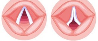
Compression and decompression
Mothers who are preparing for childbirth on their own or in courses for pregnant women have probably seen illustrations of the birth canal and imagine what a difficult path a child has to go through before being born. Nature has provided for everything: the structure of a baby’s skull is completely different from that of an adult. He has fontanelles, the bones of the skull are mobile due to the fact that all their joints are quite elastic, and thanks to this, during the birth process, the baby’s head is easily configured, adapting to the birth canal. Compression occurs. Of course, displacement of the skull bones is possible in this case, but, fortunately, nature has also provided the opposite mechanism - decompression, which turns on immediately after birth.
Head and symptoms
The spots that you can notice on the baby’s head look like birthmarks, but gradually disappear. They say that strong pressure was applied to the baby's head in this place. Most likely, the baby will cope with the problem on his own, however, the coincidence of a spot in a certain part of the head and some clinical symptoms may indicate that it is worth contacting an osteopathic doctor, since the baby needs help.
Neck injuries are usually accompanied by the following symptoms:
- sucking disorder. Despite the fact that the baby is correctly applied to the breast, he cannot latch on normally or is uncomfortable sucking;
- profuse and frequent regurgitation;
- with severe lesions, problems with speech and vision, torticollis and descending scoliosis may occur in the future.
Damage to the sphenoid bone can cause:
- strabismus;
- intracranial pressure;
- motor speech disorder (it is difficult for the child to control the articulatory apparatus).
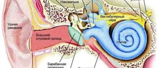
Damage to the temporal bone can cause:
- hearing impairment;
- problems with coordination of movements.
Damage to the frontal bone leads to:
Of course, with all these problems you can and should consult a doctor. Even if you do this when the baby has already grown up and the spots have disappeared, keep in mind such facts as postpartum spots, dilated veins in any part of the head, and peculiarities of the course of labor. An experienced doctor will always correlate the baby’s well-being and behavior with how the birth went and the results of a visual examination of his head. Quite often, parents attribute to their parental incompetence or the difficult nature of the baby those troubles that actually indicate displacement of the skull bones. But this can be easily corrected in the first months after childbirth.
What it is
The frontal bone of the skull consists of two parts. They are connected by a metopic suture. Normally, it remains completely open in infants up to 1 year of age. Subsequently, the seam begins to heal, this process continues until the age of 8 years.
However, sometimes the connection of the frontal bones heals during the prenatal period or in the first year of life. In this case, the baby has a noticeable metopic suture on the forehead.
This formation looks like a convex ridge that runs from the brow ridge to the large fontanel. In this case, the child has an irregular skull shape.
Doctors call this anomaly metopic suture synostosis or trigonocephaly.
Causes
Why does the suture between the frontal bones close prematurely? The following reasons can provoke its early overgrowth:
- Heredity. In approximately 2-8% of cases, this disease is transmitted from parents. The cause of metopic synostosis is damage to the gene responsible for the growth of connective tissue.
- Chromosomal abnormalities. Premature healing of the metopic suture may be just one of the manifestations of serious genetic pathologies. This deviation is observed in children with Jacobsen syndrome and Opithia trigonocephaly syndrome. In this case, not only synostosis and deformation of the skull are observed, but also anomalies of internal organs, as well as delayed mental development.
- Fetal hypoxia. Lack of oxygen during the prenatal period can cause premature healing of the suture.
- Prematurity. Babies born prematurely have an increased risk of developing trigonocephaly.
Symptoms
The metopic suture in a child is a convex groove in the middle of the forehead. It starts on the bridge of the nose or between the eyebrows and ends at the crown of the head. At the same time, in children the volume of the frontal lobes of the skull decreases and the growth of the parietal bones is noted. Because of this, the baby's head has a triangular shape.
A photo of the metopic suture can be seen below.
Parents often confuse the manifestations of synostosis with an ordinary bump on the forehead. In trigonocephaly, the groove has a hard structure. A hematoma formed as a result of a bruise is a soft formation.
Close-set eyes are one of the signs of trigonocephaly. Doctors call this defect hypotelorism. The distance between the eyeballs decreases due to deformation of the skull.
A convex metopic suture in an adult patient can also be a manifestation of synostosis. If trigonocephaly was not treated in childhood, the groove on the forehead, the triangular shape of the skull and hypotelorism persist throughout life. This disease can be easily corrected in children. Correcting such a defect in an adult is much more difficult.
However, it is important to remember that a bulge in the middle of the forehead does not always indicate illness. In some people this is an anatomical feature. If a person has a suture on his forehead, but there is no deformation of the skull, then there is no reason for concern.
Consequences
The metopic suture is not only an aesthetic problem. In advanced cases, deformation of the skull leads to pinching of the optic nerve and eye muscles. This leads to the appearance of ophthalmological pathologies:
- strabismus;
- exophthalmos (bulging of the eyeballs);
- decreased visual acuity.
In addition, early closure of the suture between the frontal bones leads to increased intracranial pressure. This is expressed in frequent headaches, dizziness, and nausea. Many babies with metopic synostosis suffer from hydrocephalus, a pathological increase in the volume of the head.
If trigonocephaly is not treated in early childhood, then in 15-20% of cases this disease leads to mental retardation. Intellectual impairments appear only at the age of 7-8 years.
The child has problems with memorization, it becomes difficult for him to assimilate new material, which creates serious problems in learning.
Mental retardation is more often observed in children suffering from high intracranial pressure.
Diagnostics
A neurologist treats trigonocephaly. If the pathology in a child or adult is accompanied by damage to the organ of vision, then consultation with an ophthalmologist may be required.
You can notice the external manifestations of metopic synostosis already during the examination of the patient. As already mentioned, trigonocephaly can be either a separate disease or one of the signs of chromosomal abnormalities. Therefore, the patient needs to undergo a comprehensive diagnosis. The doctor may order the following tests:
- Spiral computed tomography of the skull in 3D. This is the most accurate method for detecting metopic synostosis. The procedure is carried out while the child is sleeping.
- Neurosonography (ultrasound of the skull). This test is usually given to children under 1 year of age. The procedure is absolutely painless and harmless. It shows anomalies in the structure of the cranial bones and the condition of the sutures.
- MRI of the head. This study is often prescribed to children of primary school age to identify the cause of cognitive impairment.
In addition, the distance between different parts of the face is measured. This helps to identify hypotelorism and deformations of the facial part of the skull.
Additionally, an ECG, MRI of the spine and ultrasound of the gastrointestinal tract are prescribed. This allows us to identify signs of chromosomal diseases that often accompany trigonocephaly.
Surgical intervention
The protruding seam on the forehead can only be completely eliminated through surgery. Surgery is performed in a hospital setting.
Surgery is necessary only in cases where trigonocephaly is confirmed by spiral CT in 3D. If premature healing of the suture is noticeable on the three-dimensional image, then this is considered an indication for surgical treatment.
Experts recommend performing this operation on a child aged 4-6 months. Surgery is performed under general anesthesia. The doctor cuts the fused suture and inserts special plates that support the skull bones in the correct position. The operation lasts about 4 hours.
After correction of metopic synostosis in children, the normal structure of the skull is restored and deformities disappear. In the photo below you can see the results of the surgical intervention. The left picture shows the child before surgery, and the right picture shows him 2 years after the defect was eliminated.
After operation
The recovery period after surgery lasts quite a long time. In the first 2-7 days after correction of synostosis, the child is in the hospital. After surgery, hematomas and swelling in the eye area may appear. This is a natural phenomenon that passes quickly.
There are cases when synostosis recurs after correction. Therefore, doctors recommend that the child wear a special orthopedic helmet during the postoperative period. This device helps prevent skull deformation.
In the first year after surgery, children should be under medical supervision. During this time, it is necessary to regularly visit the doctor and periodically do a 3D spiral computed tomography scan of the head. This will help to track relapses of the disease in time.
Is it possible to do without surgery?
Parents of children often ask: “Is it possible to cure synostosis without surgery?” This is possible only with mild forms of trigonocephaly, if the child has only a cosmetic defect, but no functional impairment.
In such cases, an orthopedic helmet is used. It must be worn at least 20 hours a day. The duration of treatment may vary, depending on the severity of synostosis. An orthotic helmet allows you to correct existing defects in the formation of the skull. This device compresses only deformed areas and does not interfere with the natural growth of healthy bones.
Conclusion
We can conclude that metopic synostosis not only spoils the appearance of the child, but also poses a threat to the health and development of the baby. Therefore, such a pathology must be cured in time. If the doctor suggests surgical correction of trigonocephaly, then you should not refuse the operation. This will help avoid neurological disorders and vision problems in the future.
What else should you pay attention to?
Not all problems are visible to the parent's eye, but here are the points that you can note yourself.
Sometimes parents notice cyanosis or hematoma , and sometimes a cyst-like tumor (which can resolve or calcify and turn into a lump). Usually, with such phenomena, the baby’s jaundice lasts longer - this is a kind of symptom of the body’s protective reaction, which seeks to “resolve” this neoplasm.
Visually, you can notice problems with the lower jaw , if the baby cannot suck, you need to urgently consult a doctor, however, such pathologies are usually immediately noticed in the maternity hospital.
has a tear in one eye or both , this indicates that the bones of the skull have shifted and the nasolacrimal duct is narrowed. It is best to consult an osteopathic doctor while the child is still small, because otherwise the baby will have problems with nasal breathing, adenoids, and otitis media.
Big? Small?
Some parents are concerned about the size of their baby's head. Normally, its girth at birth is Deviations from the norm do not always indicate pathology; quite often a genetic factor is triggered: one of the parents had a large or small head.
During the first month, the head circumference increases by an average of 1 month. During the first month, the head and chest circumferences become comparable, then the rate of breast growth outpaces the growth of the head. For an approximate estimate, there is an empirical calculation formula: at 6 months, the head circumference (CH) is on average 43 cm, for each month up to 6, 1.5 cm is subtracted, for each month above, 0.5 cm is added. During the first year, the CG increases on average, the head grows most intensively in a full-term baby in the first 3 months, in a premature baby - later, during the period of pronounced weight gain.
At birth, the head may be smaller - in premature babies or if the baby experienced severe compression during birth. Also, a small head occurs with microcephaly, which mothers are so afraid of. However, it must be remembered that with true congenital microcephaly, the size of the skull is already small in utero, at the birth of a child the sutures are narrowed, the fontanelles are closed or small in size with dense edges, the head is of a specific shape - the brain skull is smaller than the facial skull, the forehead is small, sloping, the line of the forehead and nose is sloping, as a rule, multiple minor developmental anomalies and severe neurological pathology are present. If your baby does not have these anomalies, there is no need to think about microcephaly.
Mothers are also afraid of hydrocephalus, however, this anomaly is accompanied by severe symptoms. A progressive excessive increase in the size of the skull is accompanied by divergence of the sutures, an increase in the size of the fontanelles, their bulging even at rest, and a pronounced venous network on the head. In this case, the cerebral skull significantly predominates over the facial skull, and the frontal part protrudes sharply. The child develops poorly and has pronounced neurological symptoms. In other words, hydrocephalus cannot be ignored either.
Head sizes larger or smaller than average are most often a constitutional feature, i.e. the child repeats one of the parents, grandparents, etc. Of primary importance, of course, is the overall development of the baby. If it is generally normal, there is no need to be afraid of dire diagnoses.
Precautionary measures
On the one hand, nature has made babies resilient. On the other hand, the baby’s head and cervicothoracic region are quite fragile. Here's what parents need to remember so as not to harm their child.
You need to take the baby in your arms so that his head does not “loop” around. Always support him under his head, do not lift him by his arms or shoulders. The fact is that the vagus nerve, which regulates many body functions, runs not far from the baby’s occipital bone. If the baby experiences a displacement in this area and the nerve is pinched, this will manifest itself in a variety of symptoms: from problems with bowel movements to problems with motor development. For the same reason, in the first two to three weeks, it is better for early swimmers not to do figure eights and other exercises with the baby that can cause displacement in the cervicothoracic region.
The baby can be carried in a sling, where his head is held securely, and for transportation in a car you need to use a special car seat. But a kangaroo backpack, the back of which does not secure the head and neck, cannot be used until the baby holds his head completely confidently, like an adult.
Remember that nature has provided all possible ways to protect the brain from possible injuries, and has also built into the crumbs a huge resource for the body’s self-healing. Breastfeeding, skin-to-skin contact, positive emotions - all this greatly helps the baby overcome the stress of childbirth.
First aid at home
To prevent infection from getting into the wound, it must be washed with hydrogen peroxide and bandaged.
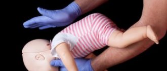
Help with cutting the forehead and any other part of the head should be provided immediately. The damaged area must be washed, the hair around it trimmed, and the wound treated with an antiseptic.
If the child loses consciousness, resuscitation assistance should be provided. Before the ambulance arrives, the baby must be placed on his back, his head secured and cardiopulmonary resuscitation performed. After the victim regains consciousness, he must be prohibited from moving, getting up, drinking and talking, otherwise the wound will hurt and bleed even more. Do not stop bleeding by pressing on the wound with your fingers. Before doctors arrive, the injury should be covered with ice around the edges.
It is forbidden to remove foreign objects from the wound, as this can cause damage to blood vessels and nerves. The bandage should be fixed to the splint. This will prevent foreign objects from moving and damaging nearby tissue. Wounds more than 1-2 cm deep should not be washed with peroxide, potassium permanganate or furatsilin.
Shape and size of a newborn's head
The head shape of newborn babies can be not only round, but also elongated, flattened, ovoid - and all these options are considered the norm. Why is this happening?
By the time they are born, the skull bones of babies are not yet very dense (they will have to completely harden during the first year of life), and the seams between them have not yet had time to heal. During birth, the bones overlap each other, allowing the baby to move out more easily. That is why, after a natural birth, the shape of the head is, as a rule, slightly elongated, while in small “Caesareans” it is smooth and round. Due to the vicissitudes of traveling through the birth canal, a baby may be born with an asymmetrical head, and sometimes also with a lump (cephalohematoma) or edema (the so-called birth edema).
At birth, the baby's head is approximately 2 cm larger in circumference than the chest. But it happens that these sizes increase even more: this happens if cerebrospinal fluid accumulates in the cranial cavity. Then the upper part becomes larger than the lower part, a heavy forehead hangs over the eyes and nose, and doctors talk about hydrocephalus. This problem can arise if during pregnancy a woman suffered a severe infection that affected the unborn baby. In this case, doctors will immediately begin treatment of the child, and in a few months his head may approach normal size.
The situation is considered more serious when the newborn, on the contrary, has a too small head (microcephaly). Sometimes this happens due to genetic disorders that will prevent the baby from developing normally. Fortunately, in many cases the reason for the unusual shape or size of the head turns out to be much simpler: the child can inherit all these features from his parents.
Only a doctor can correctly assess the baby’s head circumference, so there is no point in parents arming themselves with a centimeter themselves. But this indicator will tell specialists whether the child’s brain is developing correctly.
Normally, newborns have a head circumference of 34–36 cm. At first, the head grows quite quickly, by about 1.5 cm per month; after 3 months - by 0.5–1 cm and by 6 months it reaches 43 cm in girth. If the baby is far ahead of the norm or behind it, this may indicate problems with the nervous system.
A pit on the forehead after a bruise in a child
Children are inquisitive and restless, and therefore no one can completely avoid injuries, falls and bruises. In the process of learning about the world, babies fall quite often.
But if falling on the butt or back does not cause panic attacks in parents, then the situation changes dramatically if the child hits his head.
An authoritative pediatrician, author of numerous books and articles on children's health, Evgeniy Komarovsky, explains why such falls are dangerous and when you need to start worrying.
Features of child physiology
The head of a small child is designed in such a way that it is relatively large compared to the rest of the body, so babies most often fall on their head when they lose their balance. But there is also a positive thing: the child’s brain is quite reliably protected from injury when falling.
If a small child fell from the sofa upside down, then the greatest injury (of a psychological nature) was received by his parents, and not by himself. The bones of a baby's skull are very soft, and the "fontanelle" and dynamic "sutures" between the bones of the skull provide them with mobility. The larger the fontanel, says Evgeny Komarovsky, the less likely it is to get injured if you fall upside down.
In addition, nature has come up with another shock-absorbing mechanism - a large amount of cerebrospinal fluid.
If a child at 6-7 months, when he becomes more mobile, turns over unsuccessfully and falls from the sofa or changing table, do not immediately panic. The baby, of course, will scream heart-rendingly.
But parents must understand that he is crying not from terrible pain, but more from fear caused by a sudden movement in space.