The normal shape of a newborn’s skull is the normal circumference of the baby’s head at birth
These and other basic indicators enable doctors to get an overall picture of the health of the newborn in the first period of his life and the absence or presence of pathologies.
Normal indicators
The fact that during this period the size and shape of the baby’s head changes rapidly is considered a natural process.
Baby head shape
What determines the shape of a baby's head?
The following is considered the norm:
- Round
- Elongated
- Flattened
- Ovoid.
There is nothing to be surprised about. But why is each of these options almost the norm? There are several explanations for this.
So, in a newborn child, the bones of the skull have not yet become dense, and they will harden during the first year - the seams between them have not yet healed.
Further, the woman’s body is designed in such a way that for better passage of the baby through the birth canal, the bones overlap each other. Therefore, the shape of a newborn’s head after natural birth is somewhat elongated.
BY THE WAY : The heads of the tiny “Caesareans” are round.
Caesarean section - indications and stages of labor during cesarean section
Video: Birth tumor and skull shapes
Normal baby's head circumference at birth
A child's head is larger in circumference than the chest, by about a couple of centimeters. However, these dimensions can be either smaller or larger, say, due to the accumulation of cerebrospinal fluid in the cranial cavity. Fortunately, this doesn't happen very often.
ATTENTION : A pediatrician must monitor all parameters of the newborn’s development.
In general, the normal head circumference for a newborn baby is usually 34-36 centimeters . Moreover, this number may have deviations in the region of 32-38 cm due to the peculiarities of the anatomical structure and development of the fetus.
- At first, the growth is significant - an increase is observed every month, and the head circumference is 1.5-2 cm larger than the chest circumference.
- After 3 months the girth increases by 0.5-1 cm, and both circumferences become the same.
- Then the process is not so intense, and by six months the skull reaches 43 cm in girth - i.e., by 15-16 weeks. The chest circumference is already larger than the head circumference.
The growth of premature babies is more active, so the first data will gradually become equal to the usual parameters.
Video: Fontana and head size in children from 0 to 12 months
IMPORTANT : Strong advance or lag behind the norm indicates problems of the nervous system. Therefore, it is necessary to carefully monitor the growth - it must meet the standards.
What should a newborn's fontanel look like?
A small dimple on the crown of the child - the fontanel - performs an important task during the birth of the baby. And even after birth, she is assigned a serious role, and along with this, special attention from mothers and doctors.
Fontanas are areas at the junctions of the cranial bones, covered instead of bone tissue with soft elastic membranes. Thanks to them, the baby's head is plastic and during childbirth can adapt to the curves of the mother's pelvis. The volume and size of the baby’s head decreases at the time of birth, which helps protect both the baby’s brain and the mother’s organs from damage.
There are six fontanelles in total, but in full-term babies at the time of birth, as a rule, only one remains open, in the area of the crown - the so-called large fontanel. Normally, its size ranges from 0.5 to 3 cm, and its shape resembles a diamond. After birth, it helps the baby adapt to a changing external environment: maintain body temperature, regulate fluctuations in intracranial pressure.
We have been involuntarily trying to get around this large fontanel all year, when we stroke the child’s head, take off his cap, and comb it. Just under the skin, thin and shiny, there is a strong but elastic membrane, which will later be replaced by bone, and beneath it a fairly large vein pulsates. It is she who swells, transmitting vibrations of the arteries and heart when the baby cries, screams or takes a deep breath.
The large fontanel overgrows gradually and finally closes between 6 and 18 months. When exactly this happens depends primarily on the characteristics of the baby’s body. Although too slow or, conversely, rapid overgrowth of the fontanel can be a sign of illness, not by itself, but together with other symptoms. So, most often the “dent” heals too slowly due to rickets. It also happens that the fontanel disappears already in the first six months of the baby’s life - the reason for this is a violation of the metabolism of calcium and phosphorus in the body.
The “hollow” does not require special care. You can touch the fontanel with your hand or with a comb - although, of course, you shouldn’t put too much pressure on it, as well as on any other part of the child’s body.
By the appearance of the fontanel, you can assess the condition of the baby. Normally, it should neither swell nor sink; touching the fontanel with your fingers, you can easily feel the pulsation.
You should consult a doctor if the fontanel becomes hard to the touch, no pulsation can be felt inside it, it swells or sinks, and the baby is worried or, conversely, looks lethargic (normally, the fontanel can swell when the baby cries, but then quickly returns to its original form). When the fontanel is pulled inward, this may indicate severe dehydration of the child: he should be seen by a doctor immediately.
Types of deformation of the infant's skull in various pathologies and injuries at birth
It is known that during childbirth the fetal skull bears the main load. And therefore, sometimes its deformations occur.
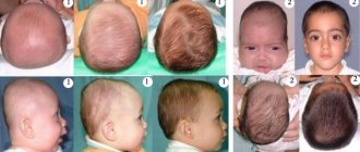
The dolichocephalic shape of the head is the norm during natural childbirth with normal fetal presentation
The brachycephalic shape of the baby's head occurs during natural childbirth, when the baby in the womb is positioned upside down and facing the mother's stomach.
Types of fetal presentation before childbirth - choice of delivery tactics
The same can happen with various pathologies.
Let us highlight the main reasons for the development of abnormalities in a child:
- Genetics . If there was no pathological change in the skull in the family, then the parents or close relatives could have larger or smaller head sizes than normal. This could be inherited by the newborn.
- Birth injury .
It may happen that, traveling along the birth canal, the child encounters a variety of vicissitudes (in the form of bones and tissues of the mother), and therefore is born with an asymmetrical head, with a bump (so-called edema). Physiological parameters are disrupted during induced labor. The process could be slow or too fast. The newborn's head is under pressure for a long time in the birth canal or has difficulty adapting to the narrow passage. In some cases, the obstetrician has to extract the fetus using various methods and means (even the use of instruments). A child may be born with a pear-shaped head, etc. - Congenital pathologies . These are often dangerous pathologies - hydrocephalus (with dropsy of the brain) and microcephaly.
BY THE WAY : Babies born by caesarean section have slight changes.
What does a newborn's skull look like?
The anatomy of newborns is such that immediately after birth they may have an unusual appearance of the skull. In pediatrics, it is believed that whatever the method of birth, the head will be like that: elongated, flattened or even. In order not to worry again, you need to know why this is the norm:
- The baby's skull has soft bones;
- They are mobile, during birth they are self-imposed, facilitating the movement of the child through the birth canal;
- During the natural process of childbirth, a child’s skull is elongated; during a caesarean section, it is flat.
Important! When characterizing the baby's head, its disproportion with the body is noted, so it looks disproportionately large. This is the difference between the body proportions of a baby and an adult.
In addition, the child’s head appears disproportionate due to the fact that its circumference is approximately 2 cm larger than the size of the chest. This is considered an age-related feature of the skull of a newborn child and an indicator of his health.
The pediatrician monitors the dynamics of development, comparing indicators using the anthropometric table.
Large and small fontanelles on the head of a newborn - why are they?
Not only the size of a child’s head matters, but also its shape and fontanel.
This invisible and constantly pulsating island on the baby’s head always scares mothers. And this is understandable - won’t ordinary touching the fontanel affect the baby’s health?
Therefore, you need to know why fontanelles are needed.
So, in short, this is a reliable protector of the baby, who must gradually adapt to our world.
And you also need to know that there is not one, but two fontanelles - large and small:
- Big spring. This is the “breathing” place on the top of the head, approximately 2x2 cm in size. It shrinks as the baby grows and becomes overgrown around the age of one year.
- Small fontanel . It is located on the back of the head, and it is much smaller (up to 1 sq. cm.) of its fellow. Many mothers do not even suspect the presence of a small fontanelle, since it is difficult to palpate (which cannot be said about premature babies).
YOU NEED TO KNOW THIS : Children's fontanelles close differently due to individual development. But, if the fontanel does not heal by the due time, you should consult a pediatrician.
Torticollis in a child at birth - causes and symptoms of torticollis in newborns
Structural features
In the first months after birth, the baby's head is disproportionately large in diameter relative to the chest. Its ratio gradually changes, and its final formation ends in adolescence. This is due to the softer bone structure and the presence of cartilage tissue, which stiffens as the child grows.
Unlike an adult, a baby’s skull has a facial to brain ratio of at least 1:8. In adulthood, the proportion is reduced to 1:2. The box is significantly increased in volume and has fontanelles on the parietal area. These are natural “windows” made of the finest cartilage tissue, which gradually turn into strong sutures.
In the first month of life, the newborn’s skull is quite soft and elastic, practically devoid of ossified areas. Within 2-4 months, bone formation begins. They gradually merge into one canvas, connected by movable seams.
The fontanelles harden by the 12th month. They move to make room for the growing brain. During childbirth, they compress painlessly, allowing the baby's head to pass freely through the birth canal without injury or rupture. They have different shapes:
- Front. The largest in size, resembles a rhombus. It starts from above in the frontal zone, extends to the crown of the child. It may not close completely until 2 years of age, allowing the baby’s brain to be examined using ultrasound.
- Rear. Paired, wedge-shaped, completely hardens by 3 months. Forms the back of the head, starts from the parietal region, is located on the side of the temporal bone.
- Mastoid. A miniature fontanelle, classified as paired, connects the occipital and parietal bones posteriorly after hardening. The closure ends by 4 months.
The shape of the baby’s head changes before our eyes, so parents should carefully monitor the position of the skull during sleep: often the soft areas become deformed when lying incorrectly on one side.
Is it possible to correct the deformation of a newborn’s skull on my own, or with traditional healers?
It must be difficult to find a person with a perfect skull. It could have become deformed while passing through the birth canal - or due to improper handling of the baby (lying on one side for a long time, etc.).
But, if deformations were noticed at first, while the bones are soft , the shape of the head can be changed.
Yes, doctors make appropriate prescriptions, and procedures are carried out under their strict supervision. Is it possible to achieve success on your own?
It is possible, in mild cases, at the initial stage.
This is achieved by:
- Periodically changing the position of the crib and the child in it.
- Turning its head or the entire body in different directions.
- Alternating the hand on which the mother holds the baby during feeding.
- Carrying a newborn in your arms while awake.
- Rolling over onto the tummy (without leaving the baby for a second!).
ATTENTION : No matter how subtle the change in the shape of the skull, consult your doctors!
MUSCULOCAL SYSTEM
SKELETAL SYSTEM
SKULL OF A NEWBORN CHILD AND AGE FEATURES OF THE SKULL
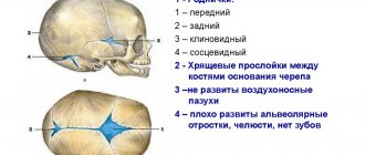
The skull of a newborn has a number of significant features (Fig. 82, 83).
The brain skull, as a result of active growth of the brain and the early formation of sensory organs, is eight times larger in volume than the facial skull, and the eye sockets are wide. The base of the skull, compared to the vault, lags behind in growth; the bones are connected to each other through wide cartilaginous and connective tissue layers. The tubercles of the frontal and parietal bones are well defined and therefore, when viewing the skull from above, it appears quadrangular. The frontal bone consists of two halves, the brow ridges are absent, and the frontal sinus is not yet formed. The jaws are underdeveloped, which causes the low height of the facial skull. The lower jaw consists of two parts (two halves). Parts of the temporal bone are separated from each other by well-defined connective tissue or cartilaginous layers, the mastoid process is not developed. Muscle tubercles and lines are not expressed on the bones of the skull. The most characteristic feature of a newborn’s skull is the fontanelles (fonticuli), which are non-ossified connective tissue (membranous) areas of the cranial vault. There are six fontanelles in total: two are located in the roof of the skull along the midline and four are located on its lateral surfaces. The largest anterior (frontal) fontanel (fonticulus anterior, s. frontalis) is diamond-shaped, located between the two parts of the frontal bone in front and both parietal bones in the back; it heals in the second year of life. The posterior (occipital) fontanel (fonticulus posterior, s. occipitalis) is triangular in shape, located between the two parietal bones in front and the occipital scales in the back. The occipital fontanel closes in the second month after birth. The anterior lateral fontanel - the wedge-shaped fontanel (fonticulus sphenoidalis) is determined at the junction of the frontal, parietal bones, squama of the temporal bone and the greater wing of the sphenoid bone. This fontanel closes in the second or third month after birth. The posterior lateral fontanel - mastoid fontanel (fonticulus mastoideus) is located in the place where the temporal, occipital and parietal bones converge, overgrows in the second or third month of life.
Thanks to the presence of fontanelles, the newborn’s skull is elastic; its shape can change as the fetal head passes through the mother’s birth canal. It is also possible to overlap the edges of the bones of the roof of the skull one on top of the other, which leads to a reduction in the size of the skull and makes childbirth easier. This is facilitated by the Budinova plate - a cartilaginous plate located in the fetus between the lateral part of the occipital bone and its scales and causing the bones to slide and pass one after another during labor. In place of the anterior fontanel, an additional bone is often formed - Vesalius bone - the bone of the anterior fontanel (os fonticuli anterioris).
The volume of the cavity of the cerebral part of the skull of a newborn is on average 385-450 cm3. In the first 6 months. After the birth of a child, the volume of the cranial cavity doubles, by the age of two it triples, in an adult it is four times larger than the volume of the cranial cavity of a newborn. The glabella is absent in a newborn; it is formed by the age of 15. The relationship between the brain and facial parts of the skull is different in an adult and a newborn. The newborn's face is short and wide. In the lateral norm, the ratio of the areas of the facial and cerebral parts of the skull in a newborn is 1:8, in a two-year-old child - 1:6, in a five-year-old - 1:4, in a ten-year-old - 1:3, in an adult woman - 1:2.5, in adult male - 1:2.
After birth, the growth of the skull occurs unevenly, therefore, in postnatal ontogenesis, three periods of its growth and development are distinguished.
The first period - from the birth of a child to 7 years - is characterized by growth, especially in the occipital region of the skull. During the first year of life, the skull grows more or less evenly. From 1 to 3 years, the skull grows especially actively at the back. This is due to the child’s transition to upright walking in the second year of life. In the second or third year of life, due to the end of the eruption of replacement teeth and the strengthening of the function of the masticatory muscles, the growth of the facial part of the skull in height and width increases significantly. From 3 to 7 years of age, growth of the entire skull, especially its base, continues. By the age of 7, growth in the length of the base of the skull basically ends, and it reaches almost the same size as that of an adult.
The second period - from 7 years to the onset of puberty (12-13 years) - is characterized by slow but uniform growth of the skull, especially in the area of its base. At this time, the vault of the cerebral part of the skull mainly grows, especially up to 8 and at 11 - 13 years. The volume of the cavity of the cerebral part of the skull reaches 1200-1300 cm3. By the age of 13, the squamous-mastoid suture is overgrown, and the fusion of parts of the individual bones of the skull, developing from independent points of ossification, ends.
Rice. 82. Skull of a newborn, fontanelles (A - front view, B - side view, right):
1 - Mandibtilarsymphysis; 2 - Milktooth; 3 — Infra-orbital foramen; 4 - Bony nasal septum; 5 - Sphenoid; Sphenoidal bone, greaterwing; 6 - Nasal bone; 7 - Maxilla, frontal process; 8 - Frontal bone; Frontal tuber; Frontal eminence; 9 - Frontal suture; Metopic suture; 10 - Anterior fontanelle; 11 - Parietal bone; 12 - Coronal suture; 13 — Supra-orbital notch/foramen; 14 - Maxilla; 15 - Temporal bone; 16 - Zygomatic bone; 17 - Mandible; 18 — Mental foramen; 19 - Occipital bone, lateral part; 20 - Mastoid fontanelle; 21 - Lambdoid suture; 22— Squamous part of occipital bone; 23—Posterior fontanelle; 24 - Temporal bone, petrous part; 25 - Parietal bone; Parietal tuber; Parietal eminence; 26 - Sphenoidal fontanelle; 27 - Piriform aperture; 28 — Temporal bone, squamous part: 29 — Tympanio ring
Rice. 83. Skull of a newborn, fontanelles (A - top view, B - bottom view):
1 — Occipital bone, squamous part of occipital bone; 2— Lambdoid suture; 3 - Sagittal suture; 4 - Anterior fontanelle; 5 - Frontal suture; Metopic suture; 6 - Frontal bone; squamous part; 7—Corona! suture; 8— Parietal bone; Parietal tuber; Parietal cminence; 9 - Posterior fontanelle; 10 - Palatine bone, horizonta! piate; 11— Vomer; 12 - Sphenoid; Sphenoidal bone, pterygoid process; 13 - Temporal bone, petrous part; 14 - Temporal bone, squamous part; 15 - Tympanio part, tympanic ring; 16 - Mastoid fontanelle; 17 - Transverse occipital suture; 18 - Occipital bone, lateral part; 19 - Foramen magnum; 20— Choana; Posterior nasal aperture; 21 - Maxilla, palatine process; 22— Incisive bone; Premaxilla; 23 - Mandible
In the third period - from 13 to 20-23 years - the facial skull grows intensively, its gender differences appear: in boys, the facial skull grows in length more than in girls, the face lengthens. If before puberty boys and girls have a round face, then after puberty in men, as a rule, the face becomes longer, while in women it remains round.
The transformation of the skull bones in old and senile age can be attributed to the fourth period. At this time, the seams between the bones of the roof of the skull heal. The healing of the sutures of the skull begins at the age of 20-30 years, and in men somewhat earlier than in women. The sagittal suture in the posterior section begins to heal first (22-35 years), then the coronal (24-42 years), lambdoid (26-42 years), mastoid-occipital (30-81 years). The scaly seam, as a rule, does not heal. The process of healing of the sutures of the skull is individual. There are cases when in old people after 80 years all the seams were well defined.
In elderly and senile people, along with healing of the sutures, changes in the facial skull occur. Due to the wear and loss of teeth, the alveolar processes of the upper and lower jaws (alveolar arches) decrease. Due to the weakening of the chewing function, partial atrophy of the masticatory muscles, the relief of the jaws changes (smoothes), they become less massive. The bones of the skull become thinner and more fragile.
Types of skull deformation in newborns
In some pathologies, the shape of the head indicates hidden anomalies in the body. The following types require increased attention:
- Plagiocephaly. Symmetry is broken, the sides are sloping, the frontal lobe becomes unnaturally flat.
- Acrocephaly. The head is strongly elongated, forming a noticeable dome.
- Scaphocephaly. The frontal or parietal lobe protrudes disproportionately relative to the other bones of the skull.
Normally, the shape of a child's head up to one year can change and take on the correct features and proportions. With the above anomalies, asymmetry provokes underdevelopment of certain parts of the brain, leading to severe mental retardation in the baby.
Deformation becomes a consequence of genetic or congenital pathologies that affect the formation of the head during pregnancy:
- intrauterine infections;
- low presentation of the embryo;
- dysplasia.
In rare cases, the cause of abnormal shape is improper care of the baby after discharge. Mom can carry the baby in one arm for a long time, without changing her body position, and apply it to the breast only on the left or right side.
Birth injuries of the skull
During a difficult delivery, the anatomical structure of the head is damaged when the skull does not fit correctly into the mother’s pelvis. In such a situation, the problem requires the help and observation of a neurologist, pediatric neonatologist and surgeon; diagnosis is carried out based on an x-ray. In rare cases, in combination with hypoxia, the brain is irreversibly damaged, basic reflexes and functions are impaired.
Doctors distinguish two types of injuries that require treatment:
- Birth tumor. It is swelling of the soft tissues, most often occurring on the face, forehead or crown of the baby. It can put pressure on the brain, reduce blood flow and provoke hypoxia.
- Cephalohematomas. In case of injury, blood accumulates between the periosteum and the skull, forming a hematoma. Often occurs during rapid labor or a large fetus, and resolves within a few months. If widespread, it can cause severe and incurable bone deformation.
In case of birth injuries, it is important to exclude an increase in intracranial pressure. For severe hematomas, the surgeon removes the contents to speed up the recovery process.
Symptoms and signs of abnormalities
After childbirth, the skull may look slightly flat and convex, but gradually returns to normal. But there are symptoms that indicate more serious damage, hypoxia and possible damage to the nervous system:
- decreased sucking reflex;
- strabismus;
- constant regurgitation after eating;
- lack of tone in the muscles of the legs and arms.
The asymmetry of the child’s face during facial movements and spontaneous lacrimation should cause alarm. The likelihood of injury increases with neonatal jaundice. Such babies are immediately prescribed a brain ultrasound or MRI.


