During childbirth, the baby passes through very narrow passages, as a result of which his body is subjected to significant stress. The head suffers first. However, nature has provided that the bones of the skull are not injured too much. They are quite mobile, soft and harden only after a few years, when the skull is finally formed. After the birth process, an uneven head in a newborn baby is a natural condition. But it is important to be able to distinguish normality from pathology.
Head shapes
A baby's skull may be injured during childbirth.
During the first months of life, a baby's head may appear too large or small, uneven or asymmetrical. Up to 6 months, the process of active growth and development affects the shape of the skull. Normally, the head circumference reaches 32-38 cm. In children under 6 months, every 4 weeks the parameter increases by 1.5-2 cm, and then the growth rate decreases to 5-10 mm.
Indicators of normal form may also differ. In pediatrics, there are several options for skull deformation in newborns:
- Brachycephaly. There is a flat forehead and widening of the bones of the crown, the occipital part protrudes forward. Common causes of the disorder include infections and trauma during childbirth.
- Acromegaly. The deformity is accompanied by a strong increase in the skull on both sides. Among the reasons are hormonal diseases due to cancer in the pituitary gland. A large nose is often observed.
- Scaphocephaly. Deformity with displacement of the frontal or occipital bone forward. Sometimes accompanied by impaired growth and development of the brain.
- Dolichocephaly. Deformation with a narrow and elongated head, “egg-shaped” crown, high forehead. Most often occurs after a rapid process of delivery. Usually it returns to normal on its own.
- Plagiocephaly. When deformed, flat side surfaces remain. The size of the skull is larger than normal, as is the weight of the fetus itself.
- Microcephaly. The head is too small, which is why brain development is quickly impaired. Often bones fuse prematurely and developmental delay occurs.
- Trigonocephaly. The deformity affects the frontal part. It becomes pointed, and the lower jaw moves forward and increases in size. It is formed due to premature fusion of the cartilage of the skull.
- Hydrocephalus. A pathology in which there is high cranial pressure and the head itself is enlarged in size. Just like microcephaly, it has dangerous consequences.
The key factor that provokes head asymmetry in a child is the characteristics of the birth process. In most cases, after a cesarean section, the head acquires a brachiocephalic shape. This is one of the indicators of the norm, which after childbirth will require a small home correction. During natural childbirth, in the first days, the newborn’s head has a dolichocephalic shape.
In the first months of life, the baby’s skull bones are characterized by softness, and their shape gradually changes. Normally it becomes round. However, pathological processes can interfere with the natural formation of contours.
A child has a flat head: is something wrong?
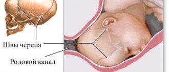
Let's start with the fact that the structure of the skull of a newborn child is different from that of an adult. In a baby, it is much larger in relation to the size of the body, and the ratio of the brain and facial parts is 1:8, while for us it is approximately 1:2. The main distinguishing feature of a newborn’s skull is that its bones are separated: the spaces between them (sutures) are filled with layers of connective tissue, or non-ossified cartilage, and the fontanelles are future sutures.
Fontanas are an extremely clever move by Mother Nature. Due to the presence of open sutures and fontanelles that can sink and protrude, a significant displacement of the skull bones themselves occurs, which makes it possible for the fetal head to pass through the narrow places of the birth canal.
Conclusion. The special structure of the skull allows the baby to be born and live.
Soft spots: counting fontanelles
- big fontanelle,
- small fontanelle,
- two wedge-shaped
- two mastoid.
It turns out 6:
- the anterior, or large, has a diamond shape and is located at the junction of the frontal and parietal bones, completely ossifies by 2 years;
- the posterior, or small, is located between the occipital and parietal bones, ossifies already in the 2-3rd month after birth;
- sphenoid fontanelle - paired, located in the anterior section of the lateral surfaces of the skull, between the frontal, parietal, sphenoid and temporal bones, ossifies almost immediately after birth;
- The mastoid fontanel is also paired, located posterior to the sphenoid, at the junction of the occipital, parietal and temporal bones, ossifies at the same time as the sphenoid.
Baby's head shape: you have to wait
Of course, a child’s head, having undergone transformation during childbirth, cannot return to its original state immediately. In the first hours, the newborn's head may be egg-shaped or slightly flattened on one side. However, these changes are always reversible and disappear by the end of the neonatal period, after 28 days.
In addition, the visualized shape of the newborn's skull is influenced by such phenomena as birth tumor and cephalohematoma. The first is swelling of the soft tissues that occurs during natural childbirth as a result of the mechanical impact of the birth canal on the tissues of the child’s head. When examining the head of a newborn at the place of presentation (the part of the skull that first passes through the mother’s birth canal), swelling is detected, soft on palpation, painful, often spreading beyond one bone. The skin over the tumor is not changed, and the child’s well-being is not affected. This phenomenon is absolutely physiological, usually does not require treatment and resolves on its own within a few days.
Cephalohematoma in a newborn is more dangerous. In essence, this is a hematoma formed during natural childbirth as a result of friction of the child’s head on the birth canal. A local detachment of the periosteum occurs, a cavity is formed between it and the bone itself, which quickly fills with blood. Most often, a cephalohematoma in a newborn does not extend beyond one bone (usually the parietal). No treatment is usually required; the hematoma resolves on its own within a few weeks.
Conclusion. Extremely rarely, with a large and long-lasting hematoma, it is possible to perform a puncture to remove accumulated blood.
Shape of a child's head: fix it yourself?
Sometimes inept child care can disrupt the initially normal shape of the newborn’s skull: lead to its curvature, flattening, or the appearance of various irregular depressions. A child's bones are still very soft due to their low calcium content. If the baby sleeps on the same side for a long time, then this half of the skull will definitely become flatter. This can be avoided by following a number of simple rules:
- alternate the side on which the baby sleeps (right side, left side, back);
- use a special orthopedic pillow for infants.
This situation is aggravated by hypovitaminosis D, which is common in our northern country - in other words, rickets. Because of it, bones are deformed: due to the initially small supply of calcium, vitamin D is less absorbed from food and deposited in bone tissue. To avoid the development of hypovitaminosis, regularly show your child to the pediatrician, give him vitamin D in a timely manner, do gymnastics and massage, and walk a lot.
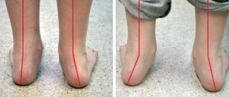
Conclusion. If, despite the parents’ efforts, the baby stubbornly turns his head to one side and always lies on his “favorite side,” you should suspect that he has torticollis and contact your pediatrician, who will refer you to an orthopedist or massage therapist.
The shape of a child's head: but this is serious
Sometimes, unfortunately, a child’s head has very strange and sometimes frightening shapes due to various diseases that disrupt the growth rate of the skull or lead to uneven growth of its individual parts. Such diseases primarily include microcephaly and hydrocephalus.
- Microcephaly, or small head. The child is already born with a reduced skull size, a head circumference of less than 34 cm, or the head stops growing as normal. With microcephaly, there is redundancy of scalp skin and increased bone density.
- Macrocephaly, or large head. The head circumference at birth exceeds the upper limit of normal - 36 cm. If the head is of normal size at birth, but in subsequent months it begins to grow excessively, then this condition is called hydrocephalus.
Conclusion. In order to notice certain changes in the size of the child’s head as early as possible, each parent should regularly show the child to the pediatrician, and if this is not possible, be able to independently measure the size (circumference) of the skull.
By inheritance
There are also genetic diseases that change the shape of a child’s head:
- acrocephaly - “tower skull”, a high conical skull, somewhat flattened in the anteroposterior direction. Occurs as a result of premature fusion of sutures. It occurs in a number of genetic syndromes, the most common of which is Crouzon syndrome. With this disease, the baby is born with prematurely closed fontanelles and sutures. This leads to very sad consequences: an actively growing brain, which does not have enough space, sharply increases intracranial pressure. This, in turn, is accompanied by a whole bunch of symptoms: increased excitability of the baby, regurgitation and vomiting, excessive protrusion of the eyeballs - exophthalmos, convulsions;
- scaphocephaly - a scaphoidally elongated skull with a protruding crest in place of a prematurely overgrown sagittal suture;
- plagiocephaly – oblique head, oblique skull. Asymmetry of the skull caused by premature ossification of part of the coronal suture.
Conclusion. These extreme deformities are very rare, are detected immediately after birth or in the first month and require urgent medical attention.
Looking in the mirror
And the shape of the child’s head is also inherited. And alas, with mom, dad or grandparents it is not always ideal. There are two options for the norm:
bracheocephalic - elongated across and seemingly flattened on top
dolichocephalic - elongated in the anteroposterior direction, but flattened on the sides.
Doctor's advice
Measure your baby's head every 4 weeks. Place the measuring tape along the most protruding parts of the skull: at the back in the area of the occipital bone, at the front along the brow ridges. Write down the number you get. Normal: newborn – head circumference 34–36 cm, 1–3 months – plus 2 cm per month, 3–6 months – plus 1 cm per month, 6–12 months – plus 0, 5 cm per month. Does not match the indicators, be sure to contact a pediatrician, neurologist or surgeon.
What factors provoke pathologies?
Minor irregularities in the skull can go away on their own within 2-3 months.
Minor irregularities and deformations are straightened out when the child is 2-3 months old and begins to sit up. However, some deviations can accelerate bone fusion, which often causes the development of pathologies. The reasons for these processes include:
- bone dysplasia caused by genetic changes;
- hereditary deviations;
- abnormal position of the fetus during pregnancy;
- intrauterine infections.
Less common factors leading to skull deformation in a newborn include:
- combination of a large fetus with a narrow birth canal;
- atony of muscle tissue in a woman in labor;
- narrow or flat pelvis;
- 2 or 3 fruits;
- expulsion of the fetus too quickly;
- incorrect insertion of the head into the pelvic ring.
There is also a group of causes of acquired asymmetry, which occurs when the child is incorrectly positioned in the crib in the first weeks of life. Acquired deformity can occur when the baby is not latched to the breast correctly and is carried incorrectly in the arms.
If a baby has a sloping head that is stretched upward, this in 99% of cases indicates an incorrect position in the crib. A flat back of the head can be a sign of rickets. Sometimes the bevel is observed to the left or to the right. This occurs due to the child remaining on one side for a long time.
Causes of pathology
External manifestations of plagiocephaly
Genetic pathologies and dysplasia (abnormal development of organs, tissues and body parts) can be provocateurs of skull deformation. In some cases, the trigger is trauma during childbirth and a deficiency of vitamins and microelements in the child’s body.
Every person from birth has cartilaginous layers between the bones. They are soft tissues that compress as the baby passes through the birth canal. In some cases, the fragments heal before the baby is born or do not develop at all. For this reason, during the birth of a baby, deformation of the skull occurs.
Sagittal plagiocephaly
Other provocateurs of anomalies include:
- pathological development of the fetus during the first trimester of pregnancy;
- congenital defects due to infections or viral diseases of a pregnant woman;
- morphological disorders of fibrous cartilaginous sutures in the skull;
- infectious diseases of the child causing complications;
- pathologies of the development of the skeletal system: osteoarthrosis, osteoarthritis, osteoporosis, osteopathy, osteomalacia, osteodystrophy, osteodysplasia, rickets;
- diseases of the blood and blood vessels: leukemia, leukopenia, anemia, marble disease;
- circulatory disorders, causing insufficient oxygen supply to the brain;
- pathological processes in the area of the upper bone fragments of the orbit, optic nerve and auditory receptors;
- Paget's disease, Albers-Schoenberg;
- Albright, Dzerzhinsky, Van Buche syndrome;
- familial Krause-Riese dysplasia;
- xanthomatosis;
- dysraphic syndrome;
- hypoplastic chondrodystrophy.
Most often, skull deformation is caused by the passage of the baby's head through the birth canal. This condition usually resolves without treatment after 6 weeks.
The risk of plagiocephaly increases with torticollis.
The acquired disease is observed in children who sleep a lot, in babies with large heads, and in infants who were born prematurely. If a child is diagnosed with torticollis and often turns his head to one side, the likelihood of developing pathology increases.
Positional plagiocephaly usually develops after the baby is born. The supine position causes pressure on one side of the skull from the pillow or mattress. Less commonly, the pathology is caused by restriction of fetal movement in the uterine cavity. For example, this happens when a pregnant woman is carrying more than one child. The deformity can also develop when a baby is born feet first.
Signs of a deformity causing problems
Persistent deformations require medical or surgical treatment.
A visually uneven skull in a child of 3-6 months does not always make one think that this is a pathology. And even minor deformations can cause compression of blood vessels, nerve endings and brain tissue. They can be suspected by the following symptoms:
- strabismus develops;
- the child spits up during and after meals;
- during feeding, an incorrect sucking process is observed;
- torticollis develops;
- intracranial pressure increases;
- movement coordination is impaired;
- hearing problems appear;
- lethargy and muscle weakness are observed.
The most dangerous symptoms are: jaundice, soft and hard bumps on the skull, purple or bluish spots, lacrimation, asymmetry of facial expressions and jaws, bulging of the fontanel in a calm state. These signs require immediate consultation with a doctor and instrumental diagnostics.
Osteopathic treatment of skull asymmetry
Osteopathic treatment begins with testing and assessment of the entire musculoskeletal system, including assessment of the child's posture and movement. Soft palpation, corresponding to the delicate tissues of the child, allows you to detect somatic dysfunctions in the child’s body. Somatic dysfunction is defined as a limitation of movement in the human musculoskeletal system. In children, somatic dysfunction of the region of the skull, cervical, lumbosacral spine is often detected, which corresponds to a predisposition in children to musculoskeletal problems such as plagiocephaly. [7]
Osteopathic treatment is personalized and relies on precise manipulation to achieve results. In the case of brachiocephaly and plagiocephaly, special attention is paid to the treatment of torticollis, dysfunctions of the cranio-vertebral junction, cervical spine, dysfunctions of the cranial region (compression of the cranial sutures, dysfunction of the falx cerebellum and tentorium cerebellum, intraosseous dysfunctions of the occipital bone and sphenobasilar synchondrosis)
Important!
Parents often turn to an osteopath not because of plagiocephaly. The main complaints are often sleep disturbance, reflux, torticollis, restlessness of the baby, delayed motor or speech development, etc. And during examination and osteopathic testing, these complaints often reveal somatic dysfunctions of the neck and skull, corresponding to platycephaly. (Subluxation in the cervical spine, lateral strain, compression of the occipital bone condyles). Often these dysfunctions cause only minimal asymmetry and disruption of the shape of the skull, but they determine the baby’s problems. Thus, skull deformation is a fairly common problem (at least more often than is commonly believed).
Clinical case
As an example, consider a clinical case discussed in detail, described in the journal “JOURNAL OF CLINICAL CHIROPRACTIC PEDIATRICS” Volume 6, No. 1 2004, Chiropractic Care of an Infant with Plagiocephaly [12]
A mother turned to an osteopath with a 7.5 month old boy. The malformation of the baby's skull was obvious, and he held his head forward and tilted forward. This was evident from birth. The mother said that the child sleeps at 20-minute intervals, after which he wakes up and demands the breast. To ensure this regime, the mother slept with the child. At the same time, the child could only sleep on his side on his stomach. When he turned onto his back, he began to scream and tried to move his head from the back of his head.
The child also had a history of colic, which resolved. The child also refused to eat grains or solid foods. At an earlier age, the child had problems with muscle tone and in the motor sphere.
From a medical point of view, the pregnancy proceeded quite calmly, but the mother experienced severe stress due to the breakup of the family.
The birth took place with stimulation and epidural anesthesia. After giving birth, the baby experienced stress and increased heart rate.
Osteopathic examination revealed subluxation of C1 (first cervical vertebra), C5, C7. Cranial examination revealed dysfunction of the occipital bone, insertion of bones along the sagittal suture, and dysfunction of the wings of the sphenoid bone.
The child received osteopathic treatment for 12 weeks.
results
As a result of the treatment, the child's facial asymmetry decreased, he began to eat normally, and his night sleep normalized. The child may have been in a supine position for a long time. The child began to show good results in motor development.
How often and how many sessions of osteopathic treatment may be required?
Each case is individual. Much depends on the severity of the asymmetry, clinical manifestations and the desire of the parents to achieve a particular result.
On average, a course of treatment of 4-6 sessions is indicated, followed by follow-up up to the age of 1.5 - 2 years with an interval of 1 time every 4-8 weeks.
Examination by a neurologist and examination
During the examination, the doctor measures the circumference and sagittal size of the head.
During a routine visit to the pediatrician, pathological asymmetry can be detected in 99% of cases. Next, a referral to a neurologist or neurologist is written out. In difficult cases, consultation with a neurosurgeon is required. If you suspect any abnormalities within 1 month, you can immediately contact a neurologist. It is recommended to do this even in the absence of symptoms of the disorder - to exclude hidden pathologies.
If a newborn’s head deformity is confirmed, the doctor interviews the parents and prescribes the following procedures:
- X-ray. With its help, features of deformation are identified. The technique is used if there is no doubt about the presence of a pathological bone structure.
- MRI or CT. Used in cases where the diagnosis is unclear or there are doubts about its reliability.
- Vascular Doppler. Detects compression of veins and arteries.
- Ultrasound. Used to determine deformities during pregnancy.
Only after studying the test results can the doctor prescribe a suitable method of therapy.
Positional asymmetry of the skull, or when the shape of the skull is disturbed from birth.
Positional plagiocephaly and brachycephaly are a condition where skull asymmetry manifests itself from the moment of birth. According to modern research, it has been shown that such a violation of the shape of the skull in children of the 1st month of life can reach 30%. [7] *the concept of positional plagiocephaly is rarely used; we will consider it in the context of a different development mechanism from deformational plagiocephaly
There are several causes of positional plagiocephaly:
Factors in the development of positional plagiocephaly during pregnancy
During pregnancy, asymmetry can be caused by a large fetal size, multiple pregnancies, increased uterine tone, abnormalities in the structure of the uterus, and relative fixation of the fetal head. [1]
Figure 6. Baby in breech position. Daniel R. Bronfin, MD. Misshapen Heads in Babies: Position or Pathology?
The position of the child in late pregnancy also affects the mechanism of development of plagiocephaly. Thus, a prolonged position in the first position (the child’s back is turned to the left side of the mother, the child lies head down) is a risk factor for flattening of the occiput on the right side [1].
Factors in the development of plagiocephaly during childbirth
By the time of birth, the bones of the skull have not completely ossified, this ensures maximum flexibility during childbirth. But also for the same reason, if the birth process is disrupted, deformations of the skull can occur. The bones of the skull adapt to the size and shape of the mother's pelvis. This is facilitated by turning the head, as well as the overlapping of the skull bones on top of each other. This process is called skull modeling.
Depending on the insertion of the fetal head, the structural features of the mother's pelvis and the duration of labor, a disturbance in the shape of the skull in the newborn may occur.
Therefore, if there is a history of the following events, it is necessary to pay close attention to the condition of the sutures and the position of the bones in the newborn: labor stimulation, epidural anesthesia, prolonged or rapid labor, a long period of contractions, incorrect insertion of the head, the use of forceps or a vacuum. [7].
Also, these factors can cause birth trauma, increase the risk of developing cranial asymmetry, and cause somatic dysfunctions of the neck and skull region. Let's look at this in more detail...
Treatment options
Treatment of irregularities using orthoses
Depending on the type of head deformation in a child, recovery tactics are selected. These may be medications in the case of dropsy, surgical interventions for dangerous disorders, and home methods for non-pathological curvature of the bones.
Use of medications
Drugs are prescribed for hormonal disorders, especially changes in the functioning of the pituitary gland. They may also be needed for immune disorders, as well as during the recovery process after surgery.
Cranial orthoses
Special orthopedic products aimed at restoring a crooked head in a child. Selected individually. Their direct purpose is improper bone formation and asymmetry. The orthosis creates constant but slight pressure in the desired part of the head. Orthopedic devices are prescribed from 4 to 6 months of life, when the bones are still soft, but do not grow as quickly as in the first 12 weeks.
They wear such a bandage constantly, and take it off only for a while - no more than once a week.
You can use orthoses even at an older age. But in this case, their shape and period of wearing will change. If in the first year of life they are prescribed for a maximum of 4 months, then in adulthood it takes up to a year to recover.
Tips for parents with minor asymmetry
During sleep, the child's head must be turned periodically.
If the doctor does not find any pathological abnormalities, he may prescribe home treatment. It comes down to simple everyday procedures and rules:
- carry the child in your arms more often if he is not sleeping, to reduce pressure on the bones of the skull;
- when resting, change the position of the newborn’s head, tilting it to the left or to the right;
- put the baby on his stomach more often, observing safety precautions and not leaving him unattended;
- sign up for a relaxing and strengthening massage course with a professional;
- put the child in different positions more often so that he independently changes the position of his head;
- Apply alternately to the left and then to the right breast when feeding.
It is useful to have a special orthopedic pillow for newborns in your arsenal.
Performing an independent massage, especially in the head area with deformation, is strictly prohibited. Even a little incorrect pressure can make the situation worse.
Working with an osteopath
If parents turn to an expert in the first year of a child’s life, the chances of recovery increase tenfold. An osteopath helps eliminate hypertension, relieves spasms and helps the proper development of the cervical spine. If it functions correctly, all organs receive sufficient nutrition and oxygen.
Surgical intervention
Premature fusion of the skull bones is corrected surgically.
The indication for surgery is premature fusion of the skull bones. This is required in approximately 7% of all head deformity cases. The procedure is simple and carried out quickly. Children recover within 2-4 months. Also prescribed when there is a risk of brain compression.
To distinguish pathological deformation of the skull in a child from natural processes that do not require medical intervention, it is important to undergo professional diagnostics. If violations are detected, the doctor will be able to quickly select effective treatment. After it, children do not experience developmental problems.
What determines the shape of a child's head?
It must be said right away that in most cases the shape of the head does not affect the formation of functions and the development of the child.
There are conditions in which the shape of the skull is changed - and this is associated with serious problems. As a rule, doctors do not miss such conditions, and we will not talk about them.
In all other cases, this is largely a “cosmetic” problem. The shape of the baby's skull gradually changes and returns to normal. The reason for this is the peculiarity of the structure of the skull of a newborn child. The baby's skull bones are soft. This is a whim of nature and a necessity, since during passage through the birth canal the skull is compressed, and the mobility of the skull bones plays a large role in this process. For this, we have everything at birth, for example, fontanelles. All this serves us well during childbirth, but can cause anxiety later.
If a child is often in one position, then the shape of his skull may change, as long-term pressure is placed on the bones. If a child lies on his back a lot, the back of his head may become flattened. If there is asymmetry in muscle tone in a child, the head may be turned to one side, and this also leads to a change in the shape of the skull. This happens with torticollis. Hematomas after childbirth can also lead to changes in the shape of the skull.
Torticollis in a child
If a child has torticollis, then you need to contact a specialist: an osteopath or a massage therapist. In this case, it is recommended to frequently change the child’s position so that he is in one position as little as possible.
In this case, I can give some recommendations to parents.
- It is very important to frequently change the baby's position during the day. If you decide to do this at night, it will result in neither you nor the child getting enough sleep. Night is a time for sleep, not for correction. During the day you can carry your baby in your arms a lot, and this is already a prevention. Take your child to objects in which he is interested more often. Moreover, it is precisely the side in which he turns his head with difficulty. This will be the most effective way. Remember that when the child is in your arms, there is no static situation: after all, you are constantly changing the position of the child, and this is exactly what experts recommend to do.
- You can lie with the baby on your side. If the child is lying on his back, then place him in your arms so that the head is in the midline - and the child is looking forward. It is visual fixation that best teaches the child to keep his head in the midline. Dynamics, styling in combination with massage or osteopathic manipulations give good and quick results.
Flat back of the head in a child
A flattened occiput most often occurs in a child when he often lies on his back and rests on the back of his head. This often happens with increased back muscle tone. Then there is prolonged and powerful pressure on the bones of the skull, and the back of the head flattens.
In this case, the recommendations do not differ from those given above for torticollis: massage, osteopath and frequent change of position, which is easiest to ensure when carrying a child in your arms.
Changing the shape of the skull does not affect the development of the child and the formation of his brain. Another thing is the habit of turning your head. The child develops and learns to fix his gaze and reach for toys.
Does head shape affect the overall development of a child?
So, if he is used to looking in one direction, then most likely he will use one hand more often. He will develop the habit of doing everything on one side. This feature is fixed in the child’s motor skills and behavior. All sensory systems “record” this method as correct, and the child tends to the usual position.
Even if parents have dealt with the baby’s muscle problems, this does not guarantee that he will turn his head in both directions.
Therefore, simultaneously with styling and massage, you also need to teach the child to fix his gaze.
It is not enough to place the child on the rollers; it is also important that he shows interest in the process and is active. Then after a while he will independently change position and choose different poses. Initially, this requires more attention from adults, but then it gives excellent results: the child will roll over in both directions, crawl symmetrically, and it will be easier for him to master all motor skills. By the way, parents will not have to worry about spinal curvature during school years.
To summarize, the altered shape of a child's skull is a symptom. The reason needs to be found out and actions taken to help the child.
I have considered only a few reasons that lead to changes in the shape of the skull, but there are others: rickets, endocrine problems, genetic abnormalities, metabolic disorders.
In all these cases, you need to consult a doctor, and in most cases you cannot do without medication. You may need to create a special diet for a nursing mother. And in such situations, I recommend visiting a pediatrician, who will refer you to the right specialist.
The article was published on the website letidor.ru>>Letidor.ru - an online project for modern and smart parents with an active lifestyle.
The first year of a baby’s life is the foundation for a successful future. Surprisingly, it is at this age that we get our first experience of success, make our first decisions, and begin to believe in ourselves and our strengths.
For parents of little ones, based on the most common questions, tasks and experiences of mothers and fathers, I created a series of mini-lectures about children from 0 to 3 years old “Online ABC. Really important."
Online ABC is a large collection of lectures that includes more than 60 videos about motor skills, psyche, toys by age, orthopedic diagnoses and much more that is really important for parents to know.
Find out details
about the author
Oleg Leonkin, children's rehabilitologist, hippotherapist, specialist in sensory integration and children's massage, husband and father of 5 children.
For more than 25 years I have been working at the intersection of 3 sciences: medicine, pedagogy and psychology, 18 of which have been with “special children”.
When observing a child, I analyze about 40 factors, see the stage of development, feel the child’s needs and know about his problems. I listen to my parents and believe them. I think, draw conclusions, connect and explain. I translate from children's language to adult language.
Features of the structure of the infant's skull
Important! The development and shape of the skull, which contains the brain, the most important organ of the human body, is very important. Therefore, pediatricians very carefully assess the size and shape of the baby’s head in all examinations.
The bones of a newborn's skull are extremely plastic. They are not as hard as in adults and are not rigidly connected to each other. Between them there are cartilages and fontanelles, which will subsequently heal. Thanks to this, the cranial bones easily adapt to pressure from within (during the growth of the brain) and to external influences.
Newborn skull
This feature of the bone structure ensures the passage of the head through the birth canal. It can also cause changes in the shape of the child’s head. Postpartum changes, as a rule, do not require any treatment and disappear during the first month of the baby's life. However, the shape of the head can change not only as a result of childbirth.
Preventive actions
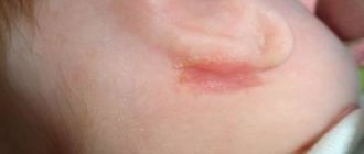
Red spots on the face of a newborn baby
Some tips on what parents can do to prevent their child from developing a sloping head:
- If the baby prefers to sleep on one side, then it is necessary to change his orientation in the crib (head-foot). Children love to look at familiar stationary objects, windows or walls; any change in orientation causes them to turn their heads;
- The recommendation to lie on your stomach while awake (starting from a few minutes and increasing to 30-60 minutes a day) also turns out to be very useful in the prevention of plagiocephaly, especially if practiced from the first weeks of life;
- Changing the position of toys in the crib to encourage the baby to turn his head in different directions;
- Alternating feeding sides. This is especially true for artificial feeding. Do not give the bottle while lying on your back;
- Reducing the time when the child rests his head on flat surfaces: in car seats, carriers and other devices.
According to recent studies, almost half of all newborns experience some degree of deformation of the skull bones, but in the vast majority of cases this is not a serious problem and is eliminated during the physical development of the child and the growth of his brain. By taking preventive measures, you can reduce the risk of developing a flat nape to almost zero.