Home › Services › For children › Urology › Cystography for children
Direction: Urology
Make an appointment
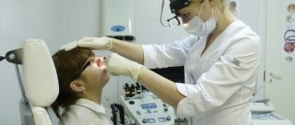
Cystography is a radiopaque diagnostic method that allows you to evaluate the anatomy of the bladder. With the help of cystography, it is possible to study the position, shape and size of the organ, as well as visualize pathological processes.
For cystography, safe water-soluble contrasts are used, which are approved for use in pediatric practice. The study is performed using modern equipment, and the images are interpreted by experienced radiologists. At all stages of diagnosis, the child is accompanied by the comfort and attentive attitude of the staff.
Preparation for the procedure
Before performing a cystography procedure, you must:
- Get a consultation with a urologist (indications for the study and the range of necessary examinations)
- Get an ultrasound of the kidneys and bladder.
- Take a general blood test.
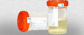
- Take a general urine test.
- Submit a urine culture to determine sensitivity to antibiotics.
- During intravenous administration of contrast, blood urea and creatinine levels are measured.
Preparation begins 2-3 days before the study.
Foods rich in fiber (dark bread, durum pasta, vegetables, herbs, fruits, etc.) should be excluded from the diet, otherwise swollen intestinal loops will disrupt the visualization of the organ being examined. On the day of cystography, a light breakfast is allowed. You must have a bowel movement 2-3 hours before the test. To do this, it is recommended to perform a cleansing enema. Immediately before cystography, you should empty your bladder.
Before X-ray contrast diagnostics, it is advisable to undergo a general clinical urine test. If there is a chronic infection of the urinary system, then 2-3 days before contrast and for 3-5 days after, prophylactic use of uroseptics is indicated (the doctor prescribes the optimal drug).
What is cystography and how is it performed?
Cystography is an endoscopic diagnostic method that is based on x-ray examination of the bladder to visualize the anatomical features and pathological changes of the organ. To conduct the study, the bladder is filled with gas or a contrast agent. During cystography, the doctor receives detailed information about the shape, size and location of the bladder. Also, with the help of a cystographic examination, you can detect various disorders of the urinary tract, ruptures and thickening of the organ, reflux, sand, stones, tumors and other dysfunctions of the urinary system.
Cystography of the bladder is prescribed only for special indications, when other diagnostic methods (cystoscopy, ultrasound) did not provide a complete picture of the possible pathology. Cystography is not recommended for preventive purposes or as part of screening. The procedure is unpleasant and accompanied by painful sensations.
Cystography is an x-ray procedure for studying the anatomical and physiological features of the bladder. To obtain an x-ray image, a contrast agent is used, which is injected in a descending or ascending manner.
Indications and contraindications for cystography
Cystography of the kidneys and bladder is prescribed by a urologist after a preliminary consultation if the following indications exist:
tumor neoplasms of the urinary tract;
- Suspicions of urolithiasis and the presence of stones;
- Traumatic bladder injury;
- Some inflammatory pathologies of the urinary system;
- Various neoplasms in the prostate area;
- The presence of pathological changes in the genitourinary system;
- Vesicoureteral reflux (return of urine to the kidneys);
- Bladder rupture.
Like any method of radiopaque diagnostics, cystographic examination has a number of contraindications:
- Previously undergone surgical interventions;
- Intolerance, allergic reaction to drugs used during the study;
- Acute nephrological diseases;
- Urinary obstruction;
- Pregnancy;
- Infectious pathologies of the bladder in the acute stage;
- Full intestines.
Preparation for cystography
To obtain reliable diagnostic results, you should first prepare for cystography of the bladder. Preparatory measures for the study include eliminating excessive gas formation and cleansing the intestines. To do this, you need to adhere to a strict diet for 2-3 days before the procedure. Any foods and drinks that cause fermentation in the intestines should be excluded from the diet. These include strong tea and coffee, carbonated water, legumes, white cabbage, fermented milk products, corn, whole milk, etc. To empty the intestines of feces in the morning, on the eve of the examination, a cleansing enema is given.
Before undergoing cystography, the patient should consult a nephrologist, radiologist and urologist. They will prescribe the necessary examination to exclude possible contraindications and provide recommendations, thanks to which the diagnostic results will be as informative and accurate as possible.
By contacting the Rainbow Health clinic , you can be confident in the accuracy of the results, safety, effectiveness and quality of the examinations performed. We employ experienced specialists using modern equipment.
Make an appointment and consultation
- need by phone 8 (831) 214-16-93
Order of conduct
There are 2 options for performing cystography.
- Ascending (retrograde) technique, in which contrast is administered through a catheter installed in the bladder. In young children, filling is carried out to physiological volume, and in children of the older age group - until the urge to urinate appears.
- Descending (antegrade) technique, in which contrast is administered intravenously, after which some time is waited for the solution to enter the bladder.
Regardless of the contrast technique, the next step is to take a series of x-rays.
In some cases, to assess the functional activity of the bladder, voiding cystography may be performed when the patient urinates while on the X-ray table. An x-ray is taken at the time of urination. After the test is completed, the child should consume more fluid to speed up the removal of the contrast from the body.
Radionuclide diagnostics
A study that allows you to reliably and objectively assess the presence of vesicoureteral reflux (VUR) both in the phase of filling the bladder and in the phase of emptying it, as well as accurately determine the functional volume at which reflux occurs.
In addition, this method allows you to determine, in addition to the height of reflux, its characteristics such as intensity and duration. The undeniable advantages of direct radionuclide cystography compared to voiding x-ray cystography are: significantly lower radiation exposure for the patient, as well as the possibility of long-term and continuous image recording.
Indications:
suspicion of vesicoureteral reflux.
Preparation for the study: No special preparation is required. Immediately before the test, you must empty your bladder.
The drug used. 99mTs-Tehnefit.
Method of performing the study. The patient is placed supine on the gamma camera table. The bladder is catheterized using a sterile disposable catheter of age-specific diameter. Under visual control on the gamma camera screen, saline solution is slowly injected into the bladder until a persistent urge to urinate appears in older children, or until independent urination in younger children.
In older children, after maximum filling of the bladder, the urethral catheter is removed. Next, the patient is seated on a bedpan with his back to the gamma camera detector and asked to strain and urinate. Very young children urinate in their diapers while remaining on the table.
During the entire duration of the study, continuous dynamic recording is performed.
Research time. The total time for performing the diagnostic procedure from the moment of administration of the radiopharmaceutical to obtaining the final images does not exceed 20-30 minutes.
The processing time for results ranges from 15 minutes to 40 minutes and depends on the specific clinical situation.
Research data is provided in printed form. At the request of the patient, the conclusion on the results of the study can be received by e-mail, or picked up independently the next day.
Additional features.
Patients who do not have the opportunity to get a consultation with a pediatric urologist at their place of residence can get a consultation at our Center with Professor A. Yu. Pavlov and other highly qualified specialists in the Department of Pediatric Uroandrology
Contraindications
Descending cystography has virtually no contraindications, except in cases of severe renal failure.
Ascending cystography is not performed if the patient has urinary tract infections in the acute stage, because when inserting a catheter, there is a risk of spreading pathogenic microorganisms to the upper parts of the urinary system. Also, this method is not applicable if there are obstacles in the urethra and neck of the bladder in the form of large scars and tumors.
Any type of cystography is contraindicated in case of intolerance to X-ray contrast agent.
Cystography allows assessment of normal and pathological anatomy of the bladder. Using this study, the following are diagnosed:
- filling defects, which are characteristic of stones, tumors, foreign bodies;
- reflux of urine into the upper parts of the urinary system - vesicoureteral reflux;
- leakage of urine into the abdominal cavity due to traumatic injury;
- leakage of urine into nearby genital organs, which indicates fistula.
Cystography also allows one to suspect the presence of a posterior urethral valve - these are folds of mucous membrane in the urethra that make it difficult to empty the bladder and lead to chronic urinary retention.
The duration of the study is about 20 minutes. The radiologist's conclusion on the presence or absence of pathology is issued immediately.
Low-dose fluoroscopic cystography compared with direct isotope cystography in children
Introduction
Both fluoroscopic cystography (fVCUG) and direct isotope cystography (DIC) are frequently used in pediatric urology. Both methods can detect vesicoureteral reflux (VUR) and have high sensitivity. While the advantages of fVCUG include the ability to determine the anatomical features, anomalies and structural details of the maxillofacial lesion, the main advantage of DIC is the low radiation dose. However, over the past 10 years there have been significant technical advances regarding fluoroscopy, which, according to the literature, have reduced the radiation dose of fVCUG.
Target
To answer the question: is it true that when performing direct isotope cystography, the radiation exposure is less than when performing fluoroscopic voiding cystography?
Study design
Direct isotope cystography was performed in 92 children and an additional 7 children; after adapting the protocol, a comparison was made with fluoroscopic voiding cystography performed in 51 children. The studies were carried out in accordance with the protocol of our clinic. Published physics models adjusted for age and sex were used to estimate effective radiation dose [mSv] for both methods. For DIC, the model of Stabin et al., 1998 was used, for fVCUG, two different physical models were used (Schultz et al., 1999, Lee et al., 2009).
results
The radiation dose with direct isotope cystography turned out to be significantly higher than with fluoroscopic voiding cystography, the average effective dose was 0.23 mSv (±0.34 m, median 0.085 mSv) versus 0.015 mSv (±0.013, median 0.008 mSv, Schultz et al. model) - 0.024 mSv (±0.018, median 0.018 mSv, model by Lee et al.), respectively. After adapting the protocol to change the duration of the DIC examination, which was associated with filling the bladder to the intended capacity, the mean dose was .07 mSv (median 0.07 mSv) and the values were more uniform.
Discussion
As we expected, at the current stage of technology development, the radiation exposure when performing fluoroscopic cystography turned out to be even less than during DIC. In our protocol, according to nuclear medicine standards, the bladder was filled to the calculated capacity. This led to longer examination times in patients with high functional bladder capacity and, accordingly, to greater radiation exposure. However, even if the protocol were changed or only patients with relatively rapid bladder emptying were examined, the radiation exposure with DIC was still higher (at least 2.9 times compared with the “worst” fVCUG data and the “best” DIC). The absolute value of radiation exposure for any of these examination methods is lower compared to other medical radiation examination methods, as well as compared to background radiation in the environment. Accordingly, the differences that may have been identified are not significantly important for the possible risk of future development of cancer. However, during repeated examinations, in order to adhere to the ALARA principle (as little as possible within the necessary limits), one should strive to use fVCUG more often. Moreover, fVCUG provides more information about the anatomical structure of the urinary tract compared to DIC.
Conclusion
Contrary to popular belief, it turned out that the radiation exposure with fluoroscopic voiding cystography is significantly lower than with direct isotope cystography. A prerequisite for our study was the use of modern equipment to optimize the examination protocol. When performing both methods of examination, the radiation exposure is small and is important only when performing repeated examinations. However, our data should be kept in mind when choosing an examination method to minimize radiation exposure to the patient.
Key words: direct isotope cystography; pediatric urology; radiation load; PMR; cystography at the time of urination
Lower radiation burden in state of the art fluoroscopic cystography compared to direct isotope cystography in children
Bernhard Haid, Tanja Becker, Mark Koen, Christoph Berger, Werner Langsteger, Bernhard Gruy, Ernst Putz, Stephanie Haid, Josef Oswald
Journal of Pediatric Urology, 2015 Feb; Vol.11, Is.1, P.35.
Translator: Garmanova Tatyana Nikolaevna
Magazine
Journal of Pediatric Urology (Journal of Pediatric Urology) 2015 February
Comments
To post comments you must log in or register