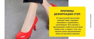
There are probably no parents who would not be concerned about the shape of their child’s legs. Often parents, and especially grandparents, complain: “They are completely crooked” or: “He has club feet!” and so on. Less than half of humanity has an ideal foot. Flat feet is one of the common diseases of the musculoskeletal system. According to scientists, from 40 to 60% of the entire world population suffer from flat feet. Studies conducted by domestic scientists over the years have revealed flat feet in 20-30% of the children examined. According to statistics, flat feet occur in 7-8% of cases in 6-7 year old children. According to the City Medical and Physical Education Dispensary of St. Petersburg, in preschool age, 80% of children have poor posture, 40% have flat feet.
Let's start with flat feet. All children are born with a flat foot, but when the child begins to stand up and then walk, loading the foot more and more, all the elements of the foot are formed correctly. After 6 years of age, a child begins to place his foot on his toe when running, and more correct elements of walking are formed. After 7-8 years, muscle strength and endurance increase. By the age of 10, the structure of the foot is formed and approaches the structure of the foot in adults.
Reduced muscle tone of the legs (with concomitant neurological pathology, rickets, etc.), excessive mobility in the ankle joint (which is often observed due to unprofessional massage), calcaneal-valgus position of the feet are signs that should alert the specialist and parents. The task of an orthopedic doctor during this period of a child’s life is to determine in time whether there is a tendency to develop flat feet or not. Therefore, visiting an orthopedist before the child begins to walk is one year old is very important.
Answers to frequently asked questions
Is it possible to completely cure a deformity?
It is possible if you consult a doctor in time (up to 2 years) and undergo treatment, strictly following all medical recommendations.
Then the child will go to school healthy. When starting treatment after two years and in advanced situations, it is difficult to completely cure the deformity. However, it is possible to significantly improve the condition of the foot.
What could be the consequences if the hallux valgus defect is not treated?
Foot deformities do not correct themselves. Treatment is necessary. Otherwise, the changes will affect not only the ankle joint, but also the upper legs and spine.
This will not only lead to an incorrect gait. The child will be limited. Sports, active games, and running will become unavailable to him. Your legs will hurt and swell after exertion.
In severe cases, the cartilage and bones of the knee and hip joints will begin to deform and collapse, and the spine will become bent. The baby may become disabled.
About the disease
In medicine, valgus is a deformation of the feet in which they are in a cruciform position in relation to each other, reminiscent of the Latin X. Most often, the pathology becomes noticeable when the child tries to step on the feet and take the first steps - the pathology is expressed in the fact that when When walking, the baby rests on the inside of the foot.
It is extremely difficult for such a baby to take steps - he gets tired quickly, sometimes experiences pain, and the steps themselves are shaky and uncertain. Orthopedists describe this condition in terms of the processes occurring in the feet - the toes and heels are turned outward, the middle part of the foot is slightly lowered. If the legs are straightened and pressed against each other in the knee area, the distance between the ankle bones will be more than 3-4 centimeters. If the height of the arch of the foot is significantly reduced, then orthopedists will say that the child has planovalgus feet. Valgus flatfoot is considered the most common diagnosis in pediatric orthopedics.
There are two types of such curvature of the feet: congenital and physiological (acquired). In the first case, the legs are bent even during the period of intrauterine development of the fetus under the influence of certain factors about which medicine still does not know much. Congenital foot pathologies are usually quite severe, and they can be seen in the first 2-3 months of a child’s independent life.
Acquired deformity is often associated with errors in the development and functioning of the musculoskeletal system, ligaments, and tendons. It is precisely such violations that become obvious closer to the age of one year. At risk are babies with weakened muscles, premature babies, those suffering from frequent and severe viral infections in the first year of life. The legs are at risk of becoming bent in obese children, since the load on the lower limbs with excess weight is very significant.
Sometimes parents themselves are to blame for the occurrence of pathology. Thus, putting the baby on his feet too early may well “trigger” the mechanism of foot deformation, and insufficient load on the foot, walking exclusively on a flat floor can cause acquired flat feet or planovalgus foot.
Flat feet scare parents no less. However, Komarovsky advises not to panic, because from birth absolutely all children have flat feet, this is a feature of babies. The arch of the foot will form gradually as the load on the legs increases, and here everything is in the hands of the parents, with the exception of congenital flat feet, which can only be corrected surgically.
Flat valgus feet as a type of deformity
Plano-valgus foot in children is an orthopedic disease, curvature of the lower extremities in the case of support on the inside of the feet when taking a standing position. In this case, the heel and toes are turned outward. According to ICD-10, it has code M21.0 and Q66.6.
More often, the disease is diagnosed in children at 4-5 years of age, when improper formation and positioning of the legs when walking begins to appear. This is a fairly rare pathology of the musculoskeletal system. If treatment is neglected, by the age of 6 the child may develop flat feet.
As pathology develops on the sole, a depression in the arch area can be observed under the influence of gravity when walking. In children, the heels and toes begin to bend (turn outward), and the middle begins to sag towards the inside of the foot.
Flat valgus foot
Foot valgus in children is easy to recognize visually; it is enough to place two feet side by side, as you can see when viewed from above that the legs resemble the letter X.
What is foot varus: characteristics and symptoms
There are two approaches to foot correction:
- conservative physiotherapeutic treatment;
- corrective surgery.
An important therapeutic measure is walking barefoot. At home and on the lawn, on a special orthopedic mat or without it - the main thing is as much as possible. Choosing the right shoes is equally important. To do this, parents need to seek help from an orthopedist. Basic requirements for orthopedic shoes:
- shoes must be strictly in size, you cannot buy them “for growth”, the length of the sole must exceed the length of the foot by 0.5 cm;
- slippers, shoes and sandals must have a solid back;
- It is necessary to have orthopedic insoles, instep supports and correctors that do not allow the foot to turn outward.
Physiotherapy exercises (PT) for this disorder are always prescribed. Parents need to learn a set of exercises with their child and repeat them daily for at least 10 minutes (preferably several times a day). Simple examples of exercises that promote proper development of the feet:
- sit on a chair and alternately bend and straighten your feet;
- external circular movements;
- sitting pose between the heels;
- squats with emphasis on the foot (you can use a chair for balance);
- exercises with heels - stand, toe rolls, descent from steps;
- various walking options (herringbone, single file, etc.).
With persistent conservative treatment, by the age of 5-7 years the deformity disappears, the legs become straight and even, and the gait becomes normal. If this does not happen, the orthopedist may suggest surgical intervention to the parents. This is a complex surgical operation that is performed in several stages:
- a small fragment is cut out from the tibia;
- the extracted part of the bone is fixed with special screws on the lower leg;
- The Elizarov apparatus is installed for several months.
To determine possible foot pathologies in a child, you need to ask him to take off his shoes. If you imagine that the leg has a center line perpendicular to the floor, the heel should be located symmetrically to this axis. If there is a clearly noticeable “collapse” to the outer side and the central axis is c-shaped, there is a varus deformity of the foot. When the pathology covers two legs, a significant gap is noticeable between them, resembling the letter “o”.
If you look at the foot from the front, it will be noticeable that the forefoot is not completely parallel to the floor - there is some space between the big toe and the surface of the floor, which the ankle tries to compensate for by bending outward. The main load falls on the external arch of the foot; the child becomes clubbed when walking. This positioning of the foot often leads to flat feet (for more details, see the article: What physical therapy exercises should be performed for flat feet in children?).
Treatment methods for hallux valgus
Treatment of hallux valgus depends on the stage of development of the disease. Only a specialist can choose a truly effective option. It is worth considering commonly used types of fight against a bunion on the big toe.
Operational
Surgery helps to quickly and permanently get rid of hallux valgus. It has the following options:
- traditional;
- laser
For quite a long time, surgery has been used to treat a protruding bone. The traditional option involves the use of general anesthesia. The operation itself is performed using a large number of instruments and is accompanied by blood loss. Upon completion, the patient is given a plaster cast. For this reason, the person remains immobilized for about 2 weeks. The recovery period itself takes up to 2 years and is accompanied by some discomfort.
Laser treatment is now widely used. This operation is performed through small incisions in the area of the lump, so it leaves only barely noticeable scars. The procedure itself lasts about an hour and is performed under local anesthesia.
Laser stone removal can be of the following types:
- exostectomy involves the removal of excess bone and soft tissue, immediately after which the foot takes on its proper appearance;
- laser osteotomy partially excises the phalanx of the thumb;
- Resection arthroplasty is used in complex cases and involves excision of valgus deformity and replacement of the joint with a prosthesis.
The decision on the need for surgery and the type is made only by the doctor based on the results of a detailed diagnosis of the patient. In any case, laser treatment is more gentle and allows you to return to work after two weeks. Leg mobility is restored the very next day after surgery.
Medication
Drug treatment for hallux valgus is reduced to relieving swelling, inflammation and pain. It will not be possible to remove the lump without surgery, but ointments such as Voltaren or Indomethocin can quickly relieve unpleasant symptoms.
Doctors may also prescribe anti-inflammatory drugs, for example, Nimulid. This remedy is used internally in a course and helps to get rid of pain in the big toe area for a long time.
The use of Meloxicam and Movali gives very good results, but these drugs should only be used with a doctor's prescription. This will allow you to get rid of pain safely and quickly.
Folk remedies
Folk remedies can also help improve your condition for a while. They are used for various diseases. There are recipes aimed specifically at relieving inflammation with hallux valgus. The most popular are:
- Dandelion infusion based on iodine. 100 g of dried flowers should be filled with iodine and left for 4-5 days, then applied in the form of a grid to the problem area for 2 weeks.
- Compresses with propolis. A small piece of propolis should be kneaded in your hands and applied to the bump overnight. Repeat for a month.
- Salt and iodine bath. Every evening, it is recommended to steam your feet in warm water with the addition of 2 tablespoons of salt and 10–15 drops of iodine.
- Burdock wraps. Fresh burdock leaves should be moistened with turpentine and applied to the pine cone.
- Cabbage and honey compress. First, honey is applied to the leg, then a mashed cabbage leaf is applied and secured with a bandage. The compress should be applied before bedtime.
- Homemade ointment based on iodine and aspirin. You need to add 5 aspirin tablets to a bottle of iodine and apply it to your feet every 3 days for a month.
These methods will not help get rid of the bone, but they relieve inflammation quite well. Before starting such treatment, possible allergic reactions must be excluded.
Manifestation of pathology in adults
The procedure for foot deformities can be carried out at home, if the person first receives consultation from a specialist in this field.
When performing therapeutic massage of hallux valgus in children and adults, you must follow certain rules:
If you have bumps, it is recommended to steam your feet. It is recommended to add relaxing ingredients to the foot bath - sea salt, chamomile infusions, sage. You can use emollient creams, gels or milk. Such products moisturize the skin well, making the procedure pleasant and effective. The person should sit comfortably on a flat surface. There is a cushion under the feet (tendons). The most effective results are achieved with daily sessions. Before massaging, it is necessary to carry out warming manipulations. For children, treatment procedures are carried out comprehensively. You need to stroke, starting from the lumbosacral region, ending with your toes
It is better if the baby lies on his stomach. Rubbing should be done carefully, without stretching the skin. Otherwise, pain and discomfort may appear after the procedure. After the massage, you can do several therapeutic gymnastic exercises.
This will consolidate the effect and have a positive effect on the condition of the legs.
The procedure takes on average no more than 15 minutes. When conducting therapy for children, it is necessary to create calm conditions. The baby should not cry, be capricious, or be afraid of the procedure. Adults should understand that a positive effect can be achieved exclusively by regular sessions, which will be supplemented by exercise therapy complexes, gymnastics, the use of special mats, wearing correct orthopedic shoes, and swimming.
In more rare cases, pathology begins to appear after 5 years or in adolescence. This is already associated with increased stress on the legs, injuries, incorrect choice of shoes or the development of endocrine diseases. It is much more difficult to cure hallux valgus after 10 years; the pathology often causes complications.
Even less often - in approximately 10% of cases, the pathology is diagnosed in infants, as it is congenital. In this case, the main treatment method is immobilization of the feet in the correct position using a plaster cast.
In adults, deformities of the foot and toes are more pronounced and are much more difficult to correct.
If in children flat-valgus feet are easily corrected, since the musculoskeletal system is still developing and ossification is not complete, then in adults it is much more difficult to cure the pathology. Most often, conservative methods only help relieve discomfort and pain, and the deformity is corrected surgically.
Pediatric orthopedics: primary care options
What orthopedic problems in childhood are more physiological than pathological in nature? How can you distinguish repeated inflammation of the hip joint from septic arthritis? How should a general practitioner assess the condition of a lame child?
| Figure 1. Varus deformity of the knees. This child has a normal arch formed by his legs; legs straighten spontaneously |
The purpose of this article is to highlight both common problems of the musculoskeletal system in children, as well as more rare ones, the early recognition of which is extremely important.
When clinically assessing the condition of a child with symptoms of damage to the musculoskeletal system, the doctor usually determines the area and causes of the disease and, based on knowledge of the physiological norms of growth and development, determines how much the child deviates from the norm and whether he needs referral to a specialist.
Joint swelling, as well as an acute unexplained illness or lameness, makes one suspect an infectious lesion of the musculoskeletal system, which must be excluded by sending the child for consultation.
Grade. Often children present with symptoms that have no direct obvious explanation. In most cases, a thorough clinical assessment will identify the affected side and make a preliminary diagnosis.
A carefully collected anamnesis helps to make a differential diagnosis, which can later be clarified using a clinical examination.
| Figure 2. Valgus deformity of the knees. As in the case presented in the first picture, the deformation does not go beyond the normal limits, and over time his legs will straighten |
Normal growth and development of the skeletal system. As the child grows, the proportions change and his skeleton becomes aligned. The baby's limbs are relatively short, but they grow faster than the body until they reach adult proportions.
The child's spine looks like a long C-shaped arch. But gradually the shape of the spine changes. The position of the head is established, and along with this, cervical lordosis appears; When the child begins to sit, balance is maintained with the help of lumbar lordosis.
The limbs grow, causing angular and torsional changes. Varus divergence of the knees, which is normal in an infant, should disappear by age two.
The valgus line is detected from two to four years, and by the age of six the adult shape of the legs is established - approximately 7o valgus.
Normal infants have internal torsion of the tibia, which, along with the varus shape, probably accounts for the tendency to fall. Adult external torsion of the tibia varies from 0 to 30 degrees and is established by three years of age.
| Figure 3. Normal child with persistent femoral anteversion: the position of the patella shows that the tendency for internal rotation of the foot to originate from the hip |
Femoral torsion is described by the direction of rotation of the femoral head and neck relative to the distal plane. At birth, the femoral head and neck deviate anteriorly, forming approximately 40 degrees of femoral anteversion, which gradually decreases to an adult 10 degrees. The degree of femoral anteversion causes greater internal rotation of the hip in children and adolescents and is the most common cause of clubfoot.
If a child has wheel legs, closed knees, toes asymmetrically turned inward or outward, limbs that are inflexible or significantly different in shape, you need to think about possible pathological causes.
| Figures 4 and 5. Ultrasound images of a normal (top) and displaced (bottom) hip joint. |
Congenital anomalies. Most congenital limb anomalies are rare and are immediately visible upon examination. Such children need to be sent to a specialist. Abnormalities of the hip and spine are less obvious. Skin defects, hair spots, and anal bays may indicate spinal defects or neurological diseases.
Postural deformation. The intrauterine position can produce certain deformities, especially in combination with oligoamnion, such as plagiocephaly, torticollis, and curvature of the dorsum of the foot (tarsus varus).
- Special orthopedic conditions: hip
Intrauterine hip dysplasia (IUD). About one in 60 newborns will have some degree of hip instability.
VDB is more common in first-born girls and on the left side. Special risk factors for VDB are a family history of VDB, breech presentation, and other developmental defects. Patients with risk factors are referred for ultrasound examination at birth. Ideally, all children should be examined at birth, at 6-8 weeks, 6-8 months, and when the child begins to walk. This will reduce the number of late detections of hip dislocation.
If the hip is displaced for a long enough time, it is fixed in this position, and the clinical manifestations change. The leg may become shortened and externally rotated with asymmetrical femoral creases and the main symptom being limited abduction in flexion. When walking, the child may limp painlessly.
At the slightest suspicion of instability of a child’s hip, we recommend sending the child to a specialist for examination. If the child is older than 6 months, it is advisable to conduct an X-ray examination before referral.
Transient synovitis. Recurrent inflammation of the hip joint is a nonspecific, rapidly passing inflammatory process that leads to effusion into the cavity of the femoral joint and is often accompanied by a viral infection. It is the most common cause of acute lameness in children aged 3–8 years; it is reported that one to four children in a thousand are affected, boys are twice as likely as girls, and in 5% of cases the disease is bilateral.
| Figures 6 and 7. These radiographs demonstrate the importance of obtaining correct images. The anteroposterior view (top) appears normal, but the lateral view (bottom) shows a slipped superior femoral head. |
The child is admitted with lameness and moderately limited hip mobility, but his general condition remains satisfactory. However, the possibility of developing septic arthritis should be taken into account and excluded.
Treatment involves bed rest and observation, at home or in a hospital; recovery usually occurs within a few days.
Perthes disease. Idiopathic aseptic necrosis of the epiphyseal part of the femoral head (Perthes disease) usually affects 4-10 year old children. Boys are affected four times more often than girls, and in 10% of cases the lesion is bilateral.
The child usually presents with hip or knee pain and lameness. Examination reveals recurrent inflammation of the hip joint with a slight fixed flexion deformity and limited abduction. The diagnosis is confirmed by x-ray, but in the early stages of the disease, changes on x-rays may be absent.
If Perthes disease is suspected, the child should always be referred for consultation with a specialist.
Slipping of the upper head of the femur. SVGB usually develops during adolescence, when there is increased growth (10-15 years); Mostly boys are affected (3:1).
Obesity is a risk factor, but is not always present. This diagnosis must be remembered in all cases where a child complains of pain in the hip or knee - this is the most common complaint. If Perthes disease is suspected, an x-ray examination should be immediately performed in the anteroposterior and lateral projection. Treatment consists of surgical stabilization of the epiphysis.
- Special Orthopedic Conditions: Knee
Knee pain. Pain in the knee during mechanical load with a joint block in children under 10 years of age can be secondary, and can also be caused by congenital malformations of the menisci.
| Figure 8. Severe bilateral clubfoot equinovarus |
The most common finding is a discoid lateral meniscus. Older children are characterized by traumatic ruptures of normal menisci; they are treated in the same way as adults.
Teenagers often complain of pain in the anterior knee area. As a rule, there are no other complaints. In this situation, the pain refers to the lower surface of the patella and in some cases can be quite severe. Treatment is conservative, physiotherapeutic; surgery does not bring relief.
Another knee disorder in adolescents involves localized pain and swelling of the tibial tubercle (Osgood-Schlatter disease). The disease is limited to this and requires only symptomatic therapy.
Areas of osteochondritis are avascular changes in the subchondral region of the bone, typically the medial femoral condyle. This causes the bone to separate from the cartilage and form a loose substance that can cause pain and mechanical stiffness of the joint.
Semi-membranous bursa. In children 4-11 years old, a painless cystic swelling may appear in the popliteal fossa, while the range of motion of the knee remains almost unchanged. The cystic formation is caused by a semimembranous bursa, often communicating with the knee joint. These benign cysts go away on their own, although occasionally surgery may be required if they become bothersome or large in size.
- Special Orthopedic Conditions: Foot
Minor deformities may be associated with other congenital malformations, especially of the hip or spine. The latter must be completely excluded during clinical examination.
Varus deformity of the metatarsus. This common postural deformity (incidence 1:100) usually resolves on its own. The front surface of the foot is in a varus position (adduction) with simultaneous supination. The sole of such a foot is similar to a bean, but, unlike congenital clubfoot, there is no horse foot. The foot is flexible and amenable to passive correction.
| Figure 9. This flat foot is painless, flexible and requires no treatment. |
Most children recover without treatment. For more severe deformities, traction or splinting is sometimes required. The need for surgical correction occurs rarely and in older children.
Calcaneovalgus foot. This is a common shape change present at birth (incidence 1:300). The top of the foot is on the shin. The leg is flexible and the deformity disappears within a few weeks. No treatment is required, except for simple traction.
Clubfoot equinovarus. Clubfoot occurs in one in a thousand newborns. Half of the cases are bilateral, boys are predominantly affected (2.5:1). Sometimes there are cases of familial clubfoot. This defect is sometimes detected during antenatal ultrasound examination.
A clubfooted child needs early specialized treatment with traction and application of a bandage made of adhesive plaster or plaster, or a course of physiotherapy.
This condition varies greatly in severity. About half of the cases, mostly when there is no fixed deformity, can be treated with the simplest treatment, but the remaining children require surgical correction in the first year of life.
Table 1. Anamnesis collection for diseases of the musculoskeletal system
Determine exactly what the problem is. If the child complains of pain, determine
Rule out significant injuries that may be causing symptoms (children often have minor injuries that are not significant)
|
Characteristic signs of clubfoot are a small foot and thin calf. Flat feet. By the age of three, all children have a flattened medial arch of the foot. A painless, flexible foot in older age is also regarded as a physiological norm.
If flat feet are accompanied by pain, stiffness and spasm, especially of the peroneal muscles, a pathological cause of this condition should be sought. In such a situation, the child is referred for further examination.
Toe deformities. They usually do not require treatment unless the calluses interfere with wearing shoes.
- Special Orthopedic Conditions: Spine
Congenital scoliosis. It is caused by structural abnormalities of the vertebrae. Severe angular deformities may be detected, but the main sign is rotation. In some cases, there are concomitant anomalies of the spinal column. Such patients should always be referred to a specialist.
Idiopathic intrauterine scoliosis. This condition is manifested by abnormal rotation of the ribs along the anterior curvature. Such patients are examined to exclude possible neurological or structural lesions. If there is any suspicion of further progression of the curvature, especially during the teenage period of increased growth, the child should be referred for consultation to a specialist.
Backache. Rarely occurs in children. If there is persistent night pain or concomitant scoliosis, an examination is necessary to exclude possible causes such as a spinal tumor or infection.
- Special Orthopedic Conditions: Trauma
Fractures in newborns are rare. For long bone fractures in this age group, non-accidental injuries must be completely excluded.
However, as a child gets older and becomes more active, fractures become common, especially in boys. Typically, there is a history of trauma with severe pain, swelling and deformation of the injury site. Most often, children belonging to this age group break their upper limbs.
Significant injuries are caused by falling onto an outstretched arm; in this case, examination should exclude a fracture of the epicondyle of the humerus or the distal part of the radius. As a rule, such fractures are clearly visible and are manifested by pain, swelling and impaired limb function.
Elbow sprains, on the other hand, are caused by traction on the straightened arm. The elbow is tender, flexed, and internally rotated, but there is no significant swelling or other signs of injury. With a flexed elbow, it is possible to limit the movement of the radial head by supinating the forearm. Recovery occurs without treatment within 48 hours.
- Special orthopedic conditions: tumors
Benign bone tumors in children are usually asymptomatic and are discovered incidentally during an X-ray examination of a pathological fracture. Most often, all bone tumors affect the knee, adjacent to the plates of the most active growth.
Table 2. Differential diagnosis for joint swelling or lameness in a child
Infection
Injury
Special orthopedic conditions
| Inflammation
Non-inflammatory conditions
Malignant tumors
|
Malignant bone tumors are rare in children. They should be suspected if the pain does not stop day or night or there is local swelling and softening of the bone.
If there is any doubt about these symptoms, the child is immediately sent for an x-ray.
- Most common problems
A child with a swollen joint. The differential diagnosis in this case includes a wide range of conditions. Most do not require immediate treatment, but intra-articular infection is an orthopedic emergency and the diagnosis must be made accurately and promptly.
First of all, it is necessary to exclude septic arthritis. A child with septic arthritis has poor general condition and the joint is extremely painful. He may be rigid and resist attempts at any movement. The surface of the joint is red, hot and swollen. If septic arthritis is suspected, the child is immediately sent to the hospital for examination and treatment. Antibiotics are usually avoided until a diagnosis is made.
In a newborn or small child, local manifestations of osteoarticular infection are less pronounced. The presence of septic arthritis should be assumed in all cases where a child feels unwell for no apparent reason. It is necessary to carefully consider the medical history and examination results.
If septic arthritis has been ruled out, other possible diagnoses must be considered.
Lame child. The causes of lameness in a child starting to walk can be very different, and the pathological area is revealed only after a thorough examination of the entire limb.
Table 3. Examination of the musculoskeletal system
| Inspection | Compare with uninvolved limb (disturbances in gait, alignment, length/girth, appearance) |
| Palpation | Palpate for warmth, tension, swelling, effusions, bony landmarks, and muscle tone/volume |
| Movements | Assess the range of passive and active movements, their pain, look for signs of pseudoparalysis |
| Peripheral circulation, sensitivity and strength | Take special care if there is damage or swelling |
| Radiography | As a rule, in two projections |
| Special methods | Bone scan, MRI, electrophysiological tests, laboratory tests |
Children often have a history of minor accidental injuries; It is important not to attribute clinical manifestations to trauma without considering the possibility of other diagnoses.
However, it is possible that the cause of lameness in children who are starting to walk is a spiral fracture of the distal tibia (“toddler fracture”). In this case, there is no clear history of trauma, but there is tension over the distal tibia. The diagnosis is made by radiological method.
If trauma has been ruled out, an important differential diagnosis is osteoarticular infection, particularly septic arthritis of the hip (see above) and long bone osteomyelitis.
With osteomyelitis, the affected bone is soft to the touch, there is redness and sometimes swelling. Often the child complains of general malaise and fever. If osteomyelitis is suspected, the child is urgently referred for examination before antibiotics are prescribed. Lameness may be accompanied by VDB, transient synovitis, Perthes disease, and SVGB. If the symptoms are unclear, the child should be examined by an orthopedic surgeon.
References
1. Barlow TG Early diagnosis and diagnosis of congenital dialocation of the hip. J. Bone Joint. Surg. (Br) 1962; 44: 292 – 801. 2. MacNicol MF Results of a 25-year screening program for neonatal hip instability.
J. Bone Joint. Surg. (Br) 1990; 72B: 1057 - 1060. For further reading
1. Apley A. G, Solomon L. Apley's System of Orthopedics and Fractures (7th Edition). Butterworth-Heinemann, Oxford, 1995. 2. Benson MKD Children (Chap. 9). In: Orthopedics in Primary Care. Carr A. J., Harnden A. (Eds). Butterworth-Heinemann, Oxford, 1997. 3. Benson MKD, Fixen JA, Macnicol MF Children's Orthopedics and Fractures. Churchill Livingstone, Edinburgh, 1994.
Symptoms of the disease
Foot valgus in children begins to appear by the age of one. The child takes his first steps. Parents notice that the baby does not step on the entire foot, but on its inner edge.
When he stands, it is clear that his legs come together at the knee joints, and his heels are spaced apart (X-shape, “kissing knees”). Even when the child crosses one knee over the other, the ankles do not close. The changes may be more pronounced on one leg than on the other.
If no action is taken, the disorders will progress with age. Gradually, the arches of the feet will drop, and flat feet will appear. The placement of your legs will change. The gait will become unsteady, shuffling and clumsy.
When the child goes to kindergarten, the load on the lower limbs will increase. He will begin to complain of fatigue, pain in his legs and spine. The outer edges of the feet will bulge, and the inner edges will collapse more and more. The disease will quickly lead to irreversible changes.
Boys will avoid active games and sports. Girls may have complexes about their deformed legs and try not to show them off. By evening, your feet will swell. Cramps in the calf muscles are possible.
Such children are exempt from physical education at school. If you examine a sneaker or boot, you will see uneven wear (along the inside of the sole).
In advanced cases, with the development of complications from the ankle, knee and hip joints, corresponding symptoms will appear. It will be difficult to get rid of them.
Valgus curvature can lead to spinal pathology and disability. Therefore, parents should know what valgus looks like in order to show the child to the pediatrician in time.
Diagnostics
A pediatrician can make a primary diagnosis of abnormal foot placement in children. Then, if suspected, he will refer you to a traumatologist or orthopedist to identify the degree of modifications and deformities of the foot.
Instrumental methods for confirming the diagnosis:
- X-ray with an image obtained in 3 projections;
- computer plantography for calculating morphological parameters of deformation, obtaining an assessment of the distribution of load on different parts of the foot;
- Ultrasound of the lower leg joints to make an accurate diagnosis;
- podography to identify gait features in children;
- electromyography to determine the degree of impairment of muscle contractions.
Plantography
The simplest method of plantography is a test involving applying a special dye to a sheet of paper and then to the sole of the foot to determine the degree of deformation.
Diagnosis of pathology
Since in children the development and formation of the musculoskeletal system lasts until 6-7 years of age, if there are signs of the disease, the diagnosis sounds like “valgus feet.” The term “flatfoot” is replaced by the term “valgus”, and if changes do not occur after the age of seven, the disease will be called “flatfoot”.
To avoid the disease becoming chronic, in which it will be very difficult to correct anything, it is necessary to regularly show him to a specialized doctor during the period of growth of the baby.
There are several degrees of foot damage:
- The first degree is characterized by a deviation of the foot of up to 15 degrees. These deviations are not uncommon for many children. With the correct approach in a timely manner, they are successfully corrected, and in a fairly short time.
- The second degree is characterized by foot deviations of up to 20 degrees. Changes in the feet in this case can be successfully corrected, but for a positive result it will be necessary to make a lot of effort.
- The third degree of the disease occurs with deviations of up to 30 degrees. Treatment will require complex and long-term treatment.
- Deviations of more than 30 degrees are characteristic of the fourth stage of the disease. Such a large foot defect can only be corrected through surgery.
Diagnosis of the disease is based on a visual examination of the foot, podometry, radiography, computer plantography and ultrasound of the baby’s joints.
If treatment is not started in time and the disease is ignored, then in the future the child may develop a number of unpleasant disorders and diseases, such as arthritis, arthrosis, chronic pain in the limbs, osteochondrosis, scoliosis, clubfoot, and bowed legs.
Signs of flat-valgus foot
During the period of standing on their feet, almost all children are clumsy, so it is difficult for parents to identify a flat-valgus foot at an early stage.
Typically, starting from 1.5 years of age, in case of incorrect foot placement, the symptoms are as follows:
- swelling of the lower extremities in the evening after standing for a long time, pain in the lower legs;
- unsteadiness, uncertainty , modification of gait with clumsiness;
- presence of shuffling sounds when walking;
- rapid fatigue of the legs due to low activity of the musculoskeletal system, variability in character, tearfulness;
- thickening, hardening of the foot, incorrect positioning of the foot when walking with more emphasis on the inside.
With a valgus foot, the appearance of the letter X is obvious when two legs are placed side by side. This already indicates an advanced degree of development of the pathology. The manifestation of hallux valgus is observed in children over 1 year of age, when only the inner surface of the feet is involved when walking.
Parents should be wary of the fact that when the baby begins to bend his foot when standing on his feet. If left untreated, the pathology can lead to adverse complications in the musculoskeletal system and even disability.
What it is?
Foot deformation refers to certain deviations from the norm in its development. This pathology can be inherited, or it can be acquired. Fortunately, it is not a life-threatening condition, and sometimes it is even curable until complete recovery of the foot. The deformity can develop in the mother in utero, but don’t be alarmed, the mother has absolutely nothing to do with this and cannot in any way affect the proper development of the foot during pregnancy. Basically, the pathology develops when the toddler begins to walk.
It is also important to know that with any pathology complications can develop that are much worse to treat and affect the child’s health. Do not think that this disease is typical only for young children.
Remember that your child does not grow and develop for 5 years. And the musculoskeletal system is able to grow and develop up to 25 years.
Types of hallux valgus
The severity of valgus is determined by the angle of deviation of the heel to the axis of the leg. In case of planovalgus deformity, the angle of the longitudinal arch of the foot is also taken into account.
When installing the heel to the shin axis, the following stages are distinguished:
- Stage I, slight deviation 10 – 15°;
- Stage II, heel deviation angle 15 – 20°;
- Stage III, curvature 20 – 30°;
- Stage IV, severe degree, deviation from the norm of 30° or more.
In the first and second degrees, treatment has a favorable prognosis; the deviation can be dealt with quite quickly, mainly using conservative methods. In the third and fourth stages, longer and more serious therapy will be required.
In case of planovalgus deformity, the degree of flattening of the longitudinal arch is additionally assessed by the angle of the arch of the foot.
There are several degrees of severity of the pathology:
- I degree – longitudinal arch angle 130 – 140 °, valgus angle of the calcaneus 5 – 10 °;
- II degree – longitudinal arch angle 141 – 160 °, valgus angle of the calcaneus 10 – 15 °;
- III degree – longitudinal arch angle 161 – 180 °, valgus angle of the calcaneus more than 15 °.
Due to the occurrence of planovalgus foot deformity, it can be congenital, acquired and neurogenic. In this case, mild deformities are mobile or movable. With them, it is possible to change the shape of the foot under the influence of lateral loads. And such deformities are acquired. Severe degrees occur more often against the background of neurogenic disorders or congenital malformations. Such deformations are rigid.
Treatment methods
The problem can be eliminated conservatively or surgically. Surgical intervention is used only in exceptional cases when other methods do not give the expected effect.
Conservative treatment includes exercise therapy, special massage, and the use of orthopedic shoes or insoles. It is good if these therapeutic methods are used in combination.
How to do gymnastics?
A child suffering from foot valgus must move a lot.
In addition to the usual general strengthening exercises, it is necessary to perform a set of specially designed ones every day that allow you to correct the shape of the foot.
Exercises such as:
- Walk using the outside of your foot.
- Squats without lifting your feet off the floor.
- Walking on a relief surface (if possible, the child can walk barefoot on the ground, sand, pebbles). At home, you can pour sand or cereal into a large container, on which the child should walk for several minutes.
- A small amount of warm water (comfortable for the body) is poured into the bath, and a rubber massage mat is placed at the bottom of the bath. The child should walk on it for 5-10 minutes.
- The baby can pick up small objects scattered on the floor with his toes.
- From a sitting position, the child rolls a small ball with his feet (using the entire foot). It’s good if its surface is embossed, in this case there is also a massage effect.
What are the signs of cerebral palsy in a child? Read about it here.
Massage techniques
Massage is an effective way to correct foot valgus. It is necessary to place the child on a hard surface (changing or massage table), and place a soft cushion under the knees.
The massage begins from the lumbar region. To work through, use stroking, rubbing, kneading movements, as well as patting with fingertips.
After this, move on to the buttocks and legs
Particular attention should be paid to the back of the shin. Movements can be more intense here
Next, the inner edges of the foot are worked on.
This requires rubbing and pressing movements.
Massage movements should not cause pain or discomfort to the child, so the procedure must be carried out as carefully as possible. It is best if you initially entrust this work to a specialist.
Massage and gymnastics for flat valgus feet in this video:
Orthopedic shoes and insoles
To successfully correct foot valgus, you must wear special shoes equipped with a rigid back and arch support. Shoes should give the foot the correct position and securely fix it while walking.
In this case, shoes must correspond to the size of the child’s feet and be made of high-quality material that does not rub the skin and allows the feet to breathe.
Such shoes are slightly more expensive than usual, but their use is a prerequisite for successful treatment.
Orthopedic insoles also help to establish the correct position of the foot when walking. The insole must be selected in accordance with the individual characteristics of the foot and the stage of development of the pathology.
How to determine the shoe size your child needs
There are several ways to find out your child's foot size. To do this successfully, remember:
- It is better to take foot measurements and try on new shoes in the afternoon. Since in the evening the length and width of the leg increase slightly.
- Take measurements and fittings when the child is standing (not lying or sitting). The length of the foot increases with weight.
- The length of the inner insole should be 8–12 mm longer than the length of the child’s leg, since the foot moves slightly forward when walking.
- For measurements, do not use a measuring tape, take a simple ruler or tape measure.
Methods for measuring a child's feet
1. Place the child on a piece of paper that is on a flat, smooth surface. Trace the outlines of both feet one at a time using a pen or pencil. Determine the two points that are most distant from each other in the resulting drawing of the left foot. Measure the distance between them. Do the same with the contour image of the right leg. If the measurements of both feet do not match, we take into account the larger indicator. We round the figure to 5 mm, using the result obtained we find the shoe size corresponding to this leg length according to the table below.
kids' shoe size chart
| Foot length in cm | Shoe size | Foot length in cm | Shoe size | |
| 9,5 | 16 | 18,1 | 29 | |
| 10,5 | 17 | 18,7 | 30 | |
| 11 | 18 | 19,4 | 31 | |
| 11,6 | 19 | 20,1 | 32 | |
| 12,3 | 20 | 20,7 | 33 | |
| 13 | 21 | 21,4 | 34 | |
| 13,7 | 22 | 22,1 | 35 | |
| 14,3 | 23 | 22,7 | 36 | |
| 14,9 | 24 | 23,4 | 37 | |
| 15,5 | 25 | 24,1 | 38 | |
| 16,2 | 26 | 24,7 | 39 | |
| 16,8 | 27 | 25,4 | 40 | |
| 17,4 | 28 |
2. Take a blank sheet of paper, a ruler and a pencil. Draw a straight line approximately in the center of the sheet. Help your child stand with his foot on this sheet of paper so that the line is located under the foot: from the middle of the big toe to the middle of the heel. Make marks on the line at the big toe and where the heel ends. Measure the length of the resulting segment with a ruler. Next, determine the size according to the table.
How to measure foot length
3. The “wet footprint” method. Prepare a sheet of paper. Wet your baby's feet with plain water and help him stand on the leaf with his wet feet. Measure the parameters of the resulting print. The advantage of this method is that it can demonstrate the correct formation of the child’s foot: what kind of instep, as well as the presence/absence of flat feet.
Treatment
Treatment of the disease requires an individual approach. To study the features of the foot, its shape and the distribution of load on its surface, diagnostic methods such as computer plantography and podometry are used. Treatment must be comprehensive. The most effective methods include:
- massage;
- exercise therapy;
- swimming;
- foot baths;
- physiotherapy;
- muscle electrical stimulation.
The average number of sessions is 15 days, but the exact number must be determined by an orthopedic surgeon. It depends on the degree of the disease (1 session lasts about 20 minutes). During the year, the child must undergo at least 4 massage courses.
Massage procedures are performed by a chiropractor. But later, parents can master these manipulations themselves and do massage at home on their own. At the same time, the baby should be in a good mood and not be capricious. Otherwise, he will not allow you to complete the procedure to the end.
It is important that at least 30 minutes pass after eating and before the manipulation begins.
You should massage not only the feet, but also the legs, thighs and buttocks
Particular attention is paid to the back and lumbar area. This is where the nerves that innervate the leg muscles originate
Therefore, this area must be stimulated. If the pathology is congenital, massage is allowed from 2 months.
In addition to massage, physical therapy (physical therapy) is mandatory. First, visit a specialized complex with your child to see how professionals perform this gymnastics. After attending several classes and grasping the essence, you can do such gymnastics at home. It can be done separately during the day or combined with morning exercises. The following exercises are suitable:
- draw a narrow path (the child must walk along it without going beyond the edges);
- walking on the outer surface of the feet;
- grab various small objects with your toes;
- squat without lifting your feet;
- stand on your toes, stretching your arms, then squat completely on your feet.
There are many exercises. They can be combined at your discretion. But each link needs to be repeated several times. Physiotherapeutic procedures include electrophoresis, magnetic therapy, and acupuncture. Paraffin therapy and mud applications are also especially effective.
The temperature should be about 40, you can add sea salt. Lay a massage mat on the bottom for the baby to walk on. The duration of the procedure should be no more than 30 minutes.
All of the listed therapeutic measures are aimed at improving blood circulation and innervation of the muscles of the lower extremities, as well as strengthening the ligamentous apparatus. In situations where conservative treatment does not bring results, splints or splints are applied to give the foot the correct position. In some cases, surgery is performed. It is usually used for advanced forms of the disease, when parents sought help too late. The operation is reduced to normalizing the angle of the arch of the foot. After it, the child recovers quickly: literally the next day he can walk independently.
To learn how to do massage and gymnastics with flat-valgus feet, see the following video.
Etiology and pathogenesis of the development of hallux valgus
There are congenital and acquired foot deformities.
With a “true” congenital deformity, the cause lies in a change in the shape and relative position of the bones of the distal parts of the lower extremities during intrauterine development. The disease, caused by a genetic defect or pathologies of pregnancy, in this case is usually detected in the first months after birth. The most severe variants of this pathology are the so-called. “vertical ram” and “rocker foot”, requiring surgical correction.
Acquired hallux valgus develops due to imperfection (underdevelopment) of the ligaments and tendons of the legs, as well as low muscle tone (hypotonia). The causes are often some diseases of the musculoskeletal system, and in some cases - wearing uncomfortable or soft shoes that do not adequately support the foot. As a rule, developmental deviations become noticeable at the age of 10-12 months, that is, when the child first begins to try to walk independently.
This orthopedic pathology is more common in children with a weakened body, which in turn may be due to a lack of necessary nutrients (hypotrophy) during embryogenesis, prematurity and frequent acute respiratory viral infections at an early age.
Important: in almost 80% of cases, the cause of foot development pathology is dysplasia (structural disorder) of connective tissue! Among the reasons for its appearance are poor ecology and low-quality food products. Disorders of bone tissue, as well as muscles and ligaments, always accompany a disease such as rickets
Hallux valgus often occurs when there is excessive load on the ligamentous apparatus of the foot against the background of excess body weight (obesity) in the child. Researchers attach great importance to genetic (family) predisposition, endocrine diseases (hypothyroidism, diabetes) and osteogenesis disorders
Disorders of bone tissue, as well as muscles and ligaments, always accompany a disease such as rickets. Hallux valgus often occurs when there is excessive load on the ligamentous apparatus of the foot against the background of excess body weight (obesity) in the child. Researchers attach great importance to genetic (family) predisposition, endocrine diseases (hypothyroidism, diabetes) and osteogenesis disorders.
The cause of secondary static-dynamic pathological changes in the legs can also be:
- cerebral palsy;
- polio;
- polyneuropathy;
- muscular dystrophy.
Hallux valgus in children is possible after prolonged wearing of a plaster cast, as well as after traumatic sprains. Often the pathology develops against the background of dysplasia or congenital dislocation of the hip.
Finally, the foot tends to become deformed if adults try to teach a child to walk too early.
Due to insufficient muscle tone (hypotonia), the arch flattens under the baby’s own weight. Stretching and weakening of the muscular-ligamentous apparatus of the lower leg and toes makes it impossible to hold the foot in a normal (physiological) position.
Two types of one disease
There are two types of flat feet: physiological and static. Physiological flatfoot occurs in children under 3 years of age. This is due to the fact that children's feet have their own characteristics. It is not an exact copy of an adult foot, only a smaller one. The fragile bone structure of babies has a partially cartilaginous structure; the muscles and ligaments are still weak and susceptible to stretching. In addition, the feet of a child under 3 years of age appear flat because the arch cavity is filled with a soft fatty “pad” that masks the correct bone base. Thus, this form of flatfoot is not pathological and does not require treatment, since the arch of the foot, with proper development, normalizes independently by the age of 5-6 years of a child’s life.
Static flatfoot develops most often. The reasons for the formation of static flat feet are: ligamentous-muscular insufficiency that occurs during prolonged physical overload, congenital weakness of the ligaments, early rise of the child to his feet, weight gain over a short period of time, uncomfortable shoes, prolonged stay in a static position. The first years of a baby’s life are the most favorable for treating static flat feet with massage and therapeutic exercises. Many experts believe that muscle tone disorders are dangerous and require treatment.
What does a baby complain about with flat feet? Pain in the foot when walking, when running, pain in the lower leg, knee joints, hip, spine. The gait loses its plasticity, smoothness and lightness, and posture is often disturbed. Some children cannot clearly identify where exactly they experience discomfort or pain, so they do not complain, but simply prefer less active games.
The flat-valgus position of the feet is especially unfavorable at the age of 1-1.5 years. In this case, the main diagnostic sign is the deviation of the heel outward from the longitudinal axis of the leg.
Prevention
Exercise therapy for planovalgus feet in children is not a sufficient means of prevention. Doctors say that to prevent curvature, it is necessary to constantly engage in your physical development. Swimming, climbing a wall bars, and moving along a ladder lying on the floor are especially effective. What about shoes? If your feet are slightly curved, using orthopedic shoes (not flat) will be sufficient. And definitely - with backdrops. In addition, it is strictly forbidden to buy shoes a little larger in size (for growth). This is very harmful to children's feet.
Many parents mistakenly believe that standard exercise therapy is excellent for prevention of valgus in all patients.
If there is no curvature, then it needs to be corrected. It is necessary to work on muscle tone. And for this there is no better way than regular massage and flexion of the limbs in the joints. If an adult does not have skills in this regard, you can make an appointment with an orthopedist or massage therapist.
If we are talking about congenital curvature, then the main thing is to choose the right orthopedic shoes (in particular, insoles). They are made in such a way that they prevent the soles from becoming flat when walking, thereby giving the cartilage and joints a normal shape. As practice shows, for flat valgus feet in children, several months of active wearing of such shoes is enough for the sole to be fixed in its normal position.
But with hallux valgus in a pronounced stage, alas, orthopedic shoes are contraindicated. Conservative treatment is not effective here, so you need to think about an early operation to surgically remove the growth. Only in this way can further curvature and shortening of the cartilage be avoided. Any kind of exercises for the bump will no longer help, since it is impossible to press the growth back into the joint capsule. Attempting to do this can only further injure your leg.
Even in patients, including adults, prevention must necessarily include a specialized diet that includes a sufficient amount of proteins, vitamins and mineral trace elements. A nutritionist can tell you more about this.
How to treat?
If you suspect hallux valgus in children, contact a pediatric orthopedist or orthopedic traumatologist. Additional examinations may be needed to confirm the diagnosis and objectively assess the situation. Inspection, questioning, functional tests, X-rays, plantography (computer analysis of deformation), podometry (assessment of load distribution) are used. Tell the doctor your observations, concerns, and describe the child’s condition in detail. To exclude pathologies of the nervous system, a consultation with a neurologist is indicated.
Treatment of valgus and flat valgus feet in children is difficult and lengthy. Success depends on age, the severity of the changes, and the persistence, patience, and support of parents. The goal of therapy is to restore the normal shape and function of the legs, strengthening the muscular and ligamentous apparatus.
What to do to correct hallux valgus in a child?
Doctors prescribe:
- special shoes, orthopedic insoles, inserts;
- massotherapy;
- gymnastics, exercise therapy;
- physiotherapy (electrophoresis, magnetic therapy, electrical muscle stimulation).
In the early stages, insoles, massage, taping, and exercise therapy for children help well.
When choosing a treatment method, you always need to find out the causes and severity of the pathology. In the first and second degrees of severity, disorders are easily corrected with the help of conservative treatment. With the third degree, treatment will be long, but there is still a chance to do without surgery. In the most severe fourth degree, the deformity is eliminated surgically.
The most common reason for the development of hallux valgus in children is muscle hypertonicity, calcium deficiency in the bones and vitamin D deficiency in the body.
If we are talking about hallux valgus, then the treatment will be very simple: sun, air, water. Very often, trips to the sea, where the child will actively run, swim, and walk on the sand, help cope with the problem in children under 6–7 years old. Such rest will contribute to the proper development and strengthening of the bones, muscles, and ligaments of the foot. If there is no opportunity to go to the sea, then just walk more with your child in the fresh air on sunny days, play outdoor games. You can replenish calcium reserves necessary for the growth and strengthening of your child’s bones through nutrition. Make sure that your baby's diet always includes dairy products.
If you live in a region where sunlight is rare, discuss with your pediatrician how to prevent vitamin D deficiency. Perhaps in this case, the doctor will prescribe vitamin complexes to your child in a dosage form appropriate for his age (solution, gummy candies, tablets, capsules).
Massage
If a child has hallux valgus, massage is prescribed first. It must be performed by a certified pediatric massage therapist. Massage is done along the entire length of the legs, including the pelvis and lumbar area. It normalizes muscle tone, the condition of connective tissue, improves blood supply, and stabilizes the ankle joint. Usually the course consists of 10 - 15 sessions, repeated every two months. A massage therapist can teach parents a massage technique for a child with hallux valgus to do at home, and suggest a video of working on the feet. And check that parents perform the first massage sessions correctly.
It is important that the procedure does not cause discomfort or pain in the child. To do this, gradually increase the intensity of the massage techniques so that the body gets used to them, avoid sudden movements.
Massage is useful at all stages of hallux valgus, but the milder the degree of change in a child’s feet, the more noticeable its results will be.
Exercise therapy
Exercise therapy is prescribed in any case, both for hallux valgus and for hallux valgus, regardless of the severity. How to do exercise therapy and gymnastics can be found on YouTube via video. With hallux valgus in children, exercises that actively involve the muscles of the foot help. They need to be selected according to the age, wishes and characteristics of the child. Therapeutic exercises should not cause negative emotions, resistance, whims, or crying.
Unfortunately, it is difficult to attract children under four years of age to exercise. Therefore, parents will have to make movements with their hands while holding the leg. The most accessible exercise for this age is walking. If the child has a severe degree of deformity, and walking causes fatigue and pain, then you should not load the baby.
For positional valgus, the following exercises are recommended:
- going up and down stairs;
- squats;
- walking in single file, squatting;
- walking on uneven massage mats and special embossed playing surfaces;
- walking on the toes, heels, external and internal lateral surfaces of the feet;
- climbing up and down the stairs on your toes;
- rolling a tennis ball or massage ball with rubber spikes on the plantar surface of the foot;
- walking on uneven surfaces (pebbles, sand, special massage mats);
- lifting small toys or other objects from the floor with your toes;
- grasping and collecting a piece of fabric with your toes;
- walking along a narrow path 10–15 cm wide, without going beyond it;
- sitting in a Turkish position;
- full-foot squat with a ball pressed between the knees.
The doctor must determine which exercises are suitable for the child. They need to be done daily. First, under the supervision of an instructor, and then independently at home or in sports centers. The main thing to remember. The basis of recovery is not in the quantity and variety of exercises, but in the regularity of their implementation. This requires a comprehensive, systematic approach.
In addition to the given exercises for exercise therapy, the child should avoid prolonged standing, especially with legs wide apart. Any exercise aimed at maintaining balance will be beneficial. This includes roller skating, ice skating, and skateboarding. Outdoor games, trampolining, jumping rope, hopscotch, football, cycling, and walking will also help. They help strengthen and develop the entire musculoskeletal system. At home, install a wall bars with a rope, beams, and ladder.
The limitation for physical activity with hallux valgus is the child’s complaints of pain and leg fatigue. If the child does not experience any discomfort and feels good while running, jumping and other active games, then there will be no harm to health from them.
Taping
New methods of treating hallux valgus in children include taping. Correct installation is simulated by applying special adhesive tapes. This method is auxiliary. If you use only it and nothing else, there will be no result. However, in complex therapy it works very well and speeds up the correction of the position of the feet.
The adhesive tapes used for the procedure are made of cotton. They come in different widths from 2 to 10 cm. They have good stretchability, so they are practically not felt on the skin. When applied, tapes can stretch up to 70% of their original length. They do not require daily removal and can be worn for up to 5 days. You can swim and shower with them, since the tapes do not come off under the influence of water.
Tapes must be fixed by a specialist. Their independent use is permissible only after training, since errors during application reduce their effectiveness.
For problems in the feet, taping is carried out using the following method:
- the first 2 - 3 cm of tape are attached without tension in the area of the instep of the foot;
- the middle part is stretched by about 25 - 30% and wrapped around the ankle in the shape of a figure eight;
- the remaining part is glued to the lower leg without tension.
The therapeutic effect of tapes for hallux valgus deformity is that they:
- eliminate pain caused by compression of the muscles of the nerve fibers;
- accelerate lymph outflow, improve blood microcirculation in the foot area;
- reduce the inflammatory process;
- correct the position of the joints, fixing them in the anatomically correct position;
- improve metabolic processes due to the micromassage effect.
Shoes
When treating hallux valgus in children, special attention is paid to shoes. It should be special with a hard insole, fixation on the side and back. You cannot buy orthopedic products of your choice, as there are many varieties of shoes and insoles. If your child has hallux valgus, it is important to consider their specific characteristics. The wrong choice will make the condition worse. To avoid mistakes, strictly follow your doctor's instructions.
The main task of shoes is to unload the foot, the correct distribution of support points. When a child puts on orthopedic shoes, it becomes easier for him to keep his feet straighter and in the correct position. Depending on the severity of the deformity, your doctor may prescribe wearing shoes all the time or for several hours a day. If the child does not have a deformity, but only has a valgus alignment, then wearing orthopedic shoes is, in principle, not necessary. With a valgus position, on the contrary, you need to train and not unload the muscles. Walking on rough terrain, sand, pebbles, and barefoot is good for this. This will help the bones and joints of the foot acquire a natural, correct shape.
Treatment
It is very important to begin treating a child’s deformity as early as possible. Moreover, the choice of therapeutic methods depends on the degree of pathology and the cause of its occurrence.
But in any case, treatment should be aimed not only at correcting the deformity. It is also necessary to strengthen the ligaments and muscles of the foot and restore its functions.
Treatment is prescribed by a doctor immediately after diagnosis. If the pathology is congenital, a plaster cast may be required before applying basic therapeutic methods. This immobilization of the foot in the correct position during active growth and development of the ligamentous apparatus helps to reduce the degree of deformity.
Also read:Massage for hallux valgus in children
After the child begins to walk, other conservative methods are used to correct the pathology:
- foot baths;
- physiotherapy;
- massage;
- electrophoresis;
- ozokerite or paraffin applications;
- magnetic therapy;
- diadynamic therapy;
- electrical stimulation of muscles;
- orthopedic shoes or special insoles.
Usually such therapy is effective. But approximately 7% of cases of planovalgus deformity in children are severe and cannot be corrected. Surgery may be required to correct it and prevent complications. Most often this is a tendon transfer, arthrodesis of the talonavicular joint, as well as the use of endo- or exoprostheses.
Therapeutic gymnastics is the main method of treating hallux valgus at the initial stage
Gymnastics
Exercise therapy is always prescribed for flat-valgus feet in children. After all, the main reason for such deformation is weakness of muscles and ligaments. Therefore, strengthening them will help return the feet to normal functioning. Special exercises should be prescribed by a doctor depending on the degree of deformity. You can do them even with a child who is not walking yet. This includes turning and rotating the feet, bending the toes and soles, and grabbing toys with the feet.
When the child begins to walk, more complex exercises are used. You can learn about them from your doctor or study the complex using video lessons. These are squats, rising on the toes, walking on the outer surface of the foot, on the heels. To strengthen the muscles on the sole, the child must grasp small objects with his fingers. It is useful to walk barefoot on sand, grass, or at home on special orthopedic mats.
Massage
Massage for flatfoot in children is one of the main methods of treatment. It helps increase elasticity, as well as strengthen muscles and ligaments. Massage relieves tension and increases stamina. Thanks to this method, blood circulation and metabolic processes are improved. This ensures the normal formation of all structures of the foot.
To get rid of hallux valgus, it is necessary to massage not only the soles
It is important to pay attention to the inner surface of the lower leg, knee joints, as well as the buttocks and lumbar region. Methods such as stroking, kneading, rubbing, and vibration are used. When massaging the feet, more attention should be paid to the area of the longitudinal arch, as well as the big toe
It is necessary to return the foot to its shock-absorbing functions
When massaging the feet, more attention should be paid to the longitudinal arch area, as well as the big toe. It is necessary to return the foot to its shock-absorbing functions.
To increase the effectiveness of treatment, it is necessary to use several methods in combination. Massage and gymnastics for flat-valgus feet should be used together.
It is important that the shoes firmly fix the child’s ankle joint, preventing it from falling inward.
A little about clubfoot
Finally, let’s dwell a little on congenital clubfoot. With congenital clubfoot, the child has a pronounced bean-shaped deformity of one or both feet with the presence of contractures. The causes of the changes are scar changes in the capsule, ligaments and joints of the foot, underdevelopment and shortening of the muscles and tendons of the lower leg. The disease is usually bilateral and is more common in boys. Pathology is easily detected in the maternity hospital. Such a disease cannot be left untreated! If a child begins to walk on such legs, the deformation of the damaged foot increases, the function of the entire leg is disrupted, “corns” appear on the foot and pain when walking. Support ability, gait and posture suffer, and difficulties arise with choosing shoes. Treatment should begin as early as possible, from the first days of the child’s life. Orthopedic treatment consists of gradually moving the feet out of an abnormal position using plaster casts and long-term fixation in a position of enhanced correction. At an early age, when the child’s muscles and ligaments are pliable and plastic, there is a high probability of complete elimination of the defect without surgical intervention. Treatment is carried out in combination with therapeutic exercises and massage. The prognosis of the disease is favorable. The sooner treatment is started, the faster the recovery will occur.