Course and outcome of burns
The course and outcome of burns in children are different and more unfavorable than in adults. The severity of the disease depends on several key points:
• on the size of the burned surface;
• on the degree of burn;
• depending on the age of the sick child;
• from his condition to the disease.
Determining the area of skin burn in relation to the total surface of the body must be done, keeping in mind that the size of the skin surfaces of various parts of the body in children changes with age. In clinical practice, the most accurate calculation is made using the Land and Browder table, which is given in accordance with age-related changes.
The depth of the lesion in children plays a smaller role and manifests itself later. The severity of the clinical picture, the dynamics of the disease and its outcome depend primarily on the size of the burned skin surface.
Due to the general anatomical and physiological characteristics of the child’s body, and, in particular, children’s skin, severe forms of burns occur in infants and young children under the influence of a relatively weak thermal agent.
Children's skin is delicate and much more easily vulnerable, and in infants its stratum corneum is very thin. Its easy accessibility to microbes, as well as the frequency of exudative diathesis, characteristic of early childhood, are other factors with a negative impact on the clinical course of burns. On the other hand, insufficient morphological differentiation of the cerebral cortex and functional imperfection of the central nervous system are important factors that determine the worse reactivity in children with burns, the younger their age.
Changes in the body that occur during a burn
The mechanism of a thermal burn is as follows: high temperature has a direct effect on tissue proteins and causes them to coagulate.
• In case of first degree burns, only the protein substances of the surface layer of the epidermis coagulate.
• In second degree burns, the coagulation process reaches the papillary layer.
• With a third degree burn, in addition to coagulation of skin proteins and underlying tissues, other more significant lesions occur in the traumatic focus.
Protein coagulation is not the only local change. In the tissues located under the layer subjected to protein coagulation, major pathological changes also develop. The vascular and nervous systems are most significantly affected. Aseptic inflammation develops (in the first moments), which is accompanied by expansion of the capillaries and an increase in the permeability of their walls. Vascular thrombosis develops in the blood vessels located in the protein coagulation sector and partly in the underlying layers.
The latter has an important pathogenetic significance for the development of one of the most serious complications of burns in children - thrombotic embolism.
As a consequence of irritation of a huge number of skin receptors and a constant massive flow of pain afferent impulses, the nervous system is involved in the painful process, and it comes to the disruption of the cerebral cortex. All this is accompanied by rapid and dangerous disorganization of important systems and functions of the body, an increase in the permeability of arterial and venous capillaries occurs and conditions are created for the transition of liquid components of the blood into the surrounding tissues at the site of the lesion and the formation of edema. First degree burns are accompanied by slight swelling. In case of second degree burns, the blood plasma that accumulates in large quantities quickly permeates the surrounding tissues and lifts the stratum corneum of the skin. This creates bubbles of various sizes. In third degree burns, fluid penetrates into the interstitial spaces and forms significant swelling. The loss of plasma released from the blood vessels can assume alarming proportions, and so-called “white bleeding” occurs. With a burn that covers 16-17% of the skin surface, in the first day the body of an adult loses more than 3 liters of blood plasma. As a result, a significant thickening of the blood occurs with a relative increase in the formed elements of the blood. In the first hours after a massive burn, the number of red blood cells can exceed 10 million, and hemoglobin can be more than 150. This is the result of significant thickening of the blood. If the burn is not complicated by infection, normal capillary permeability is restored in 5-6 days, anhydremia improves and then the presence of significant anemia is detected. The latter can be detected in the first days if large amounts of fluids are introduced into the blood circulation. Consequently, anemia has a primary origin, it is caused by:
• primary hemolysis;
• violation of the regenerative ability of the bone marrow;
• bleeding from a burned area of skin;
• hypovitaminosis C;
• suppurative processes on the burned surface.
One of the most important points in the pathogenesis of a burn is the large loss of proteins that occurs in the body of a burned child. It is based mainly on the loss of plasma from the blood vessels and partly on the breakdown of protein in the affected tissues. In severe cases, the amount of protein in the blood may decrease below 5%. Taking into account the huge role of globulin fractions of plasma proteins in immune reactivity, and in general plasma proteins in maintaining oncotic pressure and water-electrolyte balance, assessment of the oncoming severe hypoproteinemia requires rapid and systematic treatment to increase it.
Burns cause important changes in the body's electrolyte balance. The content of blood chlorides decreases, and the amount of residual nitrogen increases in the first hours. Initially, hyperglycemia is observed, which in the second week is replaced by hypoglycemia.
Even in the first hours, the activity of oxidative processes in the body decreases and the respiratory coefficient decreases. The alkaline-acid balance shifts towards acidosis.
All changes in metabolism that occur in a burned child in the first hours and days after injury are consequences of a violation of neurohumoral regulation.
Memo to parents: first aid for burns
Children's curiosity is limitless. And, unfortunately, sometimes the result is burns. How to provide first aid to a child after receiving a burn injury?
Children are at risk of burns. Children most often receive burn injuries from spilling hot liquids on themselves or touching hot objects. Preschoolers and schoolchildren - if they handle fire carelessly.
Potential causes of burns also include faulty electrical appliances, electrical short circuits, improper storage of household chemicals, playing near high-voltage power lines and hazardous chemicals, and touching exposed wires.
According to the World Health Organization, burns occur in 1 person per 1000 population. They rank third among other types of injuries, and in Japan they rank second, second only to traffic injuries. In non-war times, burns account for 5–12% of all types of injuries.
Types of burns
Depending on the cause of occurrence, burns are divided into:
- thermal (caused by flame, hot steam, hot or burning liquid, contact with hot objects);
- chemical (caused by acid, alkali, quicklime);
- electrical (caused by the action of current).
Providing first aid for thermal burns
In case of a thermal burn, it is necessary to immediately extinguish and remove burning clothing, and remove the injured child from the area of high temperature. In the first minutes after injury, the burned area of skin should be cooled by all possible means: cold water, snow or ice. This will not only limit the spread of the burn in depth, but will also have a pain-relieving effect, preventing the development of severe shock.
Treatment of thermal burns in children accounts for 1/5 of all household injuries. Most often, thermal injuries occur in children under 3 years of age.
Providing first aid for a chemical burn
For burns with acids (except sulfuric acid), the burn surface must be rinsed with cold water for 15–20 minutes. Sulfuric acid burns are treated with soapy water or a 3% soda solution (1 teaspoon of soda per glass of water). In case of burns with alkalis, you should first rinse the affected surface well with water, then treat with a 2% solution of acetic or citric acid. In case of a chemical burn, the eyes are washed under running, clean water for 10–15 minutes. If they are closed, you must very carefully open your eyelids. Burns with quicklime must be treated with vegetable oil, making sure to remove all pieces of lime before doing so.
First aid for electrical burns
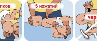
In case of electrical injury, you should immediately stop the electric current, since you should not touch the child with your hands before doing this. Afterwards, the damaged areas of the skin are covered with a bandage. If the damage is as minor as possible, it is enough to calm the victim and give him warm tea. If the child is unconscious, call an ambulance, and until it arrives, put the baby in a comfortable position, give access to fresh air, unfasten constricting clothing, open the airways, turn the head to the side, monitor the pulse and breathing.
Prohibited actions for burns
If a child receives burn injuries, it is prohibited:
- remove clothing stuck to the burn wound;
- open and puncture existing blisters;
- touch burned areas with your hands;
- apply various creams or oils to the burn wound, preventing cooling of the affected area and even, on the contrary, promoting the spread of the burn into the inner layers of the skin.
You should be wary of recommendations to cover burning clothing with thick fabric or throw snow or sand on it. This must be done very carefully, as contamination and infection of the burn wound is possible. In all cases, the burn surface must be covered with an aseptic bandage or any clean, ironed cloth.
Preventing burn wounds
Children need to be taught how to live safely, paying attention to places and situations where accidents most often occur. It is useful to teach first aid in a playful way. And it is important for parents not to lose vigilance and never leave their children unattended.
Experts: Leonid Roshal, pediatric surgeon, doctor of medical sciences, professor, director of the Moscow Research Institute of Emergency Children's Surgery and Traumatology, president of the National Medical Chamber of the Russian Federation; Razmik Keshishyan, surgeon, Doctor of Medical Sciences, Associate Professor, Deputy Director of the Moscow Research Institute of Emergency Pediatric Surgery and Traumatology
Symptoms and clinical picture
In children, there are 3 degrees of burns - the same as in adults. In them, the degree of damage depends not only on the temperature of the thermal agent, but also on the duration of action, age and general condition of the child before the burn.
Assessing the extent of injury in children is very difficult because they have burns that are considered minor but may later turn out to be second degree, and the blistered skin surface may end up with deep tissue damage in the following hours or days.
The morphological characteristics of local changes in the skin and mucous membranes in children are not particularly different from those in adults.
With burns (especially over a large area), children always experience general symptoms. Severe, almost unbearable pain accompanies burns of all three degrees. Its intensity and duration are greatest with II and III degree burns.
The temperature in the first hours never rises, and the larger the area and depth of the burn, the lower it is. By the end of the first day the temperature rises. Its height is determined by the area of the lesion and the degree of developing intoxication, the type and severity of the infection that has arisen.
All second degree burns, occupying more than 8% of the child’s skin surface, as well as third degree lesions, are always accompanied soon after the injury by general clinical phenomena. The most serious complication that develops in the early period of the disease is shock. It is the leading complication in the clinic of burns in children in the first three days. Its signs are as follows: severe general condition, apathy, accelerated and weakly filled pulse, significant pallor or cyanosis, hypothermia. Blood pressure drops progressively. Urination is weak and may even lead to anuria. At first the child is excited, but this state is quickly replaced by apathy, weakness, in some cases drowsiness and even loss of consciousness. Sick children experience extreme thirst and sometimes vomit.
In the development of shock, the area of the burn is more important than the depth of the lesion. Burns caused by flame are more likely to be complicated by shock than burns caused by hot liquids.
Many closely intertwined causes play a role in the pathogenesis of shock. The first place among them is occupied by excessive reflex excitation of the central nervous system, as a consequence of intense, prolonged and massive pain impulses entering the cerebral cortex. A significant role is played by the loss of plasma and intoxication of the body with toxic products formed from the breakdown of tissue in the lesion. As a result, mainly, changes in the central nervous system cause disturbances in hemodynamics and in the chemistry of blood and tissues.
Primary shock, occurring immediately after injury, is rarely observed - mainly with very severe and extensive lesions. Secondary shock is more common. It appears approximately two hours after the lesion, but sometimes later, on the first day.
The next period in the dynamics of the disease is the onset of the toxemic phase. It is caused mainly by the resorption of toxins resulting from the breakdown of tissue in the burn area. This period is characterized by increased temperature, increased thirst, deterioration of general condition, drowsiness, loss of consciousness, and sometimes convulsions. Kidney function is significantly impaired. Diuresis decreases. Protein, red blood cells, white blood cells and renal epithelium are found in the urine. Cardiac activity and peripheral circulation deteriorate. From the blood picture, leukocytosis with leukemia, anemia, and accelerated ROE are observed.
Then comes the transition from the toxemic to the septic phase, which occurs on the 5-6th day of the disease and is accompanied by septic temperature, a repeated increase in leukocytosis, a progressive increase in anemia, which is persistent, long-lasting and does not respond to conventional treatment; the liver and spleen are sometimes enlarged. The septic period is a consequence of purulent processes on the burned skin surface. Prolonged suppuration leads to significant exhaustion of sick children. In addition, infected burnt surfaces heal slowly, forming rough, disfiguring, keloid scars.
In the clinic, it is difficult to distinguish between the described phases of the disease. Usually they gradually transform into one another and their influence on the symptoms and syndromes of the disease is mutually intertwined.
Despite the fact that one of the anatomical and physiological characteristics of children is the rapid healing of wounds, recovery processes for burns are very often sluggish. The reasons for this are trophic changes in tissues and peripheral nerve endings that occur as a result of direct exposure to a thermal agent and as a result of changes in the central nervous system.
FIRST AID FOR BURNS
First, let's say what NOT to do.
Firstly, you should not panic. A burn in a child is always a stressful situation for parents. Don't lose your head. Take your time, what happened happened, and little depends on whether you bring the child to the medical facility a few minutes earlier or later.
Secondly, do not perform any active actions with the burn surface; you do not need to open the blisters, smear with alcohol or iodine, apply ointment bandages or otherwise actively influence it.
Thirdly, it should be remembered that a burn is not only damage to the skin, but the suffering of the whole organism. The smaller the child, the more severely he suffers this damage, and when providing first aid for a burn, the main thing is to carry out pain relief and anti-shock measures.
When helping a child with a burn, you should strictly adhere to a certain sequence of actions.
First of all, you need to stop exposing the child to the thermal agent. It is necessary to remove or pour cold water on clothing soaked in hot liquid. This needs to be done as soon as possible. Then the affected surface should be cooled by all possible means. Cold water in the form of a constantly changing compress is also suitable for this, or even better - apply some product taken from the freezer to the damaged area. The further course of the burn injury depends on how quickly the effect of the thermal agent is stopped and how effective the subsequent cooling is. It is necessary to cool the burn surface for a long time. Preferably, during transportation to a medical facility.
If the burn is large enough in area (3 times larger than the area of the child’s palm) and you are not sure that you can cope on your own, immediately, even before you start helping the child, call an ambulance.
If you decide to take your child to a medical facility yourself, give him a painkiller - analgin, and at the same time suprastin, or another similar remedy. Let us repeat once again that cooling has a good analgesic effect in this case. If the burn area is large, start feeding the child immediately, preferably warm, sweet tea.
The burn surface is covered with a sterile bandage. If you don’t have sterile materials on hand, use a simple, clean cotton cloth. Any touch to the burn surface causes sharp pain. Keep this in mind and strive to create immobility for the damaged area.
If the burn area is large, then it is better if the victim is evacuated by special transport accompanied by medical personnel.
nike kobe 2 posts on facebook account number
Complications
In severe cases of burns, children can develop a number of complications. When conditions are created for particles from clogged vessels in the affected area to break off and enter the bloodstream, they can enter various organs and cause thromboembolism there with the corresponding symptoms. Sometimes this occurs in the mucous membrane of the duodenum or stomach with the clinical picture of an ulcer. Toxic hepatitis, toxic nephritis, toxic gastritis, abscess pneumonia, otitis, acute dilation of the stomach, etc. may be observed. As a result of prolonged suppuration of the burned surface, degenerative changes sometimes occur in a number of internal organs - amyloid degeneration.
A known complication of burns in children is the appearance of scarlet fever. Indeed, in some cases, real scarlet fever is observed, which appears as a result of the weakening of the resistance of burned children and its poor reactivity. In these cases, the question concerns “wound” or “surgical” scarlet fever.