Questions:
- What is pyoderma?
- Causes of pyoderma
- Symptoms
- Types of pyoderma
- How is pyoderma transmitted?
- Chronic pyoderma
- Pyoderma in children
- Pyoderma in newborns
- Treatment of pyoderma
- Is it possible to treat pyoderma at home?
- At what age can Timogen cream be used in children?
- Treatment with folk remedies
- Prevention
Medical specialists answer frequently asked questions from users
What is pyoderma?
Pyoderma is a disease of purulent skin lesions, most often caused by pyogenic (pyogenic) cocci - streptococci and staphylococci, which are constantly found on the skin and mucous membranes. In addition to these pathogens, there are other microorganisms on human skin, such as pseudodiphtheria bacillus, yeast-like fungi, etc., which can also cause disease under certain conditions. The name of the disease comes from the Greek py'on - pus and de'rma - skin). This is one of the most common skin diseases.
to the begining
Symptoms and signs
The early stages of the disease have virtually no dangerous signs. The child’s health remains normal, and skin changes have not yet appeared. The condition worsens as the area of the affected skin gradually increases.
The bubbles themselves have a number of distinctive features - lethargy, easy to open, sagging skin under the liquid inside the bubble. Primary rashes are always localized in the mouth and mucous membranes of the lips. There are no painful sensations. Opened blisters form skin crusts or wet eczema, which take a long time to heal.
Gradually the affected area increases. Infection with bacteria leads to the appearance of suppuration and foci of inflammation, in place of which eczema and large erosions subsequently appear. Another distinctive feature is the lack of healing of the skin at the site of the blister rupture. Gradually, the erosions expand and unite into a single lesion.
All forms of pemphigus have a wave-like manifestation of symptoms. In the absence of a timely response, intoxication syndrome begins - nausea and headaches. Ultimately, the death of the little patient inevitably occurs. Viral pemphigus in children has symptoms similar to the true disease, and there is a pronounced viral infection.
When to see a doctor
Baby rash appears frequently and rarely poses a real threat to the child's life. Basically it becomes a reactive feature to allergens and infections. It is important for parents to know which rashes pose a mortal danger to the child. If signs of such a rash appear, you should seek help from a dermatologist.
The pediatric department of JSC "Medicine" (clinic of academician Roitberg) has been guarding children's health for many years. You can make an appointment with a dermatovenerologist by phone. The clinic has a convenient location, a two-minute walk from the Mayakovskaya metro station, as well as the possibility of placement in a hospital for examination and treatment.
Causes of pyoderma
Frequent causes of pyoderma are hypothermia and overheating of the body under production conditions.
When the body cools, blood circulation is disrupted, swelling of the skin occurs, sebum secretion and sweating decrease, as a result, the skin becomes dry and flaky, making it easily susceptible to injury and infection.
Overheating of the body leads to loss of water and chlorides (mineral salts), the latter affecting the vascular system and the functions of the kidneys and skin.
The possibility of pyogenic infection can be increased by additional irritants like oils and other contaminants.
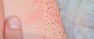
The skin is able to destroy microorganisms that fall on its surface due to the acidity of the skin (pH), the so-called “acid protective coat of the skin.” Bacterial infection occurs faster in those areas of the skin where the pH is higher - these are the armpits, interdigital folds of the feet, and inguinal folds. Therefore, pathological changes occur more often in these places.
to the begining
Kinds
Superficial staphyloderma
The most common are folliculitis, sycosis vulgaris, ostiofolliculitis, and epidemic pemphigus of newborns. Ostiofolliculitis is an abscess surrounded by a zone of mild hyperemia, penetrated by hair. With folliculitis, the abscess is located against the background of a painful bluish-pink nodule. Vulgar sycosis is called multiple, recurrent ostiofolliculitis and folliculitis against the background of thickened bluish-pink skin, often in the mustache and beard area. Epidemic pemphigus of newborns is a severe, contagious disease of the first days of life. In addition to the palms and soles, the child’s skin becomes covered with large blisters, which later form crusts and extensive erosions.
Deep staphyloderma
The most famous are boils, carbuncles, and hidradenitis. The boil develops from ostiofolliculitis or folliculitis. First, a painful node appears in the thickness of the skin, which enlarges; a large pustule is formed in its center, which opens with the subsequent separation of the purulent-necrotic core. After which, the resulting ulcer gradually scars.
A carbuncle is several boils located in one place, closely adjacent to each other, against the background of a general dense, edematous infiltrate of a bluish-purple color. The deep ulcer formed after opening the carbuncle heals with a rough scar. Hidradenitis is a purulent inflammation of the apocrine sweat glands, often recurrent. In the armpits, anogenital area, and around the nipples of the mammary glands, deep, painful nodes appear, over which the skin is colored bluish-pink. The nodes are opened with the release of liquid pus.
Superficial streptoderma
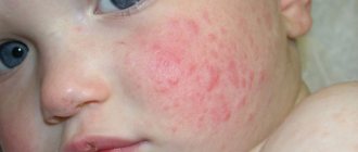
The most common is streptococcal impetigo . This disease occurs more often on the face in children and young women. Against the background of slightly hyperemic skin, phlyctens (bubbles with opalescent contents) appear, shrinking into yellowish crusts or eroding. When staphylococci are attached, the crusts become greenish-yellow or sometimes hemorrhagic ( impetigo vulgaris ). Without treatment, the disease can affect large areas of the skin. Lichen simplex , more often in children, is an abortive form of streptococcal impetigo. It is expressed by the appearance of whitish or light pink, slightly flaky spots.
Deep streptoderma
Vulgar ecthyma . With this disease, deep pustules appear, shrinking into purulent-bloody crusts, under which ulcers are found. After 2-3 weeks, the ulcers heal.
to the begining
Pemphigus symptoms
The characteristic symptoms of pemphigus are a rash of small blisters with serous contents. Their localization depends on what form of the disease develops:
- ordinary - rashes appear on the mucous membrane, accompanied by an unpleasant odor;
- leafy – blisters form on the skin, and crusts form at the same time;
- non-acantholytic - rashes are located on the mucous membrane and lips. This form of pathology is more often diagnosed in old age.
With pemphigus in children and adults, the following are also observed:
- severe weakness;
- increased drowsiness;
- deterioration or complete loss of appetite;
- losing weight even while eating high-calorie foods.
To detect pemphigus in newborns, older children and adults, the following examination methods are used:
- examination of the patient and assessment of rashes;
- Nikolsky test - with its help, differential diagnosis with other skin diseases is carried out;
- histological and cytological examination.
The symptoms of pemphigus (the second name of the disease) are often similar to the symptoms of other pathologies that will require completely different therapy. Therefore, before prescribing treatment, a thorough study of the symptoms that have arisen will be required.
Pyoderma in children
Pyoderma in children most often develops due to parents' failure to comply with the hygiene regime. That is, parents do not wash their hands before touching the child, wash the child poorly or rarely, the child’s clothes are poorly washed and not ironed, and their clothes are rarely changed. In rare cases, also if hygiene rules are not followed, it is possible for a child to become infected from relatives who have foci of chronic infection - carriers of staphylococci, streptococci, people with reduced immunity (patients with diabetes, alcoholism, old people, cancer patients).
to the begining
Pyoderma in newborns
Pyoderma in newborns most often occurs due to nosocomial infection, violation of the rules of child care and in weakened, premature children. Common pyoderma in newborns is called bullous impetigo, it is life-threatening and progresses very quickly; treatment is carried out only in a hospital. Red rashes appear on the skin, which quickly turn into hyperemia of the entire skin; detachment of the epidermis occurs with the formation of large blisters and crusts. When an infection gets into the umbilical wound, inflammation develops - omphalitis, which can quickly develop into umbilical sepsis. Symptoms of omphalitis: pus from the umbilical wound and an unpleasant odor, swelling and redness of the edges of the wound and the skin around it, increased body temperature, refusal to eat, anxiety.
to the begining
Epidemic pemphigus of newborns
Signs
The disease develops very quickly.
First, the child develops a fever and becomes restless. Within a few hours, small blisters filled with a clear yellowish liquid form on the baby’s skin in the navel, abdomen, buttocks, arms and legs, chest, back, as well as on the mucous membrane of the mouth, nose and genitals. The skin around the blisters may be slightly red and swollen, or it may be normal. After some time, the bubbles increase in size, their contents become purulent. They open, the contents expire, and an itchy bright red erosion forms in place of the bubble. Pemphigus in newborns is often accompanied by refusal to eat and weight loss.
Description
Epidemic pemphigus of newborns is caused by Staphylococcus aureus. However, some experts believe that streptococci also play a role in the development of this disease. The source of the disease is the medical staff of maternity hospitals and women in labor suffering from pyoderma, furunculosis or hidradenitis. Pemphigus can also be transmitted from child to child through the hands and clothing of medical personnel.
Most often, this disease develops in weakened children, for example, in premature babies, in children with low body weight, in those who have suffered birth trauma, as well as in children whose mothers have suffered severe toxicosis of pregnancy. It is in these newborns that the skin reacts when bacteria penetrate it by forming blisters.
If the course of the disease is favorable, it is cured in 2-4 weeks. If unfavorable, complications are possible - phlegmon, pneumonia, sepsis. Sepsis can cause the death of a child.
A particularly severe form of epidemic pemphigus of newborns is Ritter's exfoliative dermatitis. With this disease, extensive erosions form, and within a few days the entire skin is affected.
Diagnostics
The diagnosis is made based on the clinical picture. Bacteriological culture of the discharge from the vesicles must be done.
The disease is differentiated from congenital epidermolysis, syphilitic pemphigus, and chickenpox.
Treatment
Epidemic pemphigus of newborns is treated with antibiotics and sulfonamides. The blisters are opened, the wounds are treated with an ointment with an antibiotic and aniline dye or a solution of potassium permanganate. Antihistamines are used to relieve itching.
In some cases, corticosteroid ointments are prescribed to relieve inflammation and itching.
If necessary, plasma or blood transfusions are given.
Prevention
To prevent epidemiological pemphigus in newborns, sick children are immediately isolated from healthy ones.
Maternity hospital staff are regularly examined for pyoderma and furunculosis. Those who become ill are removed from working with children.
It is important to thoroughly disinfect the rooms and not neglect wet cleaning and quartz treatment.
Linen in maternity hospitals must be changed frequently and must be sterilized.
© Dr. Peter
Treatment
- Treatment in children
Treatment of omphalitis is carried out only by a doctor. In the initial stage, local use of medications is possible: washing the wound with a solution of hydrogen peroxide, treatment with brilliant green, iodine solution, methylene blue solution and various local antimicrobial drugs. Treatment of pyoderma in children involves the use of broad-spectrum antibiotics, infusion therapy, and immunomodulators. In the initial stages of pyoderma, it is possible to treat pyoderma with local means - solutions of boric and salicylic acids, sanguiritin, eucalymin, tomicide, cyminal and others in the form of solutions, lotions, under a bandage. See also our specialist’s answer to the user’s question: Pyoderma in children, how to cure it? - Treatment in adults
In adults, pyoderma is also treated by a doctor. For mild pyoderma, local medications are used (see above); for moderate severity, broad-spectrum antibiotics and sulfonamides are added to local treatment.To increase the body's resistance in the treatment of pyoderma, the immunomodulatory drug Thymogen is used. These are Thymogen cream, which is applied to the affected areas, and Thymogen solution for injection.
to the begining
Can it be treated at home?
- First of all, it is necessary to establish the nature of pyoderma. In other words, identify the causative agent of the disease and determine its sensitivity to antibiotics. After a course of antibacterial therapy (local use of antibacterial drugs in most cases is ineffective) against the background of normal multivitamin-mineral support (taking vitamins), such diseases usually go away. But pyoderma, as a rule, develops against the background of reduced immunological resistance or is provoked by diseases of the internal organs: stomach, intestines, liver, pancreas. Therefore, an in-depth examination is necessary.
to the begining
All clinical forms of pemphigus are characterized by a long, chronic, wave-like course, which, in the absence of treatment, leads to disruption of the general condition of patients, and in some cases, to death.
Pemphigus vulgaris
Pemphigus vulgaris
- the most common form of the disease, characterized by the presence of blisters of various sizes with a thin flaccid covering, with serous contents, appearing on apparently unchanged skin and/or mucous membranes of the oral cavity, nose, pharynx, genitals. The first rash most often appears on the mucous membranes of the oral cavity, nose, pharynx and/or red border of the lips. Patients experience pain when eating, talking, or swallowing saliva. A characteristic symptom is hypersalivation and a specific smell from the mouth. After 3-12 months, the process becomes more widespread with damage to the skin.
Bubbles persist for a short time (from several hours to a day). On the mucous membranes, their appearance sometimes goes unnoticed, since the thin covers of the blisters quickly open, forming long-term, non-healing painful erosions. Some blisters on the skin may shrink into crusts. Erosions in pemphigus vulgaris are usually bright pink in color with a shiny, moist surface. They tend to grow peripherally; generalization of the skin process is possible with the formation of extensive lesions, deterioration of the general condition, the addition of a secondary infection, the development of intoxication and death in the absence of therapy.
One of the most characteristic signs of acantholytic pemphigus is Nikolsky’s symptom, which is a clinical manifestation of acantholysis and is a detachment of the epidermis due to mechanical impact on the skin in the lesions located next to them and, possibly, in distant areas of the skin.
Seborrheic or erythematous pemphigus
Seborrheic or erythematous pemphigus
(Senir-Usher syndrome), unlike pemphigus vulgaris, which often initially affects the mucous membranes, begins on seborrheic areas of the skin (face, back, chest, scalp). At the beginning of the disease, erythematous lesions with clear boundaries appear on the skin , on the surface of which there are crusts of varying thickness of yellowish or brownish-brown color. The blisters are usually small in size and quickly dry into crusts, which, when peeled away, reveal a moist, eroded surface.
The blisters have a very thin, flabby covering that persists for a short time, so they often go unnoticed by patients and doctors. Nikolsky's symptom is positive mainly in the lesions. The disease may be limited in nature for many months and years. However, it is possible for the lesion to spread to new areas of the skin and mucous membranes (usually the oral cavity). When the pathological process generalizes, the disease acquires the symptoms of pemphigus vulgaris.
Pemphigus foliaceus
Pemphigus foliaceus
characterized by erythematous-squamous rashes, thin-walled blisters that reappear in the same places, when opened, pink-red erosions are exposed with the subsequent formation of lamellar crusts, sometimes quite massive due to the constant drying of the exudate that separates. Damage to the mucous membranes is uncharacteristic. Possible rapid spread of rashes in the form of flat blisters, erosions merging with each other, layered crusts, scales with the development of exfoliative erythroderma, deterioration of the general condition, and the addition of a secondary infection. Nikolsky's symptom is positive both in the lesions and on apparently healthy skin.
Pemphigus vegetans
Pemphigus vegetans
for many years it can proceed benignly in the form of limited lesions with a satisfactory condition of the patient. Bubbles most often appear on the mucous membranes of the oral cavity, around natural openings (mouth, nose, genitals) and in the area of skin folds (axillary, inguinal, behind-the-ear, under the mammary glands). At the bottom of the erosions, soft, juicy, fetid vegetations are formed, covered with serous and/or purulent plaque with the presence of pustules along the periphery. Nikolsky's symptom is positive only near the lesions. In the terminal stage, the skin process resembles pemphigus vulgaris.
Pemphigus herpetiformis
Pemphigus herpetiformis
is a rare atypical bullous dermatosis, which in some cases clinically resembles Dühring's dermatitis herpetiformis. The rash may appear as plaques with papules and vesicles surrounding the periphery, or as grouped papules, vesicles, or tense blisters, as in Dühring's dermatitis herpetiformis.
Pemphigus herpetiformis is characterized by severe itching of the skin. In the absence of adequate therapy, the disease can progress and acquire signs of pemphigus vulgaris or pemphigus foliaceus.
Paraneoplastic pemphigus
Paraneoplastic pemphigus
occurs against the background of neoplasia, and can also occur during or shortly after chemotherapy treatment for malignant neoplasms. In most cases, paraneoplastic pemphigus is combined with lymphoproliferative neoplasia, thymoma, sarcoma, carcinoma and solid cancers of various localizations.
As a rule, the clinical picture of paraneoplastic pemphigus is similar to that of pemphigus vulgaris with simultaneous damage to the skin and mucous membranes, but sometimes skin lesions atypical for the disease are observed, accompanied by itching and resembling exudative erythema multiforme, bullous pemphigoid or toxic epidermal necrolysis.
Drug-induced pemphigus
Drug-induced pemphigus
(drug-induced) may resemble the clinical picture of pemphigus vulgaris, seborrheic or pemphigus foliaceus. Its development is most often associated with taking medications containing sulfhydryl radicals (D-penicillamine, pyritol, captopril) and antibacterial drugs of the β-lactam group (penicillin, ampicillin and cephalosporins) and is caused by biochemical rather than autoimmune reactions. In cases of drug-induced pemphigus, complete recovery is possible after discontinuation of the medication.
IgA-dependent pemphigus
IgA-dependent pemphigus
is a rare group of autoimmune intraepidermal bullous dermatoses characterized by vesiculopustular rashes, neutrophilic infiltration, acantholysis and the presence of both fixed and circulating IgA autoantibodies directed to antigens of the intercellular binding substance of stratified squamous epithelium. Clinical picture of IgA-dependent pemphigus, regardless of type its manifestations are represented by flaccid vesicles or pustules located on both hyperemic and apparently “healthy” skin.
Pustules tend to merge to form ring-shaped lesions with crusts in the central part. The rashes are most often localized on the skin in the armpits, scrotum, torso, upper and lower extremities. Less commonly, the pathological process involves the skin of the scalp and behind-the-ear area, as well as mucous membranes. Patients often complain of intense itching. As a rule, IgA-dependent pemphigus is more benign than dgG-dependent pemphigus.