Recently, the number of pregnancies has increased, during which a tumor-like formation of small bubbles grows in the uterus - a royal moth or hydatidiform mole. It develops from degenerated chorionic villi - the fleecy membrane that forms the placenta.
Appointment with a gynecologist - 1000 rubles. Comprehensive pelvic ultrasound - 1000 rubles. Admission based on test results - 500 rubles. (optional)
CLICK TO MAKE AN APPOINTMENT or ultrasound tests
Most often, such a pregnancy is terminated at an early stage, but sometimes a hydatidiform mole (MH) grows, increasing in size. A woman thinks she is carrying a child, but in fact, instead of a fetus, a lump of small cysts develops in the uterus.
Where do moths come from in the uterus or why does hydatidiform mole occur?
The content of the article
The cause of the disease is improper fertilization of the egg, during which the maternal chromosomes were “lost” or the egg was fertilized by two sperm at once.
There are:
- A complete mole , whose cells are composed of 46 paternal chromosomes. Such a cell cannot form an embryo, amniotic cavity and other tissues. Instead, a polycystic formation grows, in which there is no embryo.
- Partial mole - consists of paternal, maternal and haploid cells having 69 chromosomes - 23 maternal and 46 paternal. Areas of normal chorionic villi are adjacent to vesicular ones. In such conditions, the embryo cannot develop properly, so the pregnancy will freeze or be interrupted by a miscarriage. The chance of having a baby is only 1/100.
- A destructive malignant uterine tract that grows into the wall of the uterus leads to its perforation (breakthrough) and severe intra-abdominal bleeding. Foci of drift can spread throughout the body, detecting even in the lungs.
A few words in conclusion
If the changes occurring in the uterus comply with accepted standards, neither the size nor the shape of the abdomen will in any way affect the health of the mother and her baby, and certainly not in any way affect his gender.
For parents looking forward to the birth of a baby, it doesn’t matter at all whether it’s a girl or a boy. And when the belly begins to grow during the second pregnancy with twins, there is definitely no limit to the happiness of expectant mothers and fathers
By the way, this happens quite early, and it won’t be difficult to guess why. And yet, the main thing for parents is to help every baby be born healthy and on time.
For every woman, becoming a mother means making huge changes in her life. Therefore, they want to know everything about this, especially how the fetus should develop correctly, in what month does the belly appear in pregnant women? These questions are asked quite often, especially by those who are preparing to become mothers for the first time.
First, let’s explain the definition of “tummy.” In the first months of pregnancy, a woman’s belly may grow, the reason for this is poor nutrition or gas formation. In these first months, the uterus is not yet enlarged.
Pregnant women come to a point when they begin to think: “at what stage does the belly begin to grow?” Let's try to answer this question. The uterus begins to grow only at 16 weeks from the moment of conception. Therefore, the belly will be noticeable to other people only in the 20th week of pregnancy. If a woman plays sports, i.e. if she has developed abs, then the tummy will become visible much later. Also, the appearance of the tummy depends on what kind of pelvis the pregnant woman has. If the expectant mother’s pelvis is wide enough, her tummy will become visible only by the 4-5th month of pregnancy. In obese women, it will be possible to notice the “position” even later. In women who give birth for the second time, the belly may appear earlier. The number of embryos can also affect the size of the abdomen, namely, making it larger. This is the only thing that can really affect at what month the belly appears in pregnant women. If the expectant mother has triplets or twins, then the belly may be noticeable already in the first 3 months.
In general, the answer to the question of what week of pregnancy the belly begins to grow will be as follows. If a woman has one fetus and its development is going well, then the belly should appear only at the 12th week of pregnancy. Already at week 16, your tummy will become noticeable not only to you. And by the 27th week of pregnancy, the question is: at what stage does the belly begin to grow? - will not be relevant, since by this moment it will be impossible to hide your baby under blouses and other clothes. At the same time, the expectant mother should be interested not only in what period the belly begins to grow, but also in how her baby should develop correctly. At the 8th - 10th week of pregnancy, the fertilized egg reaches a diameter of 22 mm. At the time when it is necessary to register, namely, by 12 weeks, the length of the fetus will be seven centimeters, and its weight will reach 20-25 grams. At this time, when you come for an ultrasound, you can see that the fertilized egg has almost completely filled the uterine cavity.
By the 17th week of pregnancy, the fetus will reach a length of 12 cm, while its body weight will increase to 100 grams. By this time, the question of at what time the belly begins to grow disappears on its own, since all the worries associated with it are already behind us. The tummy has noticeably grown and become rounder.
By week 20, the length of the fetus will reach 25 cm with a weight of about 300 g. At 24 weeks, your baby will have time to grow significantly. By this time, its length will be almost 30 cm, and its weight will vary from 600 to 700 grams.
By week 28, the length of the fetus is usually 35 cm with a body weight of about 1.0-1.2 kg. At the 32nd week, body weight will increase to 1.5 kg with a fetal length of 40 cm. At week 36, the baby is practically formed, and its indicators by this time will be as follows: weight - 2.5 kg, and length - about 48 cm.
However, it is impossible to say that these indicators are the same for everyone. They are quite individual, since the baby in the womb of each mother is formed in its own way. Therefore, the above data is very average, and your baby may reach large sizes or be slightly smaller due to individual characteristics. The body weight of the fetus by the date of birth is usually 2.6-5.0 kg, and its length can reach 54 cm.
Thus, it should be important for the expectant mother: to carry the child for the entire required period and provide him with everything necessary for full growth and nutrition, but the tummy and its size are not the most important thing
What should you be wary of?
As a rule, women with PD understand that something is wrong with them. Hydatidiform mole tissues, whose growth is similar to a cancerous tumor, quickly increase in size. A woman notices that her belly is too big.
Since the disease is accompanied by increased formation of hCG, tests showing weeks of pregnancy indicate a period longer than the actual one.
Many suffer from constant nausea and uncontrollable vomiting. Already in the first months, blood pressure rises and swelling occurs.
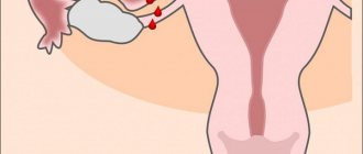
The disease is accompanied by a constant threat of miscarriage, caused by the body's desire to get rid of an abnormally developing fetus. A woman complains of dark bloody discharge from the genital tract, which may contain molar vesicles.
Alarming symptoms
Sometimes, when a large belly appears in the early stages of pregnancy, a woman notices accompanying signs:
- Tingling. They begin 3-4 days after conception. This phenomenon is associated with a change in the shape of the uterus. Its vessels fill with blood, causing the organ to become rounded. However, tingling should not cause discomfort - otherwise this symptom is alarming. If the pain intensifies and lasts more than 1 hour, you should contact an obstetrician-gynecologist.
- Pulsation in the abdomen. The sensation does not only occur in the first trimester - it can accompany the entire pregnancy. This phenomenon is considered normal. Because the inferior vena cava is located near the uterus, the vessel is sometimes compressed, creating a pulsating sensation. To eliminate discomfort, just change your body position.
- Burning in the lower abdomen. The irritating sensation of heat occurs due to the growth of the uterus. If the symptom does not go away within a few hours, you should suspect an ectopic pregnancy, threatened miscarriage, genitourinary tract infection or toxicosis.
- Hardening of the lower abdomen. The phenomenon is provoked by increased tone of the uterus - its muscles begin to contract intensively, which is dangerous for the child. The lower abdomen hardens during emotional shocks, physical overload, infections and diseases of the pelvic organs. For these reasons, in the early stages it is important to avoid stressful situations and correctly alternate work with rest.
In most cases, an urgent visit to a doctor helps save the child’s life, even in severe cases.
What can be seen on a fetal ultrasound
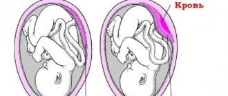
With complete degeneration of the villi, there is no embryo in the uterus. Instead, a tumor-like formation is found, consisting of tiny bubbles. All this creates a specific picture of a “snow storm” in the intrauterine space.
With a partial hydatidiform mole, the embryo or its tissues may be detected. The development of the embryo often does not correspond to the gestational age. If the changed area of the chorion is very small, the embryo can develop normally.
In case of multiple pregnancy, a developed embryo is found adjacent to the formation of the vesicular mass. Several cases have been described in which women with PZ carried twins. The twin who had a full placenta was born healthy.
Even at too short a time (4-5 weeks), while the cystic formations are extremely small, the disease can be detected in 57% of cases. And when re-examined at 10-11 weeks - almost always.
In 30% of women, the disease is accompanied by the appearance of cysts up to 6 cm in size in the ovaries, caused by an increase in hCG levels. Tumors can be multiple, especially in the second trimester. Despite their size, they rarely rupture, causing bleeding.
With a destructive hydatidiform mole, pathological tissue is visible growing into the wall of the uterus. Such women need to examine the lungs and abdominal cavity to identify distant foci of the disease.
Normal abdominal size in early pregnancy
Norms for the height of the uterine fundus and abdominal circumference
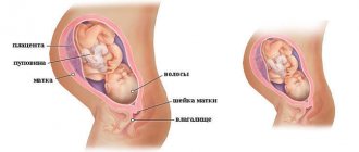
During normal pregnancy, up to 16-20 weeks, the abdomen increases slightly. The uterus in the first trimester has a small volume and takes up little space in the pelvis. By the end of 1 month it does not exceed the size of a chicken egg, and after 8 weeks it increases to the volume of a goose egg. During the first trimester, she does not have time to reach the pubic symphysis, located in the lower abdomen. In most cases, in the early stages of pregnancy, changes in the female body are not visible.
The abdominal circumference begins to increase only after the 20th week of gestation (the average is 70-75 cm). Of particular importance for diagnosis is the height of the uterine fundus (UFH) - the distance from the upper wall of the organ to the edge of the pubic joint. This figure is 2-6 cm at week 12, and by the 16th week it already reaches 10-18 cm.
In the second trimester, the baby begins to gain weight sharply. As a result, the uterus also enlarges. At 12-16 weeks, the belly is already noticeable to the woman herself, and by the 20th week it is visible to others.
Laboratory research
To confirm the diagnosis, a woman submits:
- blood for hCG, the level of which is significantly increased in PZ;
- analysis of thyroid hormones, the function of which is disrupted when pregnancy has developed;
- blood chemistry. With this disease, the functioning of the liver and kidneys is disrupted, the level of creatinine, residual nitrogen, proteins, urea, bilirubin, transaminases, and alkaline phosphatase changes.
A transvaginal ultrasound of the uterus is required.
Treatment of hydatidiform mole
The uterus is cleared of the uterus, depending on the period, using vacuum extraction or surgical curettage. The woman is prescribed antibiotics and drugs that contract the uterus. After the mole is removed, a second ultrasound is performed to ensure that nothing remains in the uterine cavity. Cysts in the ovaries do not need to be treated, because... they gradually dissolve on their own.
After getting rid of the pregnancy, the woman retains her reproductive function and can have children. However, she will have to carefully protect herself for some time, so she needs to see a doctor to choose a contraceptive method.
The destructive form of PZ is treated with surgery and chemotherapy. Women of childbearing age are trying to preserve reproductive function.
If there is a chance of carrying the pregnancy to term, the patient undergoes an unscheduled screening ultrasound once a month to find out how normally the baby is developing.
Hydatidiform mole and its consequences
After PZ, a woman may develop chorionepithelioma, an extremely malignant tumor that quickly metastasizes and leads to death. Especially often, oncology occurs in the first weeks after removal of the prostate. At this time, you need to determine the level of hCG once a week, an increase in which indicates the development of cancer. A pelvic ultrasound is performed every two weeks.
The period of occurrence of chorionepithelioma ranges from 3 weeks to 20 years, so a woman after a hydatidiform mole should periodically visit a gynecologist and undergo an ultrasound.
Where to get checked during pregnancy in St. Petersburg
To maintain your health and not lose the opportunity to have children in the future, you need to be vigilant. During pregnancy, which causes terrible early toxicosis and a rapid increase in the size of the abdomen, you need to visit a gynecologist and do an ultrasound.
You can undergo an expert examination inexpensively in St. Petersburg at the Diana gynecological clinic. The examination will not cost much, for example, an expert pelvic ultrasound using a new machine with Doppler costs only 1,000 rubles.
If you find an error, please select a piece of text and press Ctrl+Enter
Important nuances of pregnancy
The baby’s weight is one of the main indicators of his condition. That is why a very small or, conversely, large child requires close attention from a doctor. For example, delayed growth is often due to the fact that the placenta does not cope well with its function (that is, it does not transfer enough nutrients to the baby). As for large sizes, they can be “inherited” by the child from tall and portly parents, or they can be the result of metabolic disorders in the mother (including diabetes). By the way, in order to determine that the expectant mother does not have it, in the second trimester (23-25 weeks of pregnancy) you need to donate blood for analysis twice - before breakfast and after “loading” with glucose. If its level remains elevated for a long time, the doctor will recommend a special diet to the woman.
Based on the size of the abdomen during pregnancy and the rate of its growth, the doctor can judge the amount of amniotic fluid, although, of course, ultrasound provides more accurate information.
If, before giving birth, the shape of your abdomen seems unusual to the doctor, he will refer you to X-ray pelviometry to determine the internal dimensions of the pelvis (don’t worry, this method is safe for the baby). In most cases, there is no reason to be concerned, but if the pelvis is too narrow and the baby is large, the doctor may suggest a cesarean section to the expectant mother.
And finally, the most important thing: give doctors the right to perceive your stomach as an object for study, and in the meantime, communicate more with your child. The baby will definitely respond to your affection, and the dialogue with him will begin now. Yes, and don’t forget to invite dad to participate in the conversation, especially if he still cannot understand the very fact of the real existence of his son or daughter. Together, you will get to know your unborn child better, and after he is born, your already familiar voices will help him feel safe.