General information
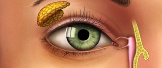
Cephalohematoma is a condition in which hemorrhage occurs, localized between the periosteum and the outer surface of the skull bones.
This tumor forms immediately after the birth of the baby, is limited to the edges of a certain bone of the skull and disappears, depending on the individual characteristics, after 3-8 weeks. This condition is referred to as birth injuries of the baby. Externally expressed by swelling in the head area. This happens, according to statistics, in 3-5 cases per 1000 babies. The disease code according to ICD-10 is P12.0. A cephalohematoma is formed during the birth of the baby's head through the birth canal due to the displacement of the skin with the periosteum. As a result, the vessels that are located between the skin and the periosteum rupture, and an area filled with blood is formed. This “pocket” can contain from 5 to 150 ml of blood, which remains in a liquid state for a long time, since the newborn does not have enough clotting factors to clot it.
Why do cephalohematomas occur in newborns?
Cephalohematoma - swelling on the head; essentially, it is a hemorrhage between the periosteum - the connective tissue that surrounds the bone - and the flat bones of the skull. The surface of the skin over the tumor is not changed, the color of the skin remains the same, but pinpoint hemorrhages may be visible on it. Cephalohematoma is observed in 0.3-0.5% of newborns.
The mechanism of injury is that the skin moves along with the periosteum and blood vessels rupture when the baby’s head moves through the birth canal due to compression of the skull bones. Cephalohematoma can be caused by obstetric forceps or vacuum extraction, which are practically not used at present. Due to the fact that the vessels have ruptured, that is, their integrity has been disrupted, a certain amount of blood accumulates in the space between the periosteum and the skull bone. The blood in a cephalohematoma does not accumulate all at once on the first day, but gradually, since the newborn has a temporary deficiency of blood clotting factors in the first days after birth and the blood does not clot immediately. Therefore, the tumor, having appeared during birth or shortly after it, continues to grow during the first 2-3 days of the child’s life. The volume of cephalohematoma ranges from 5 to 150 ml of blood.
Cephalohematoma can be located in different parts of the head: most often on one or both parietal bones, rarely on the occipital and frontal bones, and even less often on the temporal bone of the skull. The blood in the area of the hematoma remains liquid for a long time and does not clot. The periosteum is tightly fused with the bones in the area of the sutures, so the boundaries of the cephalohematoma never extend beyond the bone on which it is located.
Pathogenesis
Cephalohematoma is a consequence of soft tissue damage during childbirth . The essence of the mechanism of this injury is that as the child’s head passes through the birth canal, the bones of his skull are compressed, the soft tissues are displaced, and the vessels of the subosteum are ruptured. When the tendon helmet moves along with the skin, tension occurs on the fibrous fibers that connect it to the periosteum. Strong mechanical stress provokes damage to the small vessels of the periosteum, and blood from them pours into the subperiosteal space. As a result, a cephalohematoma is formed.
Until recently, experts believed that the main cause of this condition in infants was pathological childbirth. However, medical statistics record an increase in the number of cases of this diagnosis in recent years, and this also occurs in children born in physiological labor. Therefore, the development of cephalohematoma is currently associated with various factors, in particular with the incorrect position and presentation of the fetus, its large size, and diabetic fetopathy. Also, this pathology can be associated with various complications during pregnancy , the application of obstetric forceps during childbirth and the use of vacuum extraction.
Classification
Cephalohematomas are divided according to certain criteria.
According to the size of the subperiosteal hemorrhage:
- First degree – with a diameter of 4 cm or less.
- Second degree – with a diameter from 4.1 to 8 cm.
- Third degree – more than 8 cm.
If a baby is diagnosed with multiple cephalohematomas, the specialist evaluates the total area of hemorrhages.
In combination with other possible damage:
- with brain damage ( cerebral edema or cerebral hemorrhage, epidural hematoma);
- with a fracture of the skull bones;
- with neurological symptoms (focal and general brain symptoms).
According to the location of the cephalohematoma:
- parietal – occurs most often;
- frontal and occipital - less common;
- temporal bone - in very rare cases.
In terms of position:
- left-handed;
- right-handed;
- two-way.
By prevalence:
- focal - looks like a subperiosteal elevation, can have different sizes;
- widespread - spreads to one bone of the skull and does not extend beyond the suture line;
- mixed - one patient has several cephalohematomas.
Causes
The formation of cephalohematoma can be associated with both the fetus and the mother.
Causes related to the fetus:
- diabetic fetopathy;
- large fruit;
- presentation of the fetus, its incorrect position;
- developmental defects ( hydrocephalus );
- post-term fetus - the bones in this case become too hard and do not allow the head to transform during childbirth.
Reasons related to the mother:
- vacuum extraction of the fetus and application of obstetric forceps - such methods are used very rarely;
- discoordination of generic forces;
- protracted or rapid labor process;
- too narrow maternal pelvis and wide fetal head;
- previous pelvic injuries, exostoses of the pelvic bones;
- The age of the woman giving birth is over 35 years.
The hypoxic origin of cephalohematoma is also possible - it can occur as a result of entanglement or compression by the umbilical cord, a large amount of mucus in the child’s respiratory tract, or retraction of the tongue.
Causes of cephalohematoma
All reasons can be divided into 2 categories:
From the child's side:
- anomalies of pelvic, parietal, facial presentation and transverse position;
- large fruit;
- diabetic fetopathy;
- fetal malformations (fetal hydrocephalus);
- post-term pregnancy and others.
From the mother's side:
- pelvic anomalies (rachitic flat or narrow pelvis, the presence of exostoses on the surface of the pelvic bones (osteocartilaginous or bone growth of a non-tumor nature));
- elderly age;
- previous injuries that are accompanied by damage to the pelvic bones;
- Weakness of labor (impaired contractile function of the uterus).
Separately, we can highlight the discrepancy between the sizes of the birth canal and the fetus. The occurrence of cephalohematoma can be provoked by using obstetric forceps or vacuum extraction for delivery (at present they are practically not used).
Symptoms
Cephalohematoma in newborns becomes noticeable on the second or third day, when the birth tumor subsides.
Photo of cephalohematoma in newborns
From the first day, the size of the hemorrhage increases, since newborns lack blood clotting factors, therefore, it remains liquid for a long time, and damaged vessels are not thrombosed by blood clots. As pediatrician Komarovsky notes about cephalohematoma on the head in newborns, its size depends on how severe the bleeding .
The symptoms of cephalohematoma are especially pronounced on the second or third day after the baby is born. Its distinctive feature is precisely the gradual increase these days. The child's head becomes asymmetrical due to the appearance of a formation.
If you try this formation by touch, it will be elastic, and with slight pressure on it you can feel the liquid moving. The skin over it is unchanged, elastic.
There can be from 5 to 150 ml of blood under the periosteum. Accordingly, if the cephalohematoma is small, then it decreases approximately a week after its appearance, and no intervention is required.
If the cephalohematoma is isolated, the child’s health remains normal. If it is combined with another pathology, then the baby experiences neurological symptoms. In most cases, there is depression of the function of the central nervous system, which is manifested by a sluggish reaction to the action of pathogens and poor expression of reflexes.
In some cases, it is possible to manifest strong central nervous system excitation similar to a hydrocephalic or hypertensive complex of symptoms.
If the size of the formation is large or the child has impaired blood clotting, and the cephalohematoma does not shrink on its own, anemia , ossification , resorption hyperbilirubinemia , and infection of the hematoma are likely.
With large cephalohematomas, rapid hemolysis of red blood cells , as a result of which a yellowish tint of the skin and mucous membranes is observed in the first days of the baby’s life. In this case, jaundice persists for 10 days or more.
Sometimes, where the formation is localized, a fracture (crack) of the bone is noted.
Symptoms of cephalohematoma
The first symptoms of cephalohematoma (see photo) become noticeable on days 2–3, when the birth tumor subsides.
The size of the hemorrhage tends to increase from the first day of birth, due to a deficiency of coagulation factors in the newborn’s blood - the blood remains liquid for a long time, so it is not possible to clot damaged vessels with blood clots.
The cephalohematoma is elastic to the touch; when you press on the area of hemorrhage, you can feel the movement of fluid. If the cephalohematoma is small in size, it begins to shrink on the 7th – 8th day and disappears without a trace. If the hemorrhage is significant, the process of its resorption may take several months. Often, a bone fracture (crack) is observed in the area where the cephalohematoma is located.
Cephalohematoma always has clear boundaries in the form of a compacted ridge around the circumference of the hemorrhage. The delimitation of the cephalohematoma is associated with the tight fusion of the periosteum with the bones of the skull in the area of the sutures, so the hemorrhage is located in the area of one bone.
Tests and diagnostics
In the process of establishing a diagnosis, differential diagnosis is carried out with a birth tumor, cerebral hernia, hemorrhage under the aponeurosis . If we are talking about an uncomplicated cephalohematoma, the doctor is primarily guided by the examination data. In this case, it is not difficult to establish a diagnosis if you examine the head and detect the characteristic signs of the disease.
The clinical picture of cephalohematoma is nonspecific; symptoms of damage to the nervous system are more pronounced.
In addition, during the diagnostic process, if necessary, the following methods are used:
- computed tomography – used if there is suspicion of damage to brain tissue;
- ultrasound examination - makes it possible to find out whether there is a fracture of the cranial bone, a cerebral hernia, and also determine the size of the tumor;
- Craniography in direct and lateral projections – allows to exclude bone damage;
- multislice CT;
- neurosonography – allows you to determine the presence of damage in the brain.
Diagnostics
Differential diagnosis is carried out with a birth tumor, hemorrhage under the aponeurosis (has a doughy consistency, is flat and crosses the sutures), cerebral hernia (protrusion of the meninges through the fontanel).
Diagnosis of cephalohematoma is not difficult. Examination of the head with characteristic signs in 99% of cases allows us to establish a diagnosis.
Additionally, ultrasound of the cephalohematoma is used, which allows one to determine its size, the presence or absence of a cranial bone fracture or cerebral hernia.
It is possible to use X-ray examination if a fracture is suspected.
Prevention
To reduce the risk of this pathology in a child, the expectant mother must follow the following rules:
- her lifestyle and diet should be as healthy as possible;
- she must undergo all necessary examinations during pregnancy and not miss visits to the doctor;
- it is important to identify all risk factors and follow your doctor’s advice;
- supervision should be provided by a professional specialist who can correctly conduct labor.
Consequences and complications
As a rule, this formation goes away on its own, and the consequences of cephalohematoma do not appear later. But sometimes, if a large cephalohematoma forms on the head of newborns, consequences in the future are still noted. True, this happens only in rare cases.
The consequences of a large cephalohematoma on the head in the future may be as follows:
- lagging child in speech and mental development;
- neurological symptoms;
- cerebral palsy;
- presence of other health problems.
However, it is important to understand that such consequences are observed infrequently.
Quite serious complications of this pathology can be the following manifestations:
- anemia as a result of blood loss;
- jaundice due to bilirubin the bloodstream;
- infection of the meninges;
- suppuration, compression of the optic (auditory) nerves;
- ossification of cephalohematoma and irreversible deformation of the skull bones.
Forecast
As a rule, the prognosis for this disease is favorable.
Serious complications occur in a small number of children and are a consequence of displacement of brain structures, which are pressed by a hematoma and accumulation of blood.
Statistics show that in approximately 80% of cases, subperiosteal hematomas significantly decrease in the first week after their appearance, and completely resolve within 2-3 weeks. However, if this does not happen, then the cephalohematoma begins to ossify.
Treatment of cephalohematoma
A neonatologist and a pediatric surgeon (if indicated) are involved in the treatment of cephalohematoma. If the hemorrhage is small, the child is prescribed calcium supplements (calcium gluconate) and vitamin K for 3 to 5 days to stop the bleeding and stimulate the production of clotting factors.
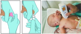
A cephalohematoma measuring 8 cm or more must be punctured (the puncture is performed by a pediatric surgeon) and the liquid blood must be sucked out. Then a pressure bandage is applied.
Mothers need to remember that a child with a cephalohematoma should not be rocked to sleep.
If the hematoma suppurates (fever, inflammation of the skin above the hemorrhage), it should be opened, pus and blood clots removed. After which the wound is drained, bandages with antiseptics and antibiotics are prescribed.
Hospital treatment for an uncomplicated cephalohematoma lasts 7–10 days, and for a complicated one – a month or more. Subsequently, the child is registered with a pediatric surgeon and neurologist for a year.
List of sources
- Obstetrics. National leadership / Ed. E. K. Ailamazyan, V. I. Kulakov, V. E. Radzinsky, G. M. Savelyeva. - M.: GEOTAR-Media, 2007. - P. 1160-1161.
- Bardeeva K.A., Pisklakov A.V., Lukash A.A. A new look at the treatment of cephalohematomas in children // Modern problems of science and education. – 2015. – No. 5.;
- Ch. 6. Children's diseases // Directory of a paramedic / Professor L.A. Isaeva. - Moscow: “Medicine”, 1975. - P. 319. - 662 p. — 280,000 copies.
- Kerchelaeva S.B. Cephalohematoma as a result of complications of childbirth / Kerchelaeva S.B., Tyagunova A.V., Kuznetsova O.V. // Attending physician - 2015. - No. 10 - p. 88–92.
- Handbook of obstetrics, gynecology and perinatology / Ed. G. M. Savelyeva. - M.: MIA, 2006. - P. 344-348.