Causes of Hirschsprung's disease
Hirschsprung's disease is a congenital disease that develops in utero; scientists believe that one of the main etiological factors is the lack of nutrients in the mother's body during intrauterine development of the fetus. Due to a lack of necessary substances, during the development of innervation of the fetal digestive system, areas of the large intestine appear that do not have nerve endings; often specialists cannot determine the cause of this disease.
The length of the area with missing innervation can vary: from a few centimeters to the entire area of the large intestine. It is not uncommon for this pathology to be transmitted hereditarily; about 50% of patients suffering from Hirschsprung's disease have a history of close relatives with the same disease.
There is an assumption that dysfunction of the development of the intestinal nervous system is provoked by a mutation in the structure of DNA genes.
In newborn boys, this disease occurs 4-5 times more often than in girls.
Causes of pathology
Although this disease was described back in 1887, it took many years to identify the causes and development of this disease.
Doctors cite several sources that provoke the development of such an unpleasant pathology as Hirschsprung's disease in children.
Causes of the disease:
- As a result of the studies, it was found that the formation of pathology occurs during intrauterine development. Nerve clusters that will perform the necessary functions in digestion are formed in the fetus from 5 to 12 weeks of pregnancy. In rare cases, nerve cells in a baby's digestive system do not fully develop. They are not formed in the large intestine. The causes of this pathology have not yet been clarified. It is believed that this is how mutations in DNA manifest themselves.
- Heredity is also noted in the occurrence of this disease. If there are cases of this disease in the family, then the likelihood that a sick child will be born is quite high. Especially if the fetus is male, since boys suffer from this disease 4-5 times more often than girls.
- In areas where there is increased radiation, children with Hirschsprung's disease are more often born.
- A factor influencing the occurrence of this disease is Down syndrome.
- Pathology can develop as a result of endocrine disorders in the mother.
Symptoms of Hirschsprung's disease
The intensity of symptoms depends on the severity of the disease (the extent of intestinal damage).
In most cases, the clinical manifestations of this disease are diagnosed in the first days of a child’s life, sometimes in adolescence or adolescence; in adulthood it is rarely detected.
In newborns, the main symptoms of Hirschsprung's disease are: lack of feces (meconium) in the first days of life, increased gas formation, vomiting (in some cases with bile), and there are also other dyspeptic disorders (diarrhea, constipation).
In older children, there may be an increase in abdominal volume, a predisposition to persistent constipation, the development of malabsorption syndrome and, as a result, retardation in growth and development. As a result of frequent loss of blood in feces, anemia may develop, infection may occur and enterocolitis may develop. In case of intestinal infection, hyperthermia, vomiting and diarrhea are observed. Severe intoxication promotes expansion of the colon.
In adults, Hirschsprung's disease is manifested by the absence of the urge to empty the rectum, spastic pain along the colon, bloating, and intoxication of the body caused by the toxicity of accumulated substances. Sometimes a clinical sign of Hirschsprung's disease is partial heterochromia - uneven coloring of areas of the iris.
How does Hirschsprung's disease manifest?
When the intestine is affected by Hirschsprung's disease, it is impossible to relax the affected area. It is characterized by constant severe muscle spasm. In addition, it lacks peristalsis, which makes it impossible for intestinal contents to move through the intestines. The intestine above the affected area expands due to the accumulation of feces and atrophy of muscle fibers.
Forms of the disease
The acute form of Hirschsprung's disease occurs with total and subtotal damage to the large intestine. It appears in the first days of a child’s life. It is characterized by a clinical picture of low intestinal obstruction - absence of meconium passage, bloating, vomiting.
The subacute form of the disease occurs with a shorter zone of aganglionosis. It is characterized by persistent constipation, stool only after an enema, an enlarged abdomen, anxiety, and retardation in neuropsychic and physical development.
The chronic form develops when only the rectum is affected. Constipation occurs more often in older children. At the beginning, constipation is short-lived and after cleansing enemas, independent acts of bowel movement are possible. As the disease progresses, fecal stones may form and obstructive intestinal obstruction may occur. These children have reduced immunity and are much more likely to get sick than their peers. A characteristic symptom of this disease is an enlarged abdomen due to an increase in the size of the large intestine.
Diagnosis of the disease
Diagnosis of Hirschsprung's disease is based on:
- clinical data;
- X-ray examination (irigoscopy);
- sphincteromanometry (study of the tone of the rectal sphincter);
- morphohistochemical method (determining the presence or absence of nerve ganglia in the submucosal layer of all parts of the colon).
Treatment of Hirschsprung's disease
The only correct treatment for Hirschsprung's disease is surgery. During surgery, the affected area of the colon is removed and the terminal section of the intestine is connected to its healthy section.
Basically, all stages of this operation are always carried out within one surgical intervention, but there are exceptions - the operation in such cases consists of two stages. At the first stage of the two-stage operation, resection (excision) of the affected area of the intestine and colostomy are performed (excision of the end of the healthy intestine through an incision in the abdomen). The stool is evacuated into a special container that the patient has to carry with him. After normalization of the patient’s intestinal function, the second stage of the operation begins - intestinal reconstruction, the task of which is to connect the free end of the healthy intestine with the rectum and close the colostomy (suturing the hole in the abdomen).
As a rule, after an operation to eliminate the affected areas of the intestine, patients gradually normalize the process of digestion and excretion of feces, that is, a complete recovery occurs. Sometimes patients may notice a tendency to constipation, but this problem can be eliminated with the help of laxatives.
Treatment protocol for Hirschsprung's disease in children
1. Code – Q 43.1
2. The name of the disease is Hirschsprung’s disease (HD):
a – acute form; b – subacute form; c – chronic form.
Depending on the volume of the lesion, HD is distinguished: 1 – rectal: a) with damage to the perineal region; b) with damage to the ampullary part; 2 – rectosigmoid: a) with damage to part of the sigmoid colon; b) with subtotal or total damage; 3 – subtotal: a) with damage to the transverse colon; b) with the spread of the lesion to the right half of the intestine; 4 – total form of HD.
3. Pre-hospital diagnostics. Complaints. The main clinical symptoms of Hirschsprung's disease are: constipation, gas retention, bloating. In some young children, manifestations of the disease may begin with diarrhea. It must be borne in mind that paradoxical emptying (after constipation), constant diarrhea in a newborn, especially if vomiting and a significant increase in the abdominal cavity are simultaneously present, should raise suspicion of Hirschsprung's disease. A significant place in diagnosis is given to data from both the medical history and life history. When collecting a medical history, it is necessary to establish the following data: at what age did constipation begin; how many days without spontaneous bowel movement; did the child experience loose bowel movements (diarrhea) after prolonged constipation? whether cleansing enemas were performed, how often, their effectiveness; whether the child has been examined before, the results of the examination; what types of therapy the child received at the time of examination, whether conservative treatment had an effect and how long it lasted. It is always necessary to analyze life history data: how the mother’s pregnancy went; whether meconium passed in the first 24 hours of the child’s life, and the time of its passage; whether the stool has changed after the introduction of complementary foods or the transfer of the child to artificial feeding; what is the dynamics of the child’s body weight during the neonatal period, was there any malnutrition; what diseases the child had at the time of examination. Subsequently, after collecting anamnesis, a general examination of the patient is carried out, which allows us to assess the mental and physical development of the child. When examining the abdomen, it is necessary to pay attention to: its size, the condition of the skin and subcutaneous tissue, blood vessels and the configuration of the abdomen (increase in volume, asymmetry, expansion of the costal arch, thinning of the anterior abdominal wall). When examining the abdominal cavity, palpation and auscultation are mandatory.
4. Prehospital therapy is not carried out; suspicion of HD is the reason for hospitalization in a children's surgical hospital.
5. Diagnostics at the hospital stage. Taking into account these complaints, medical history, life and general examination of the patient (see point 3), a detailed visual examination of the rectoanal area is carried out, which allows us to identify sphincter tone, as well as a digital rectal examination - a necessary addition to the visual examination of the rectoanal area, which is the first from special examination methods, during which the condition of the rectal ampulla and sphincter tone are assessed.
It is mandatory to conduct a general clinical examination and ultrasound to diagnose concomitant pathologies. An exclusive role is given to radiation diagnostic methods, which are mandatory. In the acute form of GD in the neonatal period, a survey radiography of the abdominal organs (ARB) and irrigography are performed, which makes it possible to differentiate GD from other types of low intestinal obstruction (the condition of the intestinal loops, the presence of microcolon and other visible changes are assessed). The leading manipulation in the diagnosis of HD is radiocontrast examination of the intestine - irrigography. The manipulation requires the presence of 3 medical specialists: a surgeon, a radiologist, an anesthesiologist-resuscitator. It is performed using a barium mixture, which is prepared in saline solution at the rate of 1 part barium: 4 parts solution. Pictures are taken in 2 projections - frontal and lateral - at the beginning of filling, when the intestine is filled with contrast, and after emptying it. The position of the patient during the examination is on the left side with the legs brought to the stomach. The amount of contrast fluid that is administered to a sick child is given in Table. 1. Table 1. Amount of contrast depending on age Age of the child Amount of contrast agent Newborn 30 – 50 ml Infants 50 – 100 ml Children 1-3 years old 100 – 200 ml Children 3-7 years old 300 – 500 ml Older children Not less than 700 ml
When interpreting the irrigogram, attention is paid to the following radiological signs of HD: • the presence of a “leukoid” transition from the distal narrowed in diameter to the proximal widened section of the intestine*.
*Note: 1 – in newborns this diagnostic method is difficult, because in the first days and weeks of life the difference in the diameter of the aganglion zone and the upper parts of the colon is insignificant; 2 – with a short aganglionic segment, expansion of normally innervated sections and a leuko-like transition to the aganglionic segment develop in infants at 2-3 months, possibly later, which requires repeated irrigography; 3 – the combination of a false negative radiological picture with clearly defined histological aganglionosis is typical for newborns and patients with total aganglionosis of the colon.
Important in diagnosis is a biopsy of the rectal wall, which is performed in two ways. Layer-by-layer rectal biopsy - performed under general anesthesia, during which a section of the muscular layer of the rectum measuring 2 x 1 cm is taken; The biopsy material must necessarily contain both muscle layers. A puncture biopsy, which is performed using biopsy clamps-nippers of a fiber colonoscope and without anesthesia, takes the mucous and submucosal layer, is performed 2-3 cm above the dentate line, then 2-2.5 cm above the first, and then 2-2 .5 cm above the second biopsy site, etc., since in more distal parts of the rectum the density of ganglion cells decreases, which makes it possible to find cells of the Meissner submucosal plexus. After the biopsy, these samples are subject to neurohistochemical examination - acetylcholinesterase (AChE) activity is determined using the Karnovsky and Roots method. Depending on the severity of the reaction to AChE, 4 variants of indicators are distinguished: patients with a sharply positive reaction - a high content of diffusely located AChE is determined in the muscular plate of the mucous membrane; patients with a positive reaction to AChE - the location of the AChE-positive substance is not so dense, and the cholinergic fibers are thinner and arranged in one stripe, less often - in several stripes; patients with a weakly positive reaction to AChE – positive fibers in the mucosal layer proper are located in one layer in the form of a thin mesh, unevenly; patients with a negative reaction to AChE - only in the muscular plate of the mucosa. A small number of AChE-positive short thick fibers are observed in the membrane of these patients. In HD, the reaction to AChE is always strongly positive or positive. 6. Surgical treatment. Treatment of Hirschsprung's disease is only surgical, and therefore both preoperative preparation and subsequent stages of treatment are important.
Preoperative preparation. The purpose of preoperative preparation is to normalize bowel movements (if possible), correct general changes in the body, prevent or treat enterocolitis, dysbacteriosis, and enzymatic disorders. Preparation begins from the day the patient is admitted to the hospital and continues during the examination of the patient until the time of surgery. The main elements of preoperative preparation for HD include: • Diet – preference is given to foods rich in fiber, vegetable oils, animal oils, fermented milk products. • After hospitalization, a diet with a minimum content of toxins, rich in protein and vitamins (protein and vitamin enpits, which contain vitamins A, E, D, C, PP, B1, B6, B5, B12, and minerals: calcium, phosphorus, potassium) is prescribed , sodium, magnesium, copper, manganese, iron, zinc, iodine, selenium, the energy value of which is 1884-2177 kJ). • Vegetable soups and fermented milk mixtures are left 2-3 days before surgery. • Active treatment of fecal intoxication: • Prescription of laxatives: Vaseline oil (in the intervals between meals) in doses: up to 3 years - 1 tsp. 3 times a day, up to 7 years – 1 des.l. 3 times a day, older children - 1 table. l. 3 times a day. • Probiotics (eubiotics). Preparations that contain microorganisms - representatives of the normal intestinal microflora or their structural components. Bifidum-bacterin-forte, bactrin, bificol, bifi-form, hilak-forte, normaze (or duphalac, portalac). The dosage of these drugs depends on the age of the child. • Multienzyme preparations: pancreatin preparations: Creon, mezim-forte, mezim, pankurmen, trienzyme, pancreatin, Kirschner's pancreas. Enzymes: festal, enzistal, digestal, polyzyme, cotazim, panzinorm-forte. • Hepatoprotectors, Essentiale, Legalon, Sirepar, etc., depending on the age of the child. • Vitamins B6, B12, E, C. • Abdominal massage, physical therapy.
Local therapy: irrigation of the large intestine with a 1% NaCl solution with mandatory monitoring of the ratio of injected and excreted fluid volumes. 3 days before surgery, an oral antibiotic of the metronidazole group (Trichopol, Metragil, etc.) is prescribed at 8.5 mg/kg body weight 3 times a day. Feeding is canceled from 16-00 (20-00) depending on the age of the child. For children under 1 year of age, the last milk feeding is at 24-00. The child is prescribed a siphon (cleansing) enema 2 times on the eve of the operation, in the morning and in the evening. On the day of surgery, a cleansing enema is performed in the morning 1-1.5 hours before surgery. 1 hour before surgery, a daily dose of a broad-spectrum antibiotic (from the group of third generation cephalosporins) is administered intramuscularly or intravenously. Surgical intervention. The main method of treatment for HD is radical surgery, which must be performed at an early age of the child - up to 10-12 months, which contributes to a faster recovery and better adaptation of the child after surgery. The choice of radical surgical intervention technique is determined by factors such as the age of the child, the presence or absence of complications, and one- or two-stage treatment tactics (three-stage is also possible). In the uncomplicated form of HD, preference should be given to a one-stage surgical intervention - resection of the aganglionic zone along with the most altered part of the intestine with the formation of a primary colorectal anastomosis using the Soave technique. It is necessary to take into account that the complication of the disease and mortality in HD is associated, first of all, with late diagnosis, and, secondly, with the irrational choice of a radical correction method. In this regard, it is necessary to take a differentiated approach to developing a treatment plan for patients in whom the course of the disease is complicated by enterocolitis, acute obstructive intestinal obstruction due to GD, severe malnutrition and anemia as a result of severe fecal intoxication, severe reaction to a siphon enema, spontaneous perforation of the large intestine, acute form of HD in young infants, as well as with concomitant developmental deficiencies. In such patients, surgical treatment should be 2- or 3-stage. The 1st stage of treatment is the application of a colostomy, the 2nd stage of treatment is radical correction of the developmental defect and the third stage is the closure of the colostomy. Surgical treatment of the acute form of HD is performed through a median laparotomy approach for a detailed examination of the abdominal organs in order to identify concomitant developmental anomalies and adequately select the location of the colostomy. Colostomy is indicated: • in newborns * • with signs of low intestinal obstruction and a pronounced transition zone • with early manifestations of enterocolitis • with perforation of the colon; • in the subacute form of GD, when it is impossible to rinse the large intestine, which is due to recurrent intestinal obstruction; with malnutrition, severe anemia, in cases of complex concomitant developmental defects, with severe and inadequate reactions to a siphon enema in the preoperative period, enterocolitis and colon perforation. • Note: in newborns with clinical signs of low intestinal obstruction, who, against the background of severe intoxication, do not have an existing organic cause of obstruction, or if it is difficult to determine the border of aganglionosis, a T-shaped ileostomy is imposed with a mandatory biopsy of the distal parts of the small and large intestines. The first stage of treatment is the formation of a colostomy. A colostomy (stoma) is formed after reliable confirmation of the diagnosis of HD and determination of the level of the zone of aganglionosis and only if conservative therapy is unsuccessful. Preference is given to the formation of a final colostomy on the proximal segment of the suprastenotic dilated section of the intestine with mandatory biopsy of the narrowed aganglionic and transition zones. Radical surgery can be either the first or the second stage of surgical treatment - after a colostomy. Performed 3-9 months after the formation of the colostomy. Methods of surgical treatment of Hirschsprung's disease: today the most widespread is the Soave operation with the imposition of a primary colorectal anastomosis. Radical surgery technique. The patient is placed in the lithotomy position. Laparotomy with a left-sided transrectal or inferomedial incision. The proximal resection border is determined and the colon is mobilized from the marking site to the transitional fold of the peritoneum. A serous-muscular sheath of the rectum is formed using the Soave technique. Then the operation continues from the perineum. Using a guide, the mobilized colon is raised through the anal canal to the marked level of the intended resection. In the case when the distal part of the colon is disconnected by a colostomy, the disconnected distal part of the colon is first evaginated and its drive loop is raised through the seromuscular sheath towards the perineum. In subtotal aganglionosis, the colon is erected through the right lateral canal without or with dislocation of the ileocecal angle. In a patient with total aganglionosis, the ileum is erected with the imposition of a primary anastomosis with the addition of the formation of a colonic-small intestinal reservoir. The mucous membrane is treated with antiseptic, circularly cut off, retreating 4-5 cm from the dentate line, all layers of the evaginate are gradually cut off in the transverse direction and interrupted sutures are applied circularly (vicryl, maxon, dexon 3/0-4/0) between all layers of the colon and the mucous membrane of the rectum, forming an extrasphinctorial anastomosis. The intercase space is drained extrasphinctorially through a separate skin incision or through a suture line. An elastic tube is inserted into the lumen of the reduced colon above the anastomosis line to drain gases and feces, and the anastomosis itself is inserted into the anal canal. From the abdominal cavity, the edge of the serous-muscular sheath is sutured to the serous membrane of the reduced colon. The application of primary anastomosis is possible both manually and using circular staplers SPTU and KTs-28. In patients with no complications during the course of the disease, surgical treatment of developmental deficiency can be carried out in one stage. 7. Postoperative treatment. During the operational period, a significant place is given to the early period. Upon completion of the surgical intervention, the child is transferred to the intensive care unit for the first stage of treatment in the early postoperative period. The intensive care program includes: compliance with all the basic principles of infusion therapy. • Antibiotic therapy – 2-3 broad-spectrum antibiotics (third generation cephalosporins + modern aminoglycosides + metrogyl). • Adequate pain relief. • Oxygen therapy. • Suction of the contents of the intercase space through tubular drainage. • Clinical and laboratory monitoring. During the postoperative period, the following complications are possible: enterocolitis; anastomotic failure; suppuration of the intercase space; local abscess; hematoma of the intercase space; anaerobic (clostridial) infection. All of the above complications can lead to the development of peritonitis, followed by the development of sepsis; relapse of constipation; adhesive intestinal obstruction rarely develops (more often in children who have undergone previous surgical interventions - stoma); encopresis of varying severity. Therapeutic tactics for the development of the following post-operative complications: • In case of post-operative peritonitis, relaparotomy, sanitation of the abdominal cavity and the formation of a colostomy on the hepatic section of the transverse colon are indicated. • For postoperative enterocolitis, infusion therapy for the purpose of detoxification, antibiotic therapy, and intestinal irrigation are indicated; in the absence of positive dynamics, total parenteral nutrition is prescribed for 1-2 weeks. • For early adhesive intestinal obstruction: relaparotomy, disconnection of connections, intubation of the intestine through the anus. • For symptoms of encopresis – conservative treatment. • If constipation recurs, examine the patient to identify their cause and promptly eliminate it (Table 2). Table 2 Typical causes of constipation in the post-operative period and their surgical correction Cause Tactics If the tone of the internal sphincter increases, perform the Lynn operation If a residual zone of aganglionic disease is identified Repeated surgery - resection of the aganglionic zone with the imposition of a primary anastomosis Stenosis of the anastomosis Pneumodilatation and bougienage are indicated Colonoptosis Colonopexy
The second stage of treatment of children in the postoperative period.
The second stage is carried out in the surgery department, where the child is transferred from the intensive care unit. In the surgical department, a gradual reduction in the volume of infusion therapy is carried out with its complete cessation within 1-2 days and the patient is gradually transferred to enteral nutrition, and antibacterial therapy is continued. The endotracheal tube will be fixed in the perianal area. The urinary catheter is removed from the bladder. On the third day, a 0.05% solution of proserin is prescribed twice a day (i.m. or i.v.) in a single dose of 0.05 ml/kg body weight for children under 1 year of age, and 0.1 ml/kg for children older at 1 year. In addition, electrophoresis with proserine is prescribed on the stomach - one procedure per day, 4-5 sessions. At the same time, from the second day after surgery, every 4 hours the rectal endotracheal tube is washed with 30-40 ml of 1% NaCl solution to decompress the intestine and stimulate its peristalsis. The drainage from the intercase space is removed on the 3-4th day, and the probe from the intestine is removed when peristalsis resumes. With the resumption of intestinal motility, patients are gradually transferred to enteral nutrition. For patients with rectal and rectosigmoid forms of aganglionosis, enteral nutrition begins from 3-4 days after surgery, for subtotal form - from 4-6 days, and for total aganglionosis - from 12 days. At this time, probiotics are continued at an age-specific dose for 3-4 weeks after surgery. The child is gradually increased enteral nutrition. Enzyme therapy is carried out. Physiotherapeutic procedures are prescribed. Sutures are usually removed on the 9th day of the postoperative period. Before the patient is discharged, control general blood and urine tests are performed. If the condition is satisfactory, the patient is discharged on the 10-12th day. 8. Rehabilitation and outpatient treatment. After discharge from the surgery department, outpatient and rehabilitation treatment is carried out. However, it should be noted that in a certain percentage of patients - every fifth patient - unsatisfactory results are noted in the form of a functional partial loss of anal control, and also in some children there are slow adaptation-compensatory processes against the background of functional inferiority of the parts of the colon that remained after its resection, especially in patients operated after a temporary colostomy. In connection with the above, the recovery period after radical surgery in a certain group of patients lasts from 6 months until puberty; on average, most patients are considered practically healthy at the end of 2 years after surgery. Rehabilitation measures are carried out for 1 month and include diet therapy, vitamin therapy (groups B, C), immunostimulating therapy (Eleutherococcus, Echinacea), eubiotics, enzyme therapy, laser magnetic therapy on biologically active points, acupuncture, electrical stimulation of the anal sphincter and perineal muscles using the apparatus " Endoton" (for encopresis), exercise therapy aimed at strengthening the pelvic floor muscles; for periodic constipation, therapeutic cleansing enemas (chamomile infusion, sea buckthorn oil) are performed 1-2 times a day for 7 days; If the effect of outpatient rehabilitation treatment is poor, hospitalization of the patient in the surgical department of the hospital is indicated for the purpose of a detailed examination and treatment of the identified pathology. After discharge from the hospital, the child comes for a follow-up examination after 1 month, then after 6 months and 1 year. 9. Referral to sanatorium-resort treatment no earlier than 3-6 months in the absence of complications (Odessa, Kuyalnik). Share
Minimizing negative consequences
To minimize constipation, diarrhea and other undesirable effects after surgery, patients are recommended to adhere to a special diet, which is based on the consumption of foods rich in plant fiber.
The risks of postoperative complications include the development of enterocolitis due to intestinal infection, therefore, if there is an increase in body temperature, vomiting, diarrhea, or signs of intestinal bleeding, you should immediately consult a doctor.
In mild cases of Hirschsprung's disease, surgical interventions can be abandoned and symptomatic treatment can be performed using siphon enemas.
Conservative treatment
Such therapy should not be relied upon. Treatment of Hirschsprung's disease in children is carried out in most cases exclusively by surgery. Conservative therapy mainly serves as preparation for further surgical intervention.
Preparatory treatment includes:
- A certain diet, adherence to a diet. Vegetables, fruits, fermented milk products, and foods that do not cause gas are recommended for consumption.
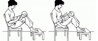
- Massage, physiotherapy, gymnastics. They stimulate intestinal motility.
- Cleansing enemas.
- Intravenous administration of protein medications and electrolyte solutions.
- Vitamins.
Consequences of Hirschsprung's disease
Complications of Hirschsprung's disease occur in the absence of adequate treatment. Impaired evacuation of intestinal contents leads to the following consequences:
- the formation of fecal stones , the removal of which by conservative methods (enemas) is ineffective, and their presence in the intestines provokes pain;
- acute enterocolitis (inflammation of the intestinal mucosa) develops against the background of general and local immunosuppression;
- anemia occurs as a result of impaired absorption of microelements and vitamins from food;
- acute intestinal obstruction is a life-threatening condition that occurs when the intestinal lumen is completely obstructed.
Types of Hirschsprung's disease
The size of the aganglionic area in Hirschsprung's disease can vary from a few centimeters to a meter or more. The severity of the child’s condition and the characteristics of the clinical picture of the disease depend on this.
Anatomical classification of congenital intestinal aganglionosis:
- Rectal form. The area of aganglionosis covers the entire rectum or part of it (perineal, ampullary or supramullary).
- Rectosigmoidal form. The non-innervated zone extends to the distal part of the sigmoid colon or covers it completely.
- Subtotal form. The ascending colon and part of the transverse colon (the left half of the intestine) are involved in the pathological process.
- Total form. The absence of intramural nerve fibers is observed throughout the intestine.
Clinical classification of Hirschsprung's disease:
- Stage of compensation (rectal form). The child has moderate symptoms of intoxication and incomplete intestinal obstruction, the problem is solved with an enema.
- Stage of subcompensation (rectosigmoid and subtotal forms). Carrying out a cleansing enema is ineffective; sometimes the child’s condition is improved by a high cleansing or siphon enema. Symptoms of intoxication increase, and signs of metabolic disorders appear.
- Stage of decompensation (all types of aganglionosis, except rectal). The pathological process progresses, which can develop into acute intestinal obstruction.