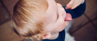
The skull of a newborn baby is different from that of an adult. The skeleton bones of the baby's head have soft spaces (sutures) and membranous non-ossified zones (fontanelles). These soft areas allow the baby's head to pass through narrow spaces in the birth canal. As a result of labor, cranial deformation occurs, which is observed in most newborns.
After birth, the baby takes his first breath in a process that expands not only the lungs, but also the cranial part of the skeleton. Then, when sucking, the wedge-shaped occipital joint acts as a kind of lever, creating pressure that can even out the asymmetry. However, there are congenital and acquired deformities that require treatment.
What head shape should babies have? Norms and deviations
Normally, a newborn's head has a circumference of 34-36 cm. A small or large skull is not always a symptom of pathology. The size of a child's head depends on heredity - a parent may have a disproportionate head. In the first month after birth, the baby's head increases by 1.5-2 cm.
Over 3-4 months, the size of the head “catches up” with the circumference of the chest, and then begins to lag behind. During the first 6 months, the skull circumference is on average 43 cm. The most intensive growth of the head of a full-term baby is observed up to 3 months, and in a premature baby - during the period of maximum weight gain. A marked advance or lag in the increase in head size may indicate an anomaly.
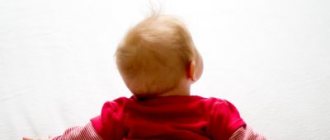
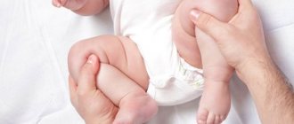
In the photo the elongated shape of the baby's head
For diagnosing pathology, the shape of the baby’s head is also of great importance. The following shape of a newborn baby’s skull is considered a normal variant:
- dolichocephalic – oval, ovoid and oblong;
- brachycephalic - round.
An elongated shape can be due to increasing the size of the distance:
- from the chin to the back of the head;
- from the forehead to the back of the head.
The shape of the head largely depends on labor and the method of birth of the baby. When the birth canal passes with the head forward, the newborn has a dolichocephalic form, and when born through a cesarean section, it is brachycephalic. But there is also a pathological deformation of the child’s head:
- plagiocephaly – sloping and asymmetrical, the forehead or back of the head is flat;
- acrocephaly – elongated head shape;
- scaphocephaly is a “curved” form in which areas of the frontal or occipital region may protrude.
Skull asymmetry in children can lead to neurological pathologies and retardation in the child’s mental/physical development.
When will the fontanelle close and what should the child’s head circumference be?
Konstantin Grigoriev, pediatrician, professor, doctor of medical sciences
- What should a newborn's fontanel look like?
- Shape and size of a newborn's head
What can the shape of a newborn’s head and its size tell parents? What “signals” does the large fontanel give about the baby’s condition? We dispel the fears and doubts of mothers.
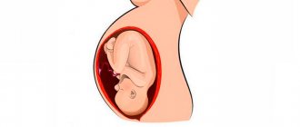
Why does skull deformation occur in infants?
Deformation of a child's head during childbirth is a physiological process. In the fetus, the brain part of the skeleton consists of movable bone plates connected by fibrous or cartilaginous tissue. During labor, the head “squeezes” through a narrow ring of pelvic bones. The pressure exerted on the skull deforms it. The displacement of the bones allows the obstacle to be passed without damaging the brain. After the child is born, the mechanism opposite to compression “turns on.” Decompression occurs due to increased intracranial pressure during breathing and sucking.
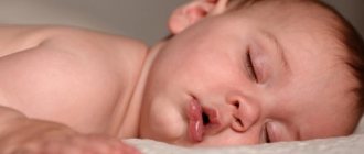
Flattened nape of a baby
The deformation disappears spontaneously. In addition to the physiological one, there may also be a pathological intrauterine anomaly of the shape of the skull, which is caused by:
- passage of a large fetus through a too narrow birth canal;
- atony of the muscles involved in childbirth;
- small (narrow) or flat pelvis;
- rapid expulsion of the fetus;
- asynclitism and synclitism - incorrect insertion of the head into the pelvic ring;
- trauma during obstetrics;
- multiple pregnancy.
The shape can be deformed by intrauterine pathology of bone formation and development at the stage of embryogenesis. Deviation from the norm occurs under the influence of:
- infections;
- violations of the position of the fetus in the uterus;
- genetic diseases;
- dysplasia.
During a difficult birth, the brain is very rarely damaged; it is reliably protected from compression, but the baby will need a little help to correct the defect.
Acquired asymmetry can be caused by incorrect:
- the position of the child in the crib;
- the position of the baby when attached to the breast, carried in the arms.
Parents should pay attention to the baby's posture and behavior during sleep and wakefulness.
Features of the structure of the newborn’s skull
In a newborn, the skull is not a monolithic bone. However, it is not like this in an adult either, but after 6 years the mobility of the bones that make up the skull is very limited, so it is so important to monitor the changes and, if necessary, correct the shape of the skull starting from the first days of the child’s life.
The baby’s head consists of two main parts - the arch, which at an early stage of life (up to 6 years), is also composite, and the base, which is formed from cartilaginous tissue and ossifies with age. The cranial vault in newborns consists of 4 parts connected by movable lamellar tissue. Over time, these parts come closer to each other and ossify in adulthood, forming a structure that is practically motionless relative to each other.
Symptoms of abnormalities in infants
Skull deformation in children can resolve spontaneously without symptoms of nervous activity disorders. With severe compression of a certain area of the brain, the following disorders may occur:
- Displacement of the bone plates in the occipital region is manifested by:
- disruption of the sucking process;
- frequent and profuse regurgitation;
- in the future – poor vision, scoliosis, torticollis and speech impediment.
- Asymmetry of the sphenoid bone leads to:
- strabismus;
- increased intracranial pressure;
- articulation disorder.
- impaired coordination of movements;
- hearing impairment.
- muscle weakness;
- lethargy;
- lagging psychomotor development.
Parents' attention should be drawn to such deviations as:
- bluish or purple spots on the baby’s skull;
- soft or hard buds;
- infantile jaundice;
- asymmetry of the jaws and parts of the facial skeleton;
- constant lacrimation;
- asymmetry of facial expressions;
- bulging of the fontanelle at rest.
If at least one sign attracts the attention of parents, then it is necessary to immediately visit a doctor and undergo an examination - MRI, ultrasound.
Others unwillingly
The first written mentions of skull deformation are found in ancient authors. In the V-IV centuries. BC e. Hippocrates, in his text “On Air, Waters and Places,” described the long-headed people, or macrocephals, living on the eastern shore of the Black Sea. Their elongated heads were a sign of belonging to the elite. However, from the text it becomes clear that macrocephals themselves voluntarily deformed the skulls of their babies.
“They consider those with the longest heads to be the noblest. ... as soon as the child is born, while his bones are still soft, his unhardened head is straightened with his hands and forced to grow in length through bandages and other suitable devices, as a result of which the spherical shape of the head deteriorates and its length increases.”
However, modern parents are often faced with congenital, unintentional deformation of the skull. And, unfortunately, it does not make their children a sign of belonging to the elite, rather the opposite...
The word "craniostenosis" comes from the Greek. cranio – “skull” and stenosis – “fusion of bones”. As recognized by the Association of Neurosurgeons of Russia, craniosynostosis (or craniostenosis) is “a disease manifested by the congenital absence or premature closure of the sutures of the skull. Premature synostosis in the area of the sutures of the skull leads to limited growth of the skull in the area of the closed suture, resulting in the development of craniocerebral disproportion. The clinical manifestation of craniocerebral disproportion is intracranial hypertension syndrome.”
How to straighten a newborn's head?
Deformation of the bones of the baby’s skull at an early stage, until the final closure of the fontanelles has occurred, can be corrected using craniosacral therapy. The doctor uses manual techniques to eliminate the asymmetry of the baby’s skull. Massage for head deformation of a child under 1 year of age has shown the effectiveness and safety of therapy, provided the doctor is highly qualified.
Parents can eliminate the anomaly on their own:
- avoid incorrect positioning of the baby during feeding;
- periodically change his position in the crib, placing him on the other side, on his tummy;
- move the crib relative to the light and sound source.
A newborn with congenital asymmetry of the skull should be periodically examined by a pediatrician or neurologist. A small child responds more easily to conservative treatment methods. If correction by such methods is ineffective, then the doctor selects an orthopedic correction helmet for the baby. It determines the size, frequency and duration of wearing the device.
Correction of a child's head using an orthopedic helmet
Parents should remember that strict adherence to the specialist’s recommendations, perseverance and consistency in performing procedures will help completely correct the injury, the child’s skeleton and avoid complications that can result from asymmetry of the baby’s head. In particularly severe cases, surgery may be required.
Deformational brachycephaly (Greek brachy, meaning “short”).
This is a flattening of the occiput and a compensatory expansion of the parietal region. Children with deformational brachycephaly have a weak or flat back of the head and their head appears disproportionately wide or “big” when viewed from the front. [2] (Figure 5)
Figure 5. Deformational brachycephaly. Gary F. Rogers, MD, JD, MBA, MPH. Deformational Plagiocephaly, Brachycephaly, and Scaphocephaly. Part I: Terminology, Diagnosis, and Etiopathogenesis
The head width to length ratio (cranial index) is normally between 0.75 and 0.8, although some range from 0.8 to 0.85 due to back sleeping. [1]
Many children with deformational brachycephaly also have other asymmetries or plagiocephaly. This combined deformity “asymmetrical brachycephaly” is the most common deformity. [1].
An interesting fact is that in 1992 in America there was a “campaign for sleeping on your back” to reduce sudden death syndrome. Although it was shown to be effective and led to a 40% reduction in the incidence of sudden death syndrome, one of the unforeseen consequences was a sharp increase in plagiocephaly and brachycephaly. [3-6]
Causes of deformational brachycephaly
Unlike plagiocephaly, when the child's head is tilted to one side, with brachycephaly the child holds his head in the center. The development of the deformity will be facilitated by increased extensor tone of the neck muscles (in this case, the child lying on his back puts more pressure on the support with the back of his head, increasing the pressure on the occipital bone, which contributes to the development of brachycephaly).
The second group of reasons is the child being in a car seat, cradle, or lying on his back for too long (when parents do not lay the child on his stomach or hold him in his arms for too long). However, it should be noted that, as a rule, even in this case, brachycephaly does not develop in all children and it would be correct to talk about other predisposing factors (impaired muscle tone, birth trauma, positional brachycephaly).
Birth injury
Birth trauma is a common cause of the development of muscular torticollis, disturbances in muscle tone, compression of the base of the skull and other problems that predispose to the development of cranial asymmetry. For this reason, in order to assess the condition and treat plagiocephaly, it is necessary to evaluate how the birth took place and conduct osteopathic testing of the cervical spine, cranio-vertebral junction, and skull. But we'll talk about this later...
Prevention of deformation of the skull bones in a child
In order not to deform the baby’s cranial skeleton, parents need to:
- change the child’s position more often during sleep and rest;
- strengthen the muscles of the back and neck;
- during wakefulness, the child should remain upright for more time;
- properly equip the sleeping place;
- Avoid soft pillows and mattresses.
It is important to remember the importance of breastfeeding. Children who feed on breast milk are less susceptible to infantile pathologies, have stronger bones and higher immunity. Skull deformation in infants can resolve on its own with breastfeeding.
Having a baby requires preparation. It is necessary to check in advance for the presence of hereditary diseases that can deform bone structures. Children should walk to synthesize vitamin D - prevention of rickets and skeletal pathologies. The birth of a baby with an asymmetrical skull is not a death sentence; most often, such children catch up and surpass their peers in development, subject to proper care and timely correction measures.
Seams that allow movement
The newborn's skull consists of five main bones: a pair of frontal bones, a pair of parietal bones and one occipital bone. These bones are connected by fibrous sutures that allow the bones to move relative to each other, facilitating birth and brain growth.
Normally, the coronal suture separates the two frontal bones from the parietal bones. The metopic suture separates the frontal bones. The sagittal suture separates the two parietal bones. The lambdoid suture separates the occipital bone from the two parietal bones.
But sometimes something goes wrong... Genetic mutations, which can be local in one area or have a systemic, widespread nature, lead to the sutures closing prematurely.
If the closure of one or another suture is accompanied by other anomalies, such craniostenosis is called “syndromic”. Syndromes characterized by premature closure of cranial sutures: Crouzon Syndrome, Apert Syndrome, Pfeiffer Syndrome, Satpre-Hotzen Syndrome, Carpenter Syndrome and others.
Craniostenosis can be a component of various hereditary syndromes. But in most cases, the hereditary nature of the disease is not confirmed, and craniostenosis does not appear in subsequent generations.
An unusually strong degree of craniostenosis (type scaphocephaly) was present in the so-called “Stettian weaver” (Bonnet, 1904). It was a worker from one of the weaving factories in Stettin. His skull was so long and narrow that the back of his head rested against his back when the unfortunate man tried to raise his head. It was said about the “Stettin weaver” that he never saw the sky and always looked gloomily at the ground. Persecuted for his ugliness by ridicule, he died of pleurisy in the 38th year of his sad life...
Sometimes not one, but several seams are subject to early overgrowth. This also leads to deformation of the skull.
In cases where sutures such as the coronal and sagittal close prematurely, the small dimensions of the upper part of the child’s skull seem to be fixed and their increase stops. Since the growth of bones in the preserved sutures of the skull continues, the so-called “tower” head appears in the adult. The skull becomes elongated upward and slightly backward.
Motor asymmetries, or when torticollis needs to be treated
Authors : Koren Oleg
Just a few years ago, I, like most of my colleagues, diagnosed every baby who had a distinct asymmetry in head position with “torticollis.” Of course, many moments confused me. First of all, the huge prevalence of the phenomenon. In extracts from maternity hospitals, “torticollis” appeared in approximately 80% of cases. In the second - unexpected spontaneous changes: for the first two weeks the child lies with his head turned to the right, and then changes priorities, turning it to the left. Thirdly, the uniformity and lack of validity of treatment approaches: electrophoresis with aminophylline, “antispasmodics”, “vascular”, massages, attempts to “puncture” something, give some pills by mouth - in the absence of a clear dependence of the results on the therapy performed.
The picture of the world began to change when I read “Lectures on developmental neurology” by Alexander Beinusovich Palchik. I will quote from there: “... in 65% of infants, the head, set in a neutral position, turns to the right, and in these same children, right-handedness is subsequently noted (he refers to Michel GF 1981). An hour-long video recording revealed that in an unstimulated state, a healthy newborn in a supine position is dominated by an asymmetrical posture, as he holds his head to the right for up to 55 minutes out of an hour. This condition is most often explained by the fact that the hemispheres of the brain in humans are unequal, and interhemispheric connections in young children are still immature. The severity of this phenomenon may vary. If such asymmetry is of a functional, non-pathological nature, then already at the stage of clinical examination one can see that neither active nor passive rotation to the “unloved” side is not mechanically difficult, or resistance is minimal and painless.” I came across information that 2% of babies prefer turning their heads to the left, and this may be a harbinger of “left-handedness.” When performing an ultrasound of the cervical vertebrae, specialists often write “signs of hypermobility of the cervical vertebrae.” But this formulation is also not a basis for establishing a diagnosis of torticollis and conducting an X-ray examination.
There is also an asymmetrical tonic cervical reflex (ASTR). What does he look like? When you turn your head to the side, muscle tone is redistributed as follows: it decreases on the front side and increases on the occipital side. The so-called “fencing pose” occurs when the front arm is extended and the back of the head is tucked. There is evidence that ASTR is most pronounced at 3 months (Zhurba, Mastyukova). But after five months it can already be regarded as pathological (by the way, it’s an interesting idea how different the terms vary among different authors). The spine also bends in an asymmetrical arc. As with many phenomena encountered during child development, genetic background is intertwined with environmental factors. There is evidence that scoliosis occurs in 70% of adults. So most of us are asymmetrical.
When we carry a child facing forward, look at how our hands are positioned. Almost always the same hand holds the baby by the chest at the top. For example, I have a left hand, and my lower hand is almost always right. At the same time, my spine is “crooked” - an arch open to the right. And if I don’t think twice, I carry the child this way all year. Well, you can analyze the position of the child’s head and the reaction of the child’s hands yourself. When placing a child in a crib, we also often do it in the same way, that is, the mother prefers to be facing the child if the crib is nearby. And even if the mother says that she is trying to turn him on one side, then on the other, the baby will still give preference to turning towards his mother.
Does this mean that torticollis does not exist? Unfortunately no! There are also subluxations and anomalies of the vertebrae (wedge-shaped vertebrae, for example). There are injuries and anomalies of the sternocleidomastoid muscles, paresis and paralysis of these muscles. It’s just that the structure itself, the very landscape of asymmetrical positions of the child’s head do not fit into a simple formula: there is torticollis / there is no torticollis.
How do motor asymmetries manifest themselves at later stages of development?
Now it's time to roll over from your back to your stomach. For most full-term babies, this is between 4 and 5 months. And if you are careful, you will find that almost all children begin to do this primarily through one “favorite” side. The next stage is crawling. Some kids start doing this right away on all fours. With this option, asymmetries are usually subtle. But children who crawl on their stomachs for some time will begin to “scare” their mothers with a phenomenon often called “crawls like a wounded soldier” on forums. That is, the baby always pushes with one foot and pulls himself up with one arm. Look carefully! The normal scenario is when the active arm and leg are positioned diagonally. This is a very ancient mechanism. Remember the gif that has been circulating on the Internet for a long time: a lizard stands on hot sand and, in order to cool its paws, raises first one two, then the other two? And these are diagonally placed limbs!
The next stage is the movement of the child at the support. In the playpen, for example, or just by the sofa. And then mothers begin to worry again. Not only does the baby often tuck his toes, or even stand on tiptoes, he often steps in one direction, while turning the toe of his leading foot to the side, like Charlie Chaplin... I’m here, sinfully, remembering one story, it seems , O. Henry wrote it about sheep that live on the side of a hill. So, some have longer left legs, while others have longer right legs. In accordance with this, some walk around the hill only clockwise, while others only walk counterclockwise. But the child is plastic and is in the process of rapid development. And, of course, in the overwhelming majority of cases these asymmetries are smoothed out. Sometimes on your own. But it’s better to pay attention to them, and then with the help of very simple exercises the motor scenarios become more symmetrical.
A short observation about the influence of external factors. Since we ourselves are mostly “crooked,” when we start leading children by the hand, we usually lead them by one, our “favorite” hand. You can try right now what comes out of this: how a child turns the toe of his front foot, how he places his back foot. And evaluate the symmetry of step movements.
So, not all of us have “PCNS”, “pyramidal insufficiency syndrome”, “muscular dystonia syndrome” and other similar passion-faces.
Source
published 26/11/2018 18:02 updated 27/04/2021 — Growth and development, Tests and examinations, Congenital and hereditary diseases, Nervous, mental and psychological diseases, Neurology and psychiatry
How is craniostenosis treated in children?
There is only one way to eliminate the pathology - surgery. It is performed to restore the shape of the skull bones. After the bones take their natural shape, they are held together with mini-screws or mini-plates and surgical wires, which are removed after a year. They are removed after a year. During the operation, doctors use modern, technologically advanced biodegradable material, which does not need to be removed surgically - it gradually dissolves on its own, and in its place, the patient’s own bone tissue grows. This greatly simplifies the patient's recovery.
The best results can be expected if the surgery is performed between 3–4 months and 2 years of age. Severe deformations of the skull can sometimes be noticed only after the bones have completed their formation, that is, at the age of 5–6 years. In such cases, the pathology manifests itself as severe headaches, blurred vision and increased intraocular pressure, increased fatigue and irritability.
If the disease occurs as a response to craniotomy or after injury, the deformed section of the bone is removed and an implant made of modern material - polymer, metal or ceramic - is installed in its place.
What is craniostenosis in children?
First of all, it depends on whether the child has other developmental defects. Sometimes pathology accompanies various syndromes - then it is called syndromic. For example, it can occur simultaneously with fusion of fingers or toes, cleft palate or lip, or cerebral hernia.
If fusion of the sutures in the bones of the skull occurs without other developmental defects, it is considered non-syndromic, that is, independent.
Doctors classify the disease into types based on which cranial sutures are fused:
- metopic,
- lambdoid,
- coronary,
- sagittal.
Synostosis - or suture fusion - can involve one to several sutures. There is a concept of pansynostosis - complete overgrowth of all seams. This type of pathology can be considered the most severe; it is less common than others.