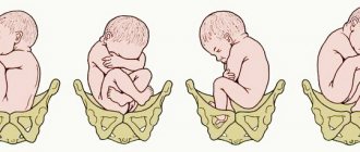
Types of cephalic presentation of the fetus
This position is divided into four types according to the area that is closest to the exit:
- Occipital presentation. This is an ideal scenario. From here, the baby moves through the birth canal with the back of the head forward with the least likelihood of birth injuries. In addition, this reduces the risk of vaginal and perineal ruptures in the mother.
- Facial. The fetal head is tilted back and the face is closest to the exit. It moves face forward along the birth canal, which is dangerous due to injuries to the face and neck.
- Frontal. This is the rarest case of cephalic presentation, occurring in 1-2%. The head is directed with the forehead towards the outlet into the mother's pelvis.
- Forecephalic. The direction of the head is with the top of the head towards the exit. Movement towards the exit with the fontanel forward.
When classifying cephalic presentation, the position of the fetus relative to the walls of the uterus and its own axis is also taken into account - close to the right or left wall, turned with its back or side. Thus, the following positions are distinguished:
- first - the back is closest to the left wall of the uterus;
- the second is the back closer to the right wall of the organ.
Extensor main presentation of the fetus
The causes of extension presentations can be:
- narrowing of the mother’s pelvis (especially the entrance to the pelvis, for example, with a flat pelvis),
- large fetal head size,
- developmental defects (anencephaly, hydrocephalus),
- decreased tone of the tissues of the birth canal and the anterior abdominal wall of the mother, especially in multiparous women,
- pelvic and fetal tumors,
- entwining the fetal neck with the umbilical cord.
The progress of labor slows down, and signs of disproportion between the fetus and the mother’s pelvis may appear.
Facial presentation of the fetus. Facial presentation is observed in 0.2% of births. In this case, the fetal head is maximally extended so that the back of the head is in close contact with the back of the fetus.
The diagnosis of facial presentation of the fetus can be determined by vaginal examination and palpation of the fetus's nose, mouth, eyes or chin. If the fetal chin is directed anteriorly, under the symphysis, vaginal delivery can be successful (posterior view). If the fetus's chin is directed posteriorly or toward the lateral wall of the pelvis, the fetus must rotate its chin anteriorly for vaginal delivery to be possible. Labor stimulation during facial presentation can lead to severe swelling of the fetal face.
Biomechanism of labor during facial presentation . Facial presentation is rarely diagnosed before labor begins. During vaginal examination, a frontal presentation may appear, which turns into a facial presentation with the onset of labor.
The head is inserted into the pelvis with a vertical dimension of 9.5 cm. The facial line is placed in the transverse or oblique dimension of the entrance to the pelvis, the chin and the anterior crown are at the same level. Then, instead of bending, the head extends as much as possible, and the fetal chin drops lower than the anterior crown, and in this position the head enters the pelvis. The internal rotation of the head occurs so that the facial line becomes the straight diameter of the pelvic outlet, and the chin fits under the pubic symphysis. If the chin returns posteriorly (anterior view), the upper limb girdle and fetal head are placed at the same level and cannot pass into the pelvic inlet unless the fetus is too small. In the latter case, labor stops if the chin does not return anteriorly.
With the formation of the posterior view of the facial presentation, the face drops until the chin is delivered, and the angle between the lower jaw and the fetal neck approaches the lower edge of the pubic symphysis. At the point of fixation - the hyoid bone - the head bends, and the forehead, crown, and back of the head of the fetus are sequentially born. The head is cut through a circle corresponding to the vertical size of the head (33 cm). Then there is an external rotation of the head, an internal rotation of the body and the birth of the fetus.
The birth tumor is localized in the area of the fetal mouth and chin; the configuration of the head is dolichocephalic.
Frontal presentation of the fetus . Frontal presentation refers to the presentation of the fetal head by the area that is located above the edge of the orbits. During a vaginal examination with the leading axis of the pelvis, the forehead of the fetus is detected, the frontal suture is placed in the transverse dimension of the entrance to the pelvis; on one side the bridge of the nose and the ocular arches are palpated, on the other - the anterior angle of the anterior fontanel.
Frontal presentation is the middle position between flexion of the head (occipital presentation) and extension (facial presentation). In the case of frontal presentation, the fetal head is inserted into the pelvis with its largest diameter. So, unless the fetal head is very small or the pelvis is not very large, frontal presentation must change to occipital (flexion of the head) or facial (extension of the head) for vaginal delivery.
If the fetus is premature or very small, after installing the fetal head with a frontal suture in the transverse size of the entrance to the pelvis, the head is extended so that the leading point becomes the center of the forehead, which first descends into the birth canal along the pelvic axis.
The internal rotation of the head is carried out by 90 °, and the frontal seam passes from transverse to oblique, and then to the straight size of the pelvic outlet. The upper jaw fits under the lower edge of the pubic symphysis, forming the first point of fixation, around which the head begins to bend and rise to the occipital protuberance. The occipital protuberance is fixed at the apex of the coccyx and forms a second fixation point, around which the head begins to extend, and the upper and lower jaws of the fetus are born. The head passes through the largest outlet of the pelvis - a large oblique diameter. The birth tumor is localized on the forehead, from the eye arches to the corner of the anterior fontanel. The configuration of the head in profile resembles a triangle with the apex at the forehead.
Parietal (anterocephalic) presentation of the fetus . Parietal (anterior cephalic) presentation is characterized by the least degree of extension of the head, compared to facial and frontal presentation.
Biomechanism of childbirth. In case of anterocephalic presentation, the head is inserted into the pelvis with a sagittal suture in the transverse, rarely - in the oblique diameter of the entrance to the pelvis with a straight size (12 cm), in contrast to the small oblique (9.5 cm) in case of occipital presentation. Then the head is slightly unbent so that its leading point becomes the anterior crown.
The anterior parietal bone descends into the pelvis first (anterior asynclitism). The frontal and occipital bones can be shifted under the parietal bones. The internal rotation of the head occurs so that the anterior crown returns to the pubic symphysis. Next, the bridge of the fetal nose fits under the lower edge of the symphysis, forming the first point of fixation.
Around this fixation point, the fetal head bends and the crown and occiput of the fetus are born. Then a second point of fixation is formed - the occipital protuberance, around which the head extends, and the forehead and face of the fetus are born. The head erupts with a straight size of 12 cm and a circumference of 34 cm. The birth tumor is localized in this case in the area of the large fontanelle; the fetal head has a brachycephalic configuration and resembles a tower.
Head presentation with low placentation
Low placentation is a dangerous complication of pregnancy with any fetal presentation, including cephalic presentation. The peculiarity of this pathology is the location of the placenta in the lowest zone of the uterus, closest to the exit. Only then, a little higher is the fetal head. The placenta covers the internal os of the uterus, which causes premature birth. An underdeveloped fetus dies as a result of hypoxia due to insufficiency of placental circulation. The probability of death is 7-25%. Bleeding during placental presentation can cause maternal death in 1-3% of cases. In some cases, the placenta changes position. Then the birth takes place on time without complications.
Low presentation of the fetus
Gynecology
At first they are related to how the baby will develop, and then more terrible experiences appear. As a rule, they are associated with premature birth, the most fear is caused by low presentation of the fetus.
Symptoms
The most common and main symptom is pain in the abdominal area. As a rule, such pain does not go away for a long time. With low presentation, bleeding may occur. Due to bleeding, the fetus is exposed to oxygen starvation. This is very dangerous, as it harms the baby's development.
However, it is worth noting that some are not aware that the fetus is in a low position. This is due to the fact that not every woman experiences symptoms.
This diagnosis can be made by a doctor during an ultrasound scan, since all possible risks become clearly visible there.
Consequences
When a girl is diagnosed with low presentation of the fetus, she must be warned about all the possible consequences that may occur.
Most often, there is a risk of labor starting early. However, they are not only associated with this diagnosis, so their onset can be prevented.
In order to prevent the onset of premature labor, a woman needs to go to the hospital. This is done in order to competently approach solving this problem and prevent all possible risks in a timely manner.
Another consequence of this diagnosis is frequent urination. This problem makes the condition worse. To prevent it, it is necessary to reduce the amount of liquid that the girl drinks.
Also, low presentation can lead to the formation of hemorrhoids. This happens because the baby's head is pressing hard. To prevent the occurrence of this disease, you should eat right and also not do much physical activity.
Adviсe
Girls with this diagnosis need to be constantly monitored by their doctor. And also report any worsening or new symptoms to your doctor so that he can provide timely assistance.
There is a prenatal bandage that helps reduce the risk of premature birth. With the help of such a device, it is possible to reduce the pressure exerted by the head, which helps to avoid premature opening of the uterus.
It is necessary to be regularly monitored by specialists in order to prevent all possible consequences and receive effective treatment in the hospital. Thanks to this, a woman will be able to maintain her own health and the health of her child.
Diagnosis of cephalic presentation of the fetus
Head presentation of the fetus is finally diagnosed during pregnancy at 30 weeks. The position of the fetus is determined at 22 weeks, but it can change before birth - the embryo changes position up to 32 weeks. Palpation and ultrasound methods are used for diagnosis.
A woman can conduct a preliminary diagnosis herself. To do this, you need to lie on your back with your legs bent and place your hand on your lower abdomen. If the position is correct, the baby's head will be slightly palpable. In this case, it is impossible to determine the type of presentation independently, as with palpation. An ultrasound is required.
Routine ultrasound allows you to evaluate the position of the embryo comprehensively. By changing the position of the device's sensor, the doctor receives a detailed picture of the fetus. The position in relation to the walls of the uterus and its pharynx is assessed, and a preliminary diagnosis is made. From this point on, the obstetrician-gynecologist checks the position of the fetus at every examination. At the slightest changes, a referral for additional ultrasound may be possible.
Fetal presentation is cephalic
The cephalic presentation of the fetus is incorrect - the longitudinal position of the fetus with the head facing the entrance to the pelvis. Depending on the presenting part of the fetal head, occipital, anterior cephalic, frontal and facial locations are distinguished. Determining fetal presentation in obstetrics is important for predicting labor. Fetal presentation is determined during examination using special obstetric techniques and ultrasound. Head presentation is the most common and desirable for spontaneous childbirth. However, in some cases (with frontal presentation, posterior type of facial presentation, etc.), surgical delivery or the application of obstetric forceps may be indicated. When the bent head begins to straighten prematurely (at the beginning of labor or even during pregnancy) and in this state moves along the birth canal, they speak of an extensor presentation of the head.
There are 3 degrees:
- I degree, anterocephalic presentation - the head passes through the birth canal in the area of the large fontanel;
- II degree, frontal presentation - the fetus passes through the birth canal with the forehead lowered below the rest of the head;
- III degree, facial presentation - the head is extended so sharply that the leading point becomes the fetal chin.
The biomechanism of labor is similar for all stages; they are characterized by their protracted nature and significantly increased trauma to the mother and fetus.
Birth tactics for various types of cephalic presentation of the fetus
In obstetrics, births that occur with an anterior occipital cephalic presentation (the back of the head is facing anteriorly) are considered correct and prognostically favorable, which helps create an optimal relationship between the size and shape of the head and pelvis of the woman in labor.
In this case, at the entrance to the pelvis, the fetal head is bent, the chin is close to the chest. When moving through the birth canal, the small fontanelle is the leading conducting point. Bending the head somewhat reduces the presenting part of the fetus, so the head passes through the small pelvis in its smaller size. Simultaneously with the movement forward, the head makes an internal rotation, as a result of which the back of the head turns out to be facing the pubic symphysis (anteriorly), and the face is facing the sacrum (posteriorly). When the head erupts, it is extended, then the shoulders rotate internally and the head externally rotates so that the baby’s face is turned toward the mother’s thigh. Following the birth of the shoulder girdle, the baby's torso and legs appear without difficulty.
In the case of labor progressing in the posterior view of the cephalic occipital presentation of the fetus, the back of the head turns toward the sacral cavity, i.e., posteriorly. The forward advancement of the head with a posterior-occipital cephalic presentation of the fetus is delayed, and therefore there is a possibility of developing secondary weakness of labor or fetal asphyxia. Such births are conducted expectantly; in case of weak labor, stimulation is performed; if asphyxia develops, obstetric forceps are applied.
The mechanism of birth with anterior cephalic presentation of the fetus in its main points coincides with the previous version. The conductive point with such a presentation of the head is the large fontanel. The tactics of childbirth are expectant; surgical delivery is undertaken in the event of a threat to the health of the mother or fetus.
With frontal cephalic presentation of the fetus, spontaneous labor is extremely rare and takes a long time with a protracted period of expulsion. With independent childbirth, the prognosis is often unfavorable: complications in the form of deep perineal lacerations, uterine ruptures, formation of vesical-vaginal fistulas, asphyxia and fetal death are common. If a frontal cephalic presentation is suspected or determined, the fetus can be rotated even before the head is inserted. If rotation is not possible, a caesarean section is indicated. In case of complicated spontaneous labor, a craniotomy is performed.
The conditions for a successful independent delivery with a facial cephalic presentation of the fetus are the normal size of the mother's pelvis, active labor, a small fetus, and an anterior view of the cephalic presentation (chin facing anteriorly). Childbirth is conducted expectantly, with careful monitoring of the dynamics of labor and the condition of the woman in labor, fetal heartbeat using cardiotocography and phonocardiography. In the posterior type of facial presentation, when the chin is turned posteriorly, a cesarean section is required; If the fetus is dead, a craniotomy is performed.
Prevention of complications during childbirth with a cephalic presentation of the fetus.
Management of pregnancy in women at risk is associated with an abnormal course of labor. Such women should be hospitalized in a maternity hospital in advance to determine the optimal tactics for childbirth. With timely diagnosis of abnormal position or presentation of the fetus, a cesarean section is most beneficial for the mother and child.
Childbirth with cephalic presentation of the fetus
If the embryo is facing the exit, many doctors recommend a cesarean section, especially if the baby is large. Natural delivery is possible only with a wide maternal pelvis and an embryo weighing up to 3200 grams. Both the activity of labor and the direction of the chin are taken into account.
Frontal presentation is a direct indication for caesarean section. The risk of severe injury to the fetus and damage to the mother is very high. The same applies to anterior cephalic presentation. Due to the fact that the child moves along the birth canal with the fontanel forward, there is a high probability of hypoxia and injury.
Low presentation is an indication for normal natural childbirth. Childbirth does not have any complications and is easy.
Head presentation
Head presentation is determined in approximately 95-97% of cases. The most optimal is the occipital presentation, when the fetal head is bent (the chin is pressed to the chest), and when the baby is born, the back of the head moves forward. The leading point (the one that goes first through the birth canal) is the small fontanelle, located at the junction of the parietal and occipital bones. If the back of the fetal head is facing anteriorly and the face is posterior, this is an anterior view of the occipital preposition (more than 90% of births occur in this position), if it is the other way around, then it is a posterior view. In the posterior form of occipital presentation, childbirth is more difficult; during the birth process, the baby can turn around, but labor is usually longer.
With a cephalic presentation, the pelvic end of the fetus may deviate to the right or left, it depends on which direction the back of the fetus is facing.
There are also extension types of cephalic presentation, when the head is extended to one degree or another. With slight extension, when the leading point is the large fontanelle (it is located at the junction of the frontal and parietal bones), they speak of an anterior cephalic presentation. Childbirth through the natural birth canal is possible, but it takes longer and is more difficult than with an occipital presentation, since the head is inserted into the small pelvis with a larger size.
Therefore, anterior cephalic presentation is a relative indication for cesarean section. The next degree of extension is frontal presentation (it is rare, in 0.04-0.05% of cases). If the fetus is of normal size, delivery through the birth canal is impossible; surgical delivery is required. And finally, the maximum extension of the head is a facial presentation, when the fetal face is born first (it occurs in 0.25% of births). Childbirth through the natural birth canal is possible (in this case, the birth tumor is located in the lower half of the face, in the area of the lips and chin), but it is quite traumatic for the mother and fetus, so the issue is often decided in favor of a cesarean section.
Diagnosis of extensor presentations is carried out during vaginal examination during childbirth.
How to achieve cephalic presentation of the fetus
The location of the embryo in the womb can be corrected. To do this, a pregnant woman needs to rest and do gymnastics. which the doctor recommends. If these measures do not work, the woman is hospitalized in advance and an external inversion of the fetus into a cephalic presentation is prescribed. This is a procedure during which the doctor directs the embryo's head down by rotating the entire body. This manipulation is possible under the following conditions:
- transverse presentation (not done with pelvic presentation);
- the abdominal wall is pliable;
- the pelvis is of normal size;
- mother and fetus are in satisfactory physical condition.
Before the procedure, the woman empties her bladder. To relieve uterine tone and pain, an injection of promedol solution is given. The patient is placed on the couch on her back with her legs and knees bent. The doctor positions himself to the woman's right and places one hand on the area where the fetal head is located. Then he gently moves it towards the cervix, while simultaneously moving the pelvic end to the bottom of the organ. After the procedure, to fix the cephalic longitudinal presentation of the fetus, two bolsters are placed on the woman’s stomach, and the stomach is bandaged on top. With this device, the patient stays in the maternity hospital until delivery.
Attention!
This article is posted for informational purposes only and under no circumstances constitutes scientific material or medical advice and should not serve as a substitute for an in-person consultation with a professional physician.
For diagnostics, diagnosis and treatment, contact qualified doctors! Number of reads: Date of publication: 08/01/2018
Gynecologists - search service and appointment with gynecologists in Moscow
Breech presentation of the fetus
Breech presentation of the fetus occurs in 3–5% of pregnant women. Usually the fetus changes its position several times during the day. In the normal course of pregnancy, by the 22–24th week the fetus is positioned head down; this position remains unstable until approximately the 35th week. If by this time the fetus has established itself in a breech position, then most likely the birth will also take place in a breech presentation. Breech presentation of the fetus is determined by external examination, vaginal examination and ultrasound.
Causes of breech presentation:
- Obstacles to the establishment of the fetal head (abnormal shape of the pelvis, uterine fibroids in the lower segment, placenta previa or its low location, tumors of the ovaries and other pelvic organs).
- Increased fetal mobility with developmental delay, polyhydramnios, prematurity.
- Limited mobility of the fetus due to a short umbilical cord, entanglement of any part of the fetal body with the umbilical cord, oligohydramnios, abnormal shape of the uterus (saddle-shaped or bicornuate uterus, presence of a septum in the uterus).
- Changes in the tone of the uterus (decreased tone of the upper segments and hypertonicity of the lower segment), which is due to the presence of a scar on the uterus and previous inflammatory processes.
Prevention
To prevent breech presentation of the fetus, women at risk are prescribed antispasmodic drugs from the 22nd week of pregnancy. A diet is also prescribed to prevent large fetuses. If a breech presentation of the fetus is detected after the 35th week of pregnancy, the woman is recommended to do special exercises to transfer the fetus to a cephalic presentation.
Breech presentation of the fetus and childbirth
At 38–39 weeks of pregnancy, the woman will be asked to go to the hospital to prepare for childbirth. After the examination, a method of delivery is selected, which depends on the weight and sex of the fetus, the type of breech presentation (breech, leg or mixed), pelvic size, concomitant diseases, and gestational age. Breech presentation of the fetus is an indication for cesarean section if the fetal weight is more than 3500 g, there is a scar on the uterus, placenta previa, anatomically narrow pelvis, foot or mixed presentation of the fetus. It has been established that boys born naturally in a breech presentation can suffer testicular injury during childbirth, so a cesarean section is also preferable for a male fetus. When choosing vaginal delivery, prenatal preparation is carried out, and the woman is prescribed restorative and antispasmodic drugs.
OSTEOPATHY AND breech presentation
The goal of the osteopath is, first of all, to identify and try to eliminate those factors that prevent the child from turning upside down.
The next stage is working with the uterus. Techniques are performed that improve the elasticity of the uterus, as well as improve blood supply, and therefore nutrition of the fetus. As a result, the uterus becomes softer and more spacious, making it easier to turn into a cephalic presentation.
Then the osteopath “listens” to where the child can move most freely, gives him a point of support and invites him to change position. If the baby floats freely, a new support point is created for him, etc.
The main condition ensuring the safety of the procedure is “no violence”; everything is done osteopathically, that is, with respect for the tissues, the body (organisms) as a whole, painlessly, without physical effort, tactfully. The movement is towards greater freedom.
The optimal timing for such a turn is 33-36 weeks of pregnancy. It is possible to do this even at the 38th week under favorable conditions.
Childbirth head first is more physiological, and not every maternity hospital will undertake pelvic birth. Therefore, if the child does not have good reasons to sit with his butt down, it is advisable to invite him to turn his head towards the exit.