Hypoxic-ischemic damage to the central nervous system of perinatal origin - This is a very common diagnosis that parents address their children with. 75-85% of diseases of the nervous system in children have their origins in the early stages of intrauterine development or problems during childbirth. Intrauterine hypoxia and hypoxia during childbirth cause disturbances in the uteroplacental circulation, which may be accompanied by a lack of oxygen in the fetus. Hypoxic disorders in children are often associated with functional imbalance, limitation of blood flow and movement of cerebrospinal fluid. Osteopathy normalizes movements in the body and helps nourish the brain, correcting the negative consequences of hypoxia.
Hypoxia and asphyxia that occur during childbirth and the neonatal period do not go away without leaving a trace and can have a negative impact on the developing brain. The outcome can be a significant loss of neurons, dystrophic changes in nerve cells in the cerebral cortex and in the subcortical structures and ventricles of the brain.
The clinical picture may include the following symptoms:
- decreased or absent sucking reflex
- nervous system damage
- belching
- cerebral palsy
- pyramidal insufficiency
- hydrocephalus
With oxygen deficiency, autoregulation of cerebral circulation is disrupted, neuronal permeability changes, metabolism in nerve cells is disrupted, and harmful amino acids and free radicals accumulate. As a result of the toxic effects of these substances, the death of neurons occurs, and programmed death of nerve cells occurs. Given the progression of neuronal deformation, clinical symptoms may appear later, after several years. This suggests that children after suffering hypoxia should be observed by a neurologist and osteopath.
Diagnosis of hypoxia
Children diagnosed with perinatal posthypoxic damage to the central nervous system are observed by a neurologist for 2 years. After this period, the diagnosis is removed or changed to a more complex one (cerebral palsy, hydrocephalus, etc.).
Methods for diagnosing post-hypoxic state:
- Ultrasound of the brain showing the anatomy of the nervous system
- electroencephalogram (EEG) characterizes neuronal function
- Dopplerography of cerebral vessels characterizes the functions of cerebral vessels, with what speed and symmetry they work and how well they deliver oxygen to the brain.
- MRI is extremely rarely performed on children in the first 2 years, as anesthesia is required for this study.
Ultrasound of the brain reveals the following post-hypoxic changes:
- Cysts (appear after 10-14 days after hypoxia).
- Deformation of the gyri and sulci of the brain.
- Periventricular leukomalacia - necrosis of brain cells around the ventricles of the brain, is an extremely unpleasant symptom in terms of prognosis, often ending in hydrocephalus and cerebral palsy.
- An increase in the size of the cerebral ventricles, interhemispheric fissure and other liquor-containing spaces.
The frequency of studies in children who have suffered hypoxia is once every 3 months to a year and once every 6 months. in the second year of life.
The Apgar score is not the only criterion for hypoxia. For example, in a fetus, in response to a lack of oxygen, the rectal sphincter opens and meconium enters the amniotic fluid, causing it to turn green. Oxygen starvation may also be indicated by the condition of the placenta (premature aging, abnormal development of blood vessels, etc.). Sometimes such children are born with a high Apgar score, but hypoxia still occurs.
The brain of children in the first 2 years of life is very plastic and gladly accepts all treatment methods, responds quickly and produces excellent results. Recently, osteopathic treatment has taken a well-deserved place in the treatment program for children who have suffered from hypoxia.
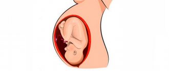
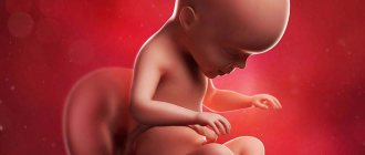
Fetal hypoxia
The resistance of the fetus to hypoxia is determined by the large cardiac output, reaching 198 ml/kg (in a newborn 85 ml/kg, in an adult 70 ml/kg); an increase in heart rate to 150-160 per minute; significant oxygen capacity of the fetal blood (on average 23%); fetal hemoglobin. Oxygen is added to the latter in a short time, and then goes to the tissues, so the tissues receive quite a lot of oxygen per unit time. The percentage of fetal hemoglobin is about 70% in fetal red blood cells.
The fetus is protected from lack of oxygen by the structure of the fetal cardiovascular system. It has 3 arteriovenous shunts: ductus venosus, interatrial foramen ovale, and ductus arteriosus. Due to the connection of arteries and veins, mixed blood enters the fetal organs. Therefore, pO2 in a fetus decreases more slowly than in an adult if a state of hypoxia develops. More than a newborn baby, the fetus uses anaerobic glycolysis. Due to this, it is provided with serious reserves of glycogen, plastic and energy metabolic products in vital organs. In this case, metabolic acidosis develops, which makes the fetus even more resistant to oxygen starvation. Such protective mechanisms are provided by nature.
If a lack of oxygen occurs for some reason, the functions of the medulla and cortical layers of the fetal adrenal glands, the production of catecholamines and other vasoactive substances, first increase. This leads to tachycardia, and the tone of peripheral vessels increases. This, in turn, leads to centralization and redistribution of blood flow:
- blood circulation in the brain increases
- decreased blood flow in the lungs, intestines, kidneys, skin, spleen
- ischemia of the above organs occurs
- cardiac output increases
- blood circulation in the adrenal glands and placenta increases
With intestinal ischemia in the fetus, the anal sphincter may open and meconium may leak into the amniotic fluid. Further, long-lasting severe hypoxia causes a sharp depression of most of the functional systems of the fetus - these are mainly the adrenal glands. Therefore, the level of cortisol and catecholamines in the blood decreases. The vital centers of the fetus are inhibited, blood pressure decreases, and the heart rate decreases.
Severe disturbances in the microcirculatory system also play a role in pathogenesis. The tone of arterioles and precapillaries decreases, which leads to vasodilation. The volume of the vascular bed increases, therefore blood flow slows down; in its extreme form, this can lead to stasis. Due to a decrease in blood flow speed and the appearance of acidosis, blood viscosity, aggregation activity of blood cells and coagulation potential become higher with the development of DIC syndrome and a decrease in gas exchange in fetal tissues.
According to recent studies, nitric oxide plays a role in the pathogenesis of disorders during fetal hypoxia. This is a universal regulator of vascular tone, which is produced by endothelial cells (including those in the umbilical cord and amnion). Nitric oxide is a vasodilator; it inhibits the process of platelet union and prevents their adhesion to the vascular wall.
Disturbances in the cellular nutrition of the vascular wall cause an increase in its permeability. Partial liquid blood and formed elements emerge from the vascular bed. These changes lead to the appearance of:
- hemoconcentration
- hypovolemia
- diapedetic hemorrhages in organs
- tissue swelling
- massive hemorrhages in the vital organs of the fetus (in some cases)
Oxygen deficiency leads to changes in metabolic processes, as a result of which more and more under-oxidized metabolic products accumulate in the body. In the literature this is called pathological metabolic or respiratory-metabolic acidosis. Severe and prolonged fetal hypoxia occurs with the activation of lipid peroxidation, which releases toxic radicals that have an inhibitory effect on enzymatic reactions, disrupt the structural and functional properties of cell membranes, reduce the activity of respiratory enzymes, and increase membrane permeability.
The balance of potassium ions changes, which leave the cellular space, causing hyperkalemia. The latter, together with acidosis and hypoxia, takes place in the structure of the development of overexcitation of the parasympathetic nervous system and the development of bradycardia in the fetus. The above-described significant changes in macro- and microhemodynamics and metabolism lead to the development of necrosis and ischemia in the tissues of the vital organs of the fetus. This primarily occurs in the adrenal gland and the central nervous system.
Consequences of hypoxia
Previously, we looked at the causes and diagnosis of post-hypoxic conditions and agreed that children born with an Apgar score of 7 and below are considered to have suffered hypoxia. This group also includes children with an umbilical cord entanglement, premature babies, etc.
It is extremely important to regularly examine such children over time at certain age periods: 1 month, 3 months, 6 months, 9 months, 1 year. The purpose of examination and observation is to prevent and reduce chronic diseases and reduce the degree of long-term consequences. About 83% of children who have suffered hypoxia have central nervous system lesions in various manifestations and combinations.
There are 3 periods during the course of the disease:
- acute period up to 1 month.
- recovery period from 2 months. up to 2 years
- outcome of the disease - after 2 years.
The most common symptoms of central nervous system damage in the acute and recovery period. Sometimes these symptoms appear in combination with each other:
- Syndrome of increased neuro-reflex excitability.
- The child is very restless, negative to examination, sleep disturbances, muscle tone disorders, etc. are noted.
- Hypertensive-hydrocephalic syndrome.
- Due to the increase in the amount of fluid in the spaces of the brain, intracranial pressure increases. It is characterized by a rapid rate of increase in head circumference, a large fontanel, restless behavior, and frequent regurgitation.
- Vegetative-visceral syndrome.
- Dysfunction of the gastrointestinal tract, frequent regurgitation, frequent bowel movements or constipation, bloating. Dysregulation of vascular tone, which manifests itself as uneven skin coloring (marbling). Irregular breathing and heart rhythm.
- CNS depression syndrome.
- Decreased motor activity, decreased muscle tone, weakened sucking and swallowing reflexes.
- Pyramid syndrome.
It is expressed by hypertnus of the calf muscles, stiffness in the ankle joints, walking on tiptoes, delayed speech development, as well as motor development.
If you have these symptoms, you should not wait for a scheduled examination; it is better to immediately contact a neurologist and osteopath for early diagnosis and treatment.
Consequences of chronic hypoxia for a child
Chronic oxygen starvation is diagnosed less frequently than the acute form, since in most cases a constant lack of oxygen develops only if a woman has an irresponsible attitude towards pregnancy. If a woman walks a lot, eats well, takes care of her health and follows all the recommendations of a specialist, hypoxia usually does not develop. Even if the expectant mother has health problems, the doctor will prescribe medication correction, including drugs to improve blood circulation in the placenta and improve metabolic processes in the fetal tissues.
The consequences of chronic fetal hypoxia are usually detected immediately after birth. The baby may be smaller and smaller than other babies born at this stage. The weight deficit can range from 10% to 30%. Children experiencing a constant lack of oxygen during intrauterine growth do not adapt well to environmental conditions; their vital reflexes (grasping, sucking, etc.) are poorly developed. Thermoregulation in such children is often impaired, so the baby's extremities may remain cold, even if socks are put on the legs and the child is wrapped in a warm blanket.
The most commonly diagnosed disease is anemia. With this pathology, the child looks pale, and blue discoloration may appear in the area of the nasolabial triangle. Other symptoms of pathology in infancy include:
- poor appetite;
- moodiness;
- frequent bouts of crying;
- sleep disorders.
Poor appetite due to low birth weight can lead to retarded physical and intellectual development, as well as diseases that develop due to a lack of certain nutrients. For example, magnesium deficiency can cause problems with the heart, seizures and other neurological pathologies. Insufficient intake of calcium and vitamin D increases the risk of rickets, and a lack of ascorbic acid can lead to diseases of the hematopoietic system.
Important!
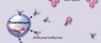
Chronic hypoxia of the fetus during pregnancy negatively affects the child’s immune system: its resistance to infectious diseases decreases, and respiratory infections occur in a more severe form than in ordinary children.
Treatment of hypoxia
Post-hypoxic damage to the central nervous system requires staged treatment after resuscitation measures (if they were necessary), the recovery period begins, the following is applied:
- Exercise therapy
- massage
- physiotherapeutic treatment
- drug therapy (nootropic drugs, vascular drugs, vitamins)
- hydrokinesitherapy (swimming)
- methods such as bobat therapy and vojta therapy are increasingly being used, which are especially useful for children with impaired muscle tone
- osteopathy.
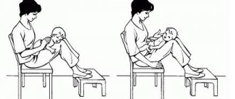
Osteopathic treatment of hypoxia during childbirth
Osteopathic treatment rightfully occupies one of the leading places in the rehabilitation of children who have suffered from hypoxia. Balancing the bones of the skull allows you to relieve tension from the dura mater and the cerebral hemispheres, thereby ensuring the most complete functioning of the central nervous system.
Osteopathic techniques allow for drainage of the venous sinuses, as a result of which cerebrospinal fluid resorption is improved and intracranial pressure is normalized. Freeing the cervical spine and eliminating torticollis promotes adequate blood supply to the brain.
Early osteopathic treatment in the first year of life allows children not only to keep up with their peers in development, but sometimes even to develop ahead of schedule. It is very important to carry out this treatment from the first months of life, as this will help get rid of long-term clinical pathological symptoms. Osteopathy sessions are conducted for children starting from the first month of life. Osteopathic treatment, creating optimal functioning of the central nervous system, stops the process of post-hypoxic changes in neurons, thereby somehow reprogramming the brain for full development.
There is no need to be afraid of hypoxia; you need to take the entire range of measures for rehabilitation after it. Moreover, modern medicine gives us many tools for this.
Future consequences
Experts believe that if a child survives the first month (the newborn period) and no consequences of hypoxia are found in him, the likelihood of their development in the future is quite small. However, this does not mean that pathological symptoms will not appear in the future. Most often, the consequences relate to behavioral characteristics. Such children are usually hyperactive, irritable, and can show causeless aggression towards peers, teachers, and kindergarten teachers.
They may have trouble sleeping. Many of these children suffer from nocturnal enuresis before adolescence. Treatment of the pathology, as a rule, gives minor improvements, but complete recovery is achieved only by the age of 12-15, and in some children this disorder persists in a mild form for life.
Advantages of treating the consequences of labor hypoxia in our Neonatus Sanus medical center
Our clinic of osteopathy and neurology on Vasilyevsky Island “Neonatus Sanus” - health from birth, has extensive practical experience in the prevention and treatment of newborns, infants and infants.
We know how and love to work with young children!
Our clinic employs experienced osteopathic doctors and neurologists. A lot of attention is paid to each child in order to understand the child, accurately assess his condition, give recommendations to parents and, if necessary, carry out effective osteopathic treatment.
In our center you can get the best examination, treatment and recommendations from leading specialists in St. Petersburg.
Clinical example of treatment of the consequences of hypoxia in our osteopathic center
Reviews from our patients about the treatment of hypoxia
Katyusha was born from her first pregnancy at 40 weeks. The condition after birth was severe due to asphyxia. Apgar score 1/4 point. After 3 hours, due to respiratory failure and convulsions, she was transferred to the intensive care unit. Artificial ventilation and brain hypothermia were performed. The result of severe brain hypoxia was the diagnosis: Cerebral Palsy, right-sided spastic hemiparesis. At the age of 1 year 6 months she first appeared in our center. The main complaint was delayed motor development; independent walking appeared only at 1 year and 4 months. When walking, she experienced great problems due to high muscle hypertonicity along the entire right side, strabismus and restless behavior were noted. After the treatment, the girl’s muscle tone was almost normalized, her gait was as close as possible to physiological, the girl has excellent speech development, she enjoys going to kindergarten and communicating with her peers. We thank the parents for providing the video material.
Watch the video review at the link
How dangerous is acute hypoxia?
Acute lack of oxygen occurs most often during labor. The cause may be the application of obstetric forceps, a prolonged period of pushing, or a discrepancy between the sizes of the mother’s pelvis and the fetal head. If acute hypoxia was diagnosed during pregnancy, the woman is immediately hospitalized in the pregnancy pathology department, since in an outpatient setting it is not possible to constantly monitor the condition and heartbeat of the fetus.
One of the most serious and dangerous consequences of acute lack of oxygen is pregnancy fading (cessation of growth and development). Pathology can occur at any stage, but the most dangerous period is considered to be from 4 to 6 weeks and from 8 to 12. It is not always possible to detect freezing immediately - it happens that a woman carries a dead child for several weeks.
You can suspect freezing at an appointment with a gynecologist, who always listens to the heartbeat using an obstetric stethoscope. If heart contractions cannot be heard, the doctor will send the woman for an ultrasound, which can reveal a discrepancy between the size of the fetus and the gestational age. After this, the woman will be prescribed an artificial termination of pregnancy (curettage or artificial birth - depending on the stage of pregnancy).
Intrauterine pneumonia
Pneumonia in a fetus can develop from inhalation of meconium, the original feces. If this happens immediately before birth, the chances of saving the newborn will be maximum. Immediately after birth, the baby will be admitted to the neonatal intensive care unit or intensive care unit. In some cases, artificial ventilation of the lungs using a ventilator may be required. After the acute process has stopped, the child will be transferred to a children's hospital.
Important!
Pneumonia of newborns is an extremely dangerous pathology with a high percentage of fatal cases, so the expectant mother must take all measures to ensure sufficient oxygen supply and reduce the risk of meconium entering the fetus’s lungs.
Intestinal necrosis
A very dangerous pathology that requires urgent surgical intervention. Intestinal necrosis is the death of tissue in a certain part of the large or small intestine as a result of insufficient oxygen supply to the cells of the organ. Even if doctors manage to save the child’s life, there is a high probability that a stoma and colostomy bag will have to be installed to pass stool.
Important!
The mortality rate of newborns from this pathology reaches 71%. In most cases, total necrosis begins to develop after an infarction of the mesentery - the ligament connecting the posterior wall of the peritoneum with the intestinal tube. If the pathology begins to develop before 28 weeks of pregnancy, there is practically no chance of saving the baby.
Pathologies of the nervous system
The most common consequence of acute oxygen starvation, which manifests itself after the birth of a child, is hydrocephalus (swelling of the brain). In most cases, the consequences of the disease are cerebral palsy (cerebral palsy) and spastic tetraparesis - limited mobility of muscle structures due to constant spasticity.
With severe damage to the nervous system, coma can be a consequence of hypoxia. The life prognosis in this case is extremely unfavorable, since almost 90% of cases of comatose lesions in infancy end in the death of the child.
Premature birth
If a child is diagnosed with acute hypoxia, the doctor may decide on the need for emergency delivery, despite the stage of pregnancy. If the gestational age of the fetus is more than 30 weeks, the risks are not so serious, but the possibility of dangerous pathologies and severe forms of developmental delay cannot be excluded. If an emergency caesarean section is performed at less than 28-30 weeks, the baby will be born with extreme prematurity, which is fraught with the following pathologies:
- intellectual development disorder;
- malformations of the heart muscle;
- neurological diseases (including cerebral palsy);
- blindness and hearing loss.
Important!
Only 20% of babies born before 28 weeks survive, and most have severe congenital conditions that affect the baby's quality of life.