According to statistics, every year for every 100,000 babies there are 14-17 sick children. Hemangioma is one of the most common pathological phenomena that occurs during this period. At risk are girls, as well as premature babies and children born with low birth weight. Vascular anomalies can be represented by extensive clinical manifestations - from small pigmentary changes in the skin to huge tumors that affect the limbs and even the internal organs of the child, thereby causing life-threatening conditions.
What is the essence of the disease?
Hemangioma is a benign vascular neoplasm, the formation of which begins during embryogenesis. Externally, it appears as a flat bluish, red or burgundy spot that rises above healthy tissue. The tumor does not have a capsule, and therefore, as it grows, it damages nearby tissues and organs. Characteristic is rapid, progressive growth and a tendency to bleed.
The presence of more than 3 hemangiomas on the skin may indicate their presence on the internal organs of the child.
The factors causing the disease are poorly understood and completely unknown. At the same time, there are a number of reasons that could serve as a predisposing factor to the development of the disease. These include:
- Genetic predisposition. According to statistics, children born into a family prone to the formation of vascular moles have a much higher chance of developing hemangiomas.
- Presence of estrogen receptors in tumor tissue. The hormone acts as a growth stimulator.
- Taking certain groups of medications by the mother during pregnancy.
Contributing factors are also unfavorable environmental conditions suffered by the mother during pregnancy, infectious diseases, and multiple pregnancies.
The neoplasm is not prone to malignancy and, if all the recommendations of the attending physician are followed, with positive dynamics of the pathology, it completely disappears by about 5-7 years. They are not dangerous and do not lead to cancer. At the same time, large vascular growths can lead to organ dysfunction or cause improper development of the body as a whole. Hemangiomas are very easily injured, and therefore there is a risk of infection of the child through an open wound. They also help reduce blood clotting.
Main services of Dr. Zavalishin’s clinic:
- consultation with a neurosurgeon
- treatment of spinal hernia
- brain surgery
- spine surgery
Treatment methods
Dr. Komarovsky writes in his books and talks about this in his programs that it is imperative to treat hemangioma in newborns. However, as he states, surgical or any other method of therapy must be resorted to in cases where the hemangioma begins to grow or does not go away on its own by the age of seven.
First, an experienced doctor chooses observational tactics, since neoplasms often regress on their own, starting to lighten in the middle. If the spot does not shrink or, worse, begins to grow, or is located on the face or near the genitals, it is removed surgically using freezing, cauterization, or other procedures. Small lesions can be treated with conservative methods.
Medication
Small hemangioma can be treated with hormonal therapy, for example, Prednisolone or Propranolol. The baby is given injections and also given hormone tablets. During therapy, the spot becomes light and disappears in eighty percent of cases.
Surgical method
This method is used if conservative therapy has not yielded results, or if the hemangioma is rapidly increasing in size. Depending on the size of the tumor, the anesthesiologist administers general or local anesthesia, after which the surgeon excises the vascular tangle with a scalpel, simultaneously cauterizing the capillaries.
Sclerosis
The method is used only for cavernous hemangiomas. An agent is injected into the middle of the tumor, which seals the vessels from the inside, leading to their death. This method is quite painful and time-consuming. Large tumors may take two to four weeks to heal.
Cryodestruction
Cold treatment is carried out for small tumors that are not localized on the face. The tumor is susceptible to exposure to liquid nitrogen or carbon dioxide. Under cold conditions, frostbite occurs on the skin and blood vessels; after a while, a crust appears at the site of the hemangioma, under which healthy skin forms. In some cases, cryotherapy leaves a small scar.
Radiation therapy
Radiation treatment is justified for large flat tumors and cavernous hemangiomas of subcutaneous localization. In addition, irradiation is used in the treatment of tumors located in the eye. Radiation therapy is not used until six months; after this age, the baby is irradiated in several sessions with breaks from a couple of weeks to six months.
Electrocoagulation
The method of cauterization of hemangioma is used to treat children after three years of age, however, after such therapy, scars on the skin are possible. The procedure is performed on areas of the body that are under clothing.
Treatment with traditional methods
Some people are against modern medicine and try to cure any disease with traditional medicine. Unfortunately, treatment of hemangioma with folk remedies is not justified. Using celandine for lotions can burn the baby’s delicate skin and lead to a secondary infection.
Self-medication is dangerous, especially when it comes to newborn children.
Classification
Depending on the histological structure, pathology is divided into:
- Capillary.
The appearance of neoplasms depends on the degree of involvement of the dermis in the process, the depth, diameter and location of the lesion. The manifestation of the disease is typical already in the first months of a child’s life, as well as complete recovery by 5-6 years (in approximately 30% of cases).
- Cavernous.
They are represented by a limited subcutaneous nodular formation with a soft-elastic consistency. The basis consists of separate caves (cavities) filled with blood. The skin on top is rarely changed. As the tumor grows, the skin acquires a blue-purple tint.
- Mixed.
According to their localization, all hemangiomas are divided into superficial and subcutaneous forms.
Hemangioma
Ulcer
54343 January 20
IMPORTANT!
The information in this section cannot be used for self-diagnosis and self-treatment.
In case of pain or other exacerbation of the disease, diagnostic tests should be prescribed only by the attending physician. To make a diagnosis and properly prescribe treatment, you should contact your doctor. Hemangioma: causes of appearance, symptoms, diagnosis and treatment methods.
Definition
Hemangioma is called vascular hyperplasia (proliferation of cells in the vascular wall) - a disease that is classified as a benign tumor (benign neoplasm) and can be either congenital or appear within a few months after the birth of the child.
Skin hemangioma occurs in adults, but not often, since most tumors resolve on their own in childhood. If a similar tumor appears on the skin of an adult, where there were no surface defects before, then such a tumor requires careful diagnosis.
If in childhood the hemangioma is weakly expressed (in the form of an abrasion or a small spot), then in adulthood under the influence of certain factors it can grow and enlarge.
Hemangiomas form, as a rule, on the scalp, face, neck and, less commonly, on the torso and limbs. Favorite locations on internal organs are the liver, bones, and lungs.
Skin hemangiomas are most often single neoplasms, but there are exceptions. It has been noticed that if there are more than five hemangiomas in visible areas of the body, there is a high probability that there is also a hemangioma of internal organs.
Causes of hemangiomas
The causes of hemangiomas are not fully understood. One theory is that the overgrown endothelial cells may be residual embryonic cells. Another assumption is that the hemangioma begins to grow in utero under conditions of chronic lack of oxygen. It is possible that the formation of a tumor can be provoked by acute infections, including respiratory viral infections, if the mother had any in the first trimester of pregnancy. This is only a theory, because hemangiomas also occur in children born to mothers with normal pregnancies.
It has been noted that hemangiomas are more common in premature babies and in multiple pregnancies.
Threats of miscarriage, placenta previa (a very low location of the placenta in the uterus, making natural childbirth difficult), abruption and inflammation of the placenta, entanglement of the umbilical cord, preeclampsia (a set of symptoms including edema, increased blood pressure and detection of protein in a urine test), maternal age (over 38 years) are also risk factors.
Classification of the disease
Hemangiomas of childhood are divided into congenital and infantile (infantile).
Congenital hemangiomas appear, grow and reach their maximum size in utero.
Often large hemangiomas can be seen during ultrasound screening. A newborn appears with a hemangioma, which can subsequently decrease in size and disappear on its own within a year and a half (rapidly involuting), decrease but not completely disappear (partially involuting) or not change at all (not involuting). If a congenital hemangioma grows, it grows only in proportion to the child’s growth. It occurs with equal frequency in both boys and girls.
Infantile (baby) hemangiomas appear during the first weeks of life.
Infantile hemangioma
This hemangioma is preceded by an area of changed skin or a red spot. The growth of hemangioma can continue up to six months of the child’s life. Then comes a period of growth cessation, which lasts on average two months, and then there is a phase of slow reduction of the tumor, which can last up to ten years.
Infantile hemangioma occurs five times more often in girls than in boys.
Infantile hemangiomas can be located on the skin (simple capillary hemangiomas), under the skin (venous and cavernous hemangiomas) and be combined.
Depending on the histological structure and the caliber of the vessels, hemangiomas are divided into:
- to capillary
- venous,
- cavernous,
- combined.
Capillary or superficial hemangioma of the skin (port-wine stain).
It is located on the surface of the skin and is a dense network of closely intertwined capillaries lined with endothelial cells. This form occurs in 90-96% of cases, is characterized by intensive proliferation of new vessels and is considered the initial stage of development of the formation.
Venous hemangiomas.
The formations are represented by veins of small and medium caliber. Such hemangiomas tend to grow deep into the skin and above its surface.
Cavernous or cavernous hemangiomas.
Cavernous formation is considered the next stage in the development of the venous form of hemangioma. It consists of cavities bounded by the endothelial layer of blood vessels and separated by septa. Such a hemangioma is formed as a result of the overflow of defective capillaries with blood and rupture of their walls with the formation of cavities (cavities) and hemorrhages (hematomas). The blood in these cavities forms small clots due to increased coagulation properties.
Combined hemangiomas.
This type of hemangiomas includes formations that have signs of several forms of varying severity.
Symptoms of hemangioma
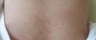
- Capillary or superficial hemangioma of the skin (port-wine stain) is prone to growing into surrounding tissues and is a voluminous elastic formation from pale pink to deep red or burgundy, with uneven outlines, slightly rising above the surface of the skin.
When pressed, the hemangioma turns pale, but then quickly returns to its original appearance. When localized on the back of the head it is called “stork bite”, when localized on the forehead it is called “angel’s kiss”. The edges of the hemangioma indicate the stage of its development: a smooth, defined edge is observed in the growth arrest phase and the tumor shrinkage phase. During active growth, the edges are blurred. - Venous hemangiomas are dark red in color with a blue or purple tint. They are not found often, but they are quite large in area.
- Cavernous or cavernous hemangiomas have the appearance of a soft bluish-purple or purple elastic subcutaneous formation with outlines clearly demarcated from the surrounding tissues and a rough surface through which small vessels are visible. A small part of the formation rises above the surface of the skin, the rest is located deep in the tissue and can penetrate not only into the subcutaneous tissue, but also into the muscles. When pressure is applied for several seconds, the tumor shrinks, its volume and color intensity decrease, which quickly recover after the pressure is removed.
- Combined hemangiomas combine the features of the superficial and subcutaneous forms, with the subcutaneous part occupying a larger volume.
- Hemangiomas of internal organs.
Small hemangiomas of internal organs often do not manifest themselves and are accidentally detected during studies related to other diseases.
In order for a liver hemangioma to cause pain, it must reach an impressive size - 5–10 cm. A hemangioma of the spine does not cause any symptoms if it is located inside a vertebra. Once it affects the periosteum (the membrane covering the bone) or ligaments, constant pain occurs. A tumor that compresses the roots of the spinal nerves can lead to various sensory disturbances in the limbs.
Some hemangiomas immediately cause symptoms, this is explained by the peculiarity of their location. As they grow, they quickly disrupt the function of the organ. These include hemangiomas of the larynx, trachea, and eye tumors.
Diagnosis of hemangioma
Diagnosis of hemangioma begins with asking the patient and/or his representatives about the course of the disease, the first manifestations, and the dynamics of events. Then a detailed examination of the vascular formation is carried out. Depending on the symptoms and location of the hemangioma, an in-depth examination by a specialist (ophthalmologist, otolaryngologist, etc.) may be required.
To clarify the size, depth, and structure of the neoplasm, an ultrasound examination with the study of blood flow is indicated. However, in some hemangiomas it may not be informative.
Features of diagnosis and treatment in children
Examination of tumor growth includes a consultation with a pediatric dermatologist and surgeon, x-ray of the affected area, ultrasound, CT, MRI, angiography and a blood clotting test. Hemostasis indicators are also important.
To treat hemangiomas, the following is carried out:
- electrophoresis with calcium chloride;
- cryotherapy;
- sclerosing therapy;
- radiation therapy;
- surgical intervention.
Depending on the degree of damage and the shape of the hemangioma, treatment tactics are determined. Initially, strict control is established over the tumor, the intensity of growth, the degree of cosmetic defect and the effect on vital organs are assessed.
The use of electrophoresis with calcium chloride in treatment allows the neoplasm to be completely removed without leaving a scar on the body. Treatment takes a long time.
Cryotherapy is carried out using carbon dioxide snow. The use is rational for small diameter tumors. Snow is applied to the affected area, capturing up to 1 cm of healthy tissue. Over time, a bubble forms, covered with a scab.
Sclerosing therapy is carried out with the injection of 70% alcohol and a solution of quinine-urethane. After this, a process similar to cryotherapy occurs. The method is effective when hemangioma is localized on the oral mucosa or upper eyelid.
Radiation therapy is successfully used to treat tumors located on the internal organs of a child. But due to the strong impact on the body, the method has many contraindications, incl. and an age limit - therapy can only be performed on children over 6 months old.
Rapidly progressing tumor growth is a direct indication for surgery. Surgical treatment is carried out only if the operation does not entail a severe cosmetic defect and does not disrupt the function of internal organs.
Treatment of hemangioma
In the vast majority of cases, treatment is surgical, which is performed as early as possible when located on the head and neck, genitals, and anorectal area.
It is necessary to quickly remove those hemangiomas that grow rapidly, increasing the affected area by 2 times within a week. Tumors are immediately removed in case of suppuration, bleeding, ulcerative process, infection, necrosis (death). The traditional scalpel is used, but less and less often. In modern pediatric medicine, the following removal methods are used:
- electrocoagulation – cauterization of the feeding vessel with electric current;
- cryodestruction – destruction by liquid nitrogen and other types of targeted cold exposure;
- laser removal – evaporation of excess tissue with a directed beam of light energy;
- sclerotherapy - administration of drugs that glue the vascular wall;
- embolization – artificial blockage of a vessel feeding a tumor;
- X-ray therapy;
- hormonal treatment.
The surgeon chooses a specific treatment method or a combination of them in order to completely destroy the tumor and obtain a minimal cosmetic defect.
Postoperative scars can completely resolve as the baby grows older. Sometimes, instead of treatment, the surgeon chooses a wait-and-see approach if obvious age-related regression of the hemangioma is noticeable.