Pediatrician, physiotherapist, head of the pediatric medical department
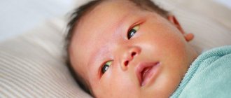
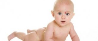
Neonatal jaundice is a change in the color of the skin, conjunctiva and sclera (whites of the eyes) in newborns to a yellowish tint due to increased levels of bilirubin (a pigment that is formed when hemoglobin breaks down) in the blood plasma during the newborn period. Neonatal jaundice is usually mild and transient. However, it is important to monitor newborns with severe jaundice or whose jaundice does not resolve naturally, as this may lead to long-term negative consequences. Severe neonatal hyperbilirubinemia is associated with a neurological dysfunction known as bilirubin-induced neurological dysfunction (BIND). In severe hyperbilirubinemia, unconjugated bilirubin crosses the blood-brain barrier, binds to the basal ganglia and brainstem nuclei, leading to acute bilirubin encephalopathy, or progresses to permanent neurological dysfunction that manifests as cerebral palsy, seizures, buckling, and sensorineural hearing loss.
Signs and manifestations
Conjugation jaundice is usually limited to yellow discoloration of the skin and sclera. This does not pose a danger to the child and goes away on its own.
Too high a level of bilirubin can cause deterioration of the child’s condition, which manifests itself in:
- Weak sucking reflex.
- Increased sleepiness.
- Vomiting.
- Cramps.
- Throwing back the head.
In this situation, a complete examination of the baby is required. Increased bilirubin levels can lead to kernicterus (bilirubin encephalopathy). This condition poses a danger to the newborn, as it provokes serious developmental defects: deafness, mental retardation, paralysis.
If pathological jaundice develops, the child will experience the following symptoms:
- darkening of urine;
- stool lightening;
- enlarged liver;
- abdominal pain when pressed;
- the appearance of hematomas on the baby’s skin.
These symptoms require immediate examination and specific treatment.
What is the nature of jaundice in newborns?
Jaundice or, scientifically speaking, unconjugated hyperbilirubinemia in newborns occurs due to physiological or pathological reasons. More than 75% of cases of neonatal jaundice are due to physiological reasons. Physiological jaundice is also called non-pathological jaundice because it is mild and disappears spontaneously. This occurs due to the peculiarities in the metabolism of bilirubin in the neonatal period. Increased bilirubin load in newborns occurs due to its increased production, which is caused by a higher mass of red blood cells (RBCs) with a decrease in the duration of their existence in newborns. At the same time, a newborn’s liver is still very inactive, and it processes water-soluble bilirubin into conjugated bilirubin, which is water-insoluble, 100 times slower than in an adult.
Physiological jaundice usually occurs between days 2 and 4 of life, peaks between days 4 and 5, and resolves within 2 to 3 weeks. Physiological jaundice never occurs in the first 24 hours of a newborn's life.
What determines the number of procedures?
The minimum value of bilirubin is an indication for the first procedure in a newborn (the child is affected by pathological factors and there is a risk of encephalopathy). Body weight determines the bilirubin level, which affects the number of phototherapy procedures prescribed. If the newborn's body weight is less than one and a half kilograms, the permissible rate of bilirubin is from 85 to 140 µmol/l. The weight of a child up to 2 kg determines the limit dose of bilirubin - 200 µmol/l. With a weight of more than 2.5 kg, the critical value of this indicator is 295 µmol/l. Average indicators may be complicated by concomitant diseases; before the procedure is prescribed, the newborn is fully examined.
Best materials of the month
- Coronaviruses: SARS-CoV-2 (COVID-19)
- Antibiotics for the prevention and treatment of COVID-19: how effective are they?
- The most common "office" diseases
- Does vodka kill coronavirus?
- How to stay alive on our roads?
What is pathological jaundice of newborns?
The causes of pathological unconjugated hyperbilirubinemia are also associated with an increase in bilirubin production, a decrease in its excretion from the body and increased blood circulation in the liver and intestines. Pathological jaundice can occur in a newborn in the first 24 hours of life and is characterized by a rapid increase in bilirubin levels of more than 5 mg/dL per day.
The causes of increased bilirubin production in pathological jaundice are immune-mediated hemolysis (destruction of red blood cells) arising due to Rh conflict with the mother, blood group incompatibility or non-immune-mediated causes such as cephalhematoma, defects in red blood cell membranes (hereditary spherocytosis and elliptocytosis), defects enzyme system (glucose-6-phosphate dehydrogenase and pyruvate kinase), which most often affects boys.
Incompatibility of blood groups according to the ABO system occurs in mothers with blood group O, who have IgG antibodies to factors A and B, which penetrate the placenta into the child’s body and cause hemolysis in newborns with blood group A or B. In case of Rh incompatibility, Rh- a negative mother exposed to Rh positive blood from a previous pregnancy becomes sensitized to such blood. This leads to hemolysis in a fetus with Rh-positive blood. To prevent Rh conflicts, anti-D gamma globulin is currently used, which reduces the incidence of hemolysis.
Decreased bilirubin clearance is also observed with hereditary diseases such as Crigler-Najjar and Gilbert syndrome, as well as with diabetes in the mother or congenital hypothyroidism in the baby. For Crigler-Najjar syndrome, newborns may require liver transplantation or long-term use of phototherapy. In Gilbert syndrome, a mutation of the UGT1A1 gene is observed, causing unconjugated hyperbilirubinemia. Gilbert syndrome is usually diagnosed during adolescence, although the first manifestations may occur as early as the neonatal period. It can be diagnosed using genetic testing.
What is it and what causes it?
If the yellowness does not go away within 2-3 weeks, then there is cause for concern.
Conjugative jaundice of newborns is a condition that arose during the process of adaptation of the child to new living conditions.
In the baby’s blood, fetal hemoglobin is replaced by hemoglobin A, which is formed as a result of the exchange; due to the immaturity of the liver, bilirubin accumulates in the tissues and turns the skin yellow.
The yellowness is most pronounced on the 3-4th day of life; gradually, as the enzyme systems work, the yellowness disappears on the 8-9th day. If jaundice does not go away within 2-3 weeks, then they speak of a protracted conjugative form.
This condition is dangerous, since excess bilirubin leads to disruption of the central nervous system.
Risk factors for the development of prolonged jaundice include:
- poor maternal nutrition during pregnancy;
- lack of iodine in the mother's body;
- bad habits of women;
- taking certain medications during pregnancy;
- poor environmental conditions;
- mother's diabetes.
Prolonged jaundice requires examination of the child by a specialist in order to differentiate it from pathological jaundice, which can be of the following types:
| Hemolytic | Occurs due to the accelerated breakdown of red blood cells in the child’s blood |
| Parenchymatous | Is a consequence of infectious liver diseases |
| Obstructive | Appears due to disruption of the bile outflow process |
If conjugation jaundice is a physiological condition and resolves within 10 days, then pathological jaundice requires immediate treatment.
Reasons for appearance
Conjugation jaundice may appear due to uncontrolled use of medications that lead to disruption of liver function. Also causes of jaundice in adults are:
- Gilbert-Meulengracht syndrome;
- liver tumors;
- gallstones.
The following pathologies contribute to the occurrence of prolonged jaundice in newborns:
- Kligler-Najjar syndrome. Characterized by hereditary deficiency of enzymes that process bilirubin.
- Breast milk jaundice. If breast milk contains an increased amount of estrogen and other substances that slow down the absorption of bilirubin. To normalize the child’s condition, it is recommended to stop breastfeeding.
- Congenital endocrine diseases associated with decreased synthesis of thyroid hormone.
- Incompatibility of the Rh factor of mother and child.
- Prescribing medications : chloramphenicol, vitamin K, salicylic acid.
Jaundice due to breastfeeding disorders
A decrease in intestinal activity in a newborn leads to increased enterohepatic circulation. Jaundice in the absence or insufficient breastfeeding and intestinal obstruction are common conditions associated with increased enterohepatic circulation, which leads to unconjugated hyperbilirubinemia. Non-breastfeeding jaundice occurs in the first week of life and is caused by insufficient breast milk intake, which leads to dehydration and sometimes high sodium levels in the baby. Lack of breastfeeding leads to decreased intestinal motility and decreased excretion of bilirubin in feces or meconium. This jaundice occurs at the end of the first week, peaks in the second, and usually resolves by 12 weeks of age.
Jaundice resulting from impaired bile transport
Conjugated hyperbilirubinemia is always a pathology and occurs due to defects in the formation or transport of bile caused by mechanical obstructions to its outflow or systemic diseases that can affect the liver. Such conditions include biliary atresia, common bile duct cysts, idiopathic neonatal hepatitis, and Alagille syndrome. Congenital metabolic disorders such as galactosemia, tyrosinemia, antitrypsin-1 deficiency also manifest themselves in the form of conjugated hyperbilirubinemia. Biliary atresia is the most common cause of conjugated neonatal hyperbilirubinemia. It affects both intrahepatic and extrahepatic bile ducts and usually appears during the first 2–4 weeks of life along with pale stools. Initial diagnosis is by ultrasound, which may show the absence of the gallbladder. Ultrasonography can also detect cysts with normal or dilated intrahepatic bile ducts, or sclerotic ducts in biliary atresia.
Jaundice due to infectious diseases
Systemic infections such as toxoplasmosis, syphilis, varicella, rubella, cytomegalovirus and herpes simplex, and systemic conditions such as sepsis, shock and birth asphyxia may also present as jaundice caused by conjugated hyperbilirubinemia. Neonatal urinary tract infections and long-term use of parenteral nutrition in preterm infants are also known causes of conjugated hyperbilirubinemia.
How common is jaundice in newborns?
Almost all newborns will have a total bilirubin level above the upper limit of normal for adults and older children of bilirubin of 1.5 mg/dL with less than 5% total conjugated bilirubin. Up to 60% of full-term infants and 80% of newborns at 35 weeks' gestation or more will have jaundice, which occurs when plasma bilirubin levels exceed 5 mg/dL. Neonatal jaundice is more common in people living at high altitudes and in people living around the Mediterranean Sea, especially in Greece.
How is jaundice diagnosed?
Diagnosis of a newborn with jaundice begins with a detailed history, including birth history, family history, and onset of jaundice. A complete examination of the newborn should include general appearance, eye examination, abdominal assessment for hepatomegaly, splenomegaly or ascites, neurological examination and assessment of skin rashes. Bilirubin levels can be assessed using a transcutaneous measurement device or blood draw to determine total serum or plasma bilirubin levels. Transcutaneous measurement reduces the frequency of bilirubin blood tests, but is not possible after phototherapy. In addition, if the transcutaneous bilirubin level is greater than the 95th percentile on the transcutaneous nomogram or the 75% of the total serum bilirubin nomogram for phototherapy, the total serum bilirubin level should be measured.
Ultrasound and additional tests such as antibody titers for infection, urine culture, viral culture, serologic titers, amino acids, and α-antitrypsin phenotype may be added to the list of investigations in the case of a suspected diagnosis of conjugated hyperbilirubinemia.
Laboratory testing of the mother's blood will also be required to distinguish between unconjugated and conjugated jaundice. The American Academy of Pediatrics recommends universal screening of all newborns for jaundice and identification of risk factors for severe hyperbilirubinemia. Major risk factors in newborns over 35 weeks include high bilirubin levels, jaundice observed in the first 24 hours of life, blood type incompatibility, gestational age between 35 and 36 weeks, previous siblings who have received phototherapy, cephalhematoma or significant bruising, absence of breastfeeding feeding and East Asian race. Prematurity is also a known risk factor for the development of severe hyperbilirubinemia.
Minor risk factors include maternal diabetes, polycythemia, male gender, and maternal age over 25 years.
Indications for phototherapy
Children who need phototherapy undergo a whole course, after which the newborn undergoes a secondary examination. Infants who are at risk of developing a hemolytic disease, as well as newborns with a perinatal risk of hyperbilirubinemia (this risk is determined during pregnancy), are subject to this treatment:
- increase in antibody titer in a woman;
- signs of hydrops fetalis, which are visible on ultrasound;
- The first blood group of the mother, creating the likelihood of developing jaundice in the newborn.
Infants with signs of immaturity (multifunctional) and premature children are required to undergo a course of phototherapy. The indication for this type of therapy is hemorrhage under the skin (extensive or multiple). Newborns who require resuscitation undergo phototherapy as prescribed by the doctor. The likelihood of hemolytic anemia appears to be a direct indication for phototherapy - for these cases, a complete history of the child’s parents is needed, which will show how dangerous anemia is for the fetus.
The indication for a treatment procedure with a phototherapy device is the level of bilirubin in the child’s blood from 50 to 68 µmol/l - such newborns include children at risk of developing anemia. Pathological jaundice is the most common reason for which a course of phototherapy is prescribed.
How is jaundice treated?
To prevent acute bilirubin encephalopathy and kernicterus, severe hyperbilirubinemia is treated with phototherapy, intravenous immunoglobulin, or exchange transfusion. There are nomograms for determining bilirubin levels at which phototherapy and replacement blood transfusion are indicated.
Phototherapy is initiated taking into account risk factors and serum bilirubin levels on a nomogram. Bilirubin optimally absorbs light in the blue-green range (460 to 490 nm) and is either photoisomerized and excreted in bile or converted to lumirubin and excreted in urine. During phototherapy, the newborn's eyes should be protected and as much of the body's surface area should be illuminated as possible. It is important to maintain hydration and urine output because most bilirubin is excreted in the urine as lumirubin. The use of phototherapy is not indicated for conjugated hyperbilirubinemia and may result in “bronze baby syndrome” with grayish-brown skin discoloration.
After cessation of phototherapy, there is an increase in total serum bilirubin levels, known as “reverse bilirubin.” The level of "reverse bilirubin" is usually lower than the level at the beginning of phototherapy and does not require restarting phototherapy.
Intravenous immunoglobulin is recommended if bilirubin levels increase as a result of isoimmune hemolysis despite phototherapy. Intravenous immunoglobin is started when bilirubin levels are between 2 and 3 mg/dL.
Replacement blood transfusion is indicated if there is a risk of neurological dysfunction, either with or without phototherapy. It is used to remove bilirubin from the bloodstream and, in isoimmune hemolysis, also removes circulating antibodies and sensitized red blood cells. Blood transfusions are performed in neonatal or pediatric intensive care units. A double exchange blood transfusion (from 160 to 180 ml/kg) is performed to replace the newborn’s blood. Phototherapy should be restarted and continued after blood transfusion until bilirubin reaches a safe level.
The clinical lecture presents the main causes and types of neonatal jaundice, stages of differential diagnosis and treatment.
Hyperbilirubinemia is the most common condition in the neonatal period [1, 4, 7, 11]. In total, there are about 50 diseases that are accompanied by the appearance of yellowness of the skin. Neonatal jaundice is most often physiological in nature, is a transient condition and does not require treatment. At the same time, this may be a symptom of a serious disease that requires timely diagnosis and therapy [3, 6]. The most dangerous complication of indirect bilirubinemia is the development of a neurotoxic effect, leading to severe neurological complications [5, 9]. This most often occurs in premature babies and children in the first days of life. The task of a pediatrician is to timely assess the child’s condition and exclude pathologies that require more detailed examination and treatment.
Bilirubin exchange
Bilirubin is the end product of heme catabolism and is formed mainly due to the breakdown of hemoglobin (about 75%) with the participation of heme oxygenase, biliverdin reductase, as well as non-enzymatic reducing substances in the cells of the reticuloendothelial system.
The natural isomer of bilirubin, indirect free bilirubin, is highly soluble in lipids but poorly soluble in water. In the blood, it easily enters into a chemical bond with albumin, forming a bilirubin-albumin complex, due to which only less than 1% of the resulting bilirubin enters the tissues. In combination with albumin, bilirubin enters the liver, where it penetrates the cytoplasm through active transport and is transported to the endoplasmic reticulum. There, under the influence of uridine diphosphate glucuronyl transferase (UDPGT), bilirubin molecules combine with glucuronic acid. Conjugated bilirubin is water-soluble, non-toxic and is excreted from the body in bile and urine. Next, direct bilirubin is excreted into the bile capillaries and excreted along with bile into the intestinal lumen. In the intestine, under the influence of intestinal microflora, further transformation of bilirubin molecules occurs, resulting in the formation of stercobilin, which is excreted in the feces.
In newborns, bilirubin metabolism has a number of features:
1) a relatively larger amount of hemoglobin per unit of body weight;
2) shorter life expectancy of red blood cells with fetal hemoglobin - 70-90 days (while in adults 120 days)
3) the binding of bilirubin to albumin is reduced, especially in premature infants;
4) UDFGT activity is sharply reduced in the first day of life;
5) increased intestinal reabsorption of bilirubin from the intestine.
The consequence of this is the development of a number of conditions characteristic of the neonatal period.
All jaundices are usually divided according to the level of block of bilirubin metabolism into:
1) suprahepatic (hemolytic), associated with increased breakdown of red blood cells, when liver cells are not able to utilize large amounts of bilirubin formed in an avalanche;
2) hepatic (parenchymal), associated with the presence of an inflammatory process that disrupts the functions of liver cells;
3) subhepatic (mechanical), associated with impaired bile outflow.
Indirect bilirubinemia
During the newborn period, several types of unconjugated (indirect) bilirubinemia may occur, which have a benign course and do not affect the development of the child in the future [1, 4, 5]. These include physiological jaundice, breastfeeding jaundice and breast milk jaundice. Physiological jaundice is observed in most newborns and has the following symptoms: the appearance of jaundice at the age of more than 36 hours of life; the hourly increase in bilirubin should not exceed 3.4 µmol/l h (85 µmol per day); the maximum bilirubin level does not rise above 204 µmol/l; the general condition of the child is not disturbed.
Early increase in physiological jaundice due to lack of breast milk is called breastfeeding jaundice. In this case, there is an increase in bilirubin levels above 184 µmol/l, and the duration of jaundice can be from 3 weeks to 3 months. Stopping breastfeeding (for 24-48 hours) leads to a decrease in bilirubin [2, 6]. It must be emphasized that bilirubin levels <354 µmol/L in healthy full-term newborns do not adversely affect the development of the child.
Rare causes of indirect bilirubinemia can be: Gilbert's syndrome, Crigler-Najjar syndrome [7]. Conjugation jaundice can be observed with hypothyroidism, polycythemia, pyloric stenosis and high intestinal obstruction, and can also be a symptom of hereditary metabolic disorders (galactosemia, fructosemia, tyrosinemia) [6, 11].
All hemolytic jaundices are characterized by the presence of a symptom complex, including jaundice on a pale background (lemon jaundice), enlarged liver and spleen, increased serum levels of indirect bilirubin, and normochromic anemia of varying severity with reticulocytosis [1, 6]. The severity of the child’s condition is always determined not only by bilirubin intoxication, but also by the severity of anemia.
Hemolytic disease of newborns occurs as a result of incompatibility of the blood of mother and child according to the Rh factor, its subtypes or blood groups. The disease occurs in the form of edematous, icteric and anemic forms. The edematous form is the most severe and is manifested by congenital anasarca, severe anemia, and hepatosplenomegaly. As a rule, such children are not viable. Jaundice and anemic forms of the disease are more favorable, but can also pose a threat to the child’s health. In the clinical picture, jaundice is either congenital or appears during the first day of life, has a pale yellow (lemon) tint, and steadily progresses, against the background of which neurological symptoms of bilirubin intoxication may appear. Hepatosplenomegaly is always noted. Changes in the color of stool and urine are uncharacteristic. Damage to the structures of the central nervous system (CNS) occurs when the level of indirect bilirubin in the blood serum of full-term newborns increases above 342 µmol/l. For premature babies, this level ranges from 220 to 270 µmol/l. However, it must be remembered that the depth of damage to the central nervous system depends not only on the level of indirect bilirubin, but also on the time of its exposure in the brain tissue and concomitant pathology, aggravating the serious condition of the child [3, 4].
The causes of hemolytic anemia in the neonatal period can be hereditary hemolytic anemia (Minkowski-Choffard anemia, infantile pycnocytosis, hemoglobinopathies).
Large hematomas in the neonatal period can also cause severe indirect hyperbilirubinemia and anemia.
Direct bilirubinemia
With conjugated bilirubinemia, the level of direct bilirubin is more than 15% of the total bilirubin level. Regardless of the cause, direct hyperbilirubinemia is accompanied by the development of cholestasis [1, 3, 11]. Cholestasis results from structural and functional damage to the hepatobiliary system and can often be a sign of severe and progressive disease. The cause of cholestasis can be both infectious and toxic liver damage and mechanical disturbances in the outflow of bile. Obstructive jaundice has a greenish tint and is accompanied by an increase in the size of the liver, a change in the color of the stool (discoloration) and urine (increasing color intensity). Factors in the development of obstructive jaundice in the neonatal period may include malformations of the biliary tract: intra- and extrahepatic atresia of the bile ducts, polycystic disease, torsion and kinks of the gallbladder, arteriohepatic dysplasia, Alagille syndrome, syndromic reduction in the number of interlobular bile ducts.
Infectious liver lesions can be caused by viruses, bacteria and protozoa: hepatitis B and C virus, cytomegalovirus, Coxsackie, rubella, Epstein-Barr, herpes simplex virus, treponema pallidum, toxoplasma, etc. [3, 5]. The clinical picture of parenchymal jaundice includes a number of common signs: children are often born premature or immature, with intrauterine growth retardation, and have signs of damage to several organs and systems. Jaundice is already present at birth and has a grayish, “dirty” tint, against the background of severe microcirculation disorders, often with manifestations of cutaneous hemorrhagic syndrome. Hepatosplenomegaly is characteristic.
Diagnostics
Diagnostic measures for neonatal jaundice should take into account a number of provisions:
1) when collecting anamnesis, it is necessary to pay attention to the possible familial nature of the disease: cases of prolonged jaundice, anemia, splenectomy in parents or relatives are important;
2) the mother’s medical history must necessarily contain information about the blood type and Rh factor of her and the child’s father, the presence of previous pregnancies and births, operations, injuries, blood transfusions without taking into account the Rh factor. During pregnancy, a woman may be diagnosed with impaired glucose tolerance, diabetes mellitus, and an infectious process. It is also necessary to find out whether the woman has taken drugs that affect bilirubin metabolism;
3) the newborn’s medical history includes determining the gestational age, weight and height indicators, Apgar scores at birth, determining the nature of feeding (artificial or natural), the time of appearance of icteric staining of the skin;
4) physical examination helps determine the shade of jaundice. The presence of cephalohematomas or extensive ecchymoses, hemorrhagic manifestations, edematous syndrome, and hepatosplenomegaly is determined. You should pay attention to the color of urine and stool. An important diagnostic point is the correct interpretation of the child’s neurological status;
5) laboratory methods include a clinical blood test with determination of hematocrit, peripheral blood smear, determination of blood group and Rh factor in the mother and child;
6) biochemical blood test at the first stage: determination of total bilirubin and its fractions, level of liver transaminases, alkaline phosphatase, concentration of total protein, albumin, gammaglutamyl transpeptidase (used to detect cholestasis) [1, 8];
7) ultrasound examination of the liver and gallbladder, as well as magnetic resonance cholangiography if indicated;
 duodenal test (absence of bile within 24 hours during duodenal intubation indicates the presence of biliary atresia) [1].
Treatment
Treatment methods for bilirubinemia can be divided into 3 groups:
1) preventing the increase of bilirubin in the blood serum;
2) promoting the excretion of bilirubin;
3) elimination of the main cause of the pathological increase in bilirubin levels.
Replacement blood transfusion is performed when conservative methods of therapy are ineffective, there is a progressive increase in bilirubin levels, and in the presence of absolute indications, that is, when there is a threat of developing kernicterus. Exchange transfusion is performed in the amount of two volumes of circulating blood, which allows replacing up to 85% of circulating red blood cells and reducing the bilirubin level by 2 times. The current indications for this procedure are: edematous-anemic form of hemolytic disease of the newborn, when transfusion is performed in the first 2 hours of life; the level of total bilirubin in umbilical cord blood is above 76.5 µmol/l; cord blood hemoglobin level is below 110 g/l; hourly increase in bilirubin above 17 µmol/l; with hemoglobin 110-130 g/l [1,10].
Phototherapy at the present stage is the most effective method of treating indirect hyperbilirubinemia. The essence of phototherapy is the photoisomerization of indirect bilirubin, that is, its transformation into a water-soluble form. Currently, there are several types of blue light lamps, with a wavelength of 425-475 nm. Phototherapy begins when there is a threat of bilirubin growth to a toxic value. The optimal phototherapy regimen is alternating 2-hour irradiation with a 2-hour break. In case of liver diseases and obstructive jaundice, phototherapy is contraindicated [2, 4].
Indications for infusion therapy are conditions that require additional fluid administration: vomiting and regurgitation syndrome, fluid loss during phototherapy, as well as other pathological losses.
The feasibility of using inducers of microsomal liver enzymes (phenobarbital) is currently a controversial issue, since enzyme induction reaches an effective value by the end of the second week of life, when the risk of bilirubin encephalopathy decreases [1, 3].
Enterosorbents (smecta, polyphepan, enterosgel, etc.) are included in therapy in order to interrupt the hepatic-intestinal circulation of bilirubin. However, they do not have a significant effect on serum bilirubin levels.
Of the choleretics and cholekinetics for cholestasis (with the exception of atresia of the extrahepatic bile ducts and impaired synthesis of bile acids due to fermentopathy), magnesium sulfate and allochol can be used. Currently, preference is given to the drug ursodeoxycholic acid - Ursofalk, which is available in the form of a suspension, is easy to dosage for newborns, and is characterized by a rapid and clear therapeutic effect [2, 6].
Thus, the main task of a pediatrician in case of neonatal jaundice is to conduct a comprehensive analysis of the causes, clinical and laboratory status and choose the optimal tactics for managing the child.
E.V. Volyanyuk, A.V. Kuznetsova
Kazan State Medical Academy
Volyanyuk Elena Valerievna - Candidate of Medical Sciences, Assistant of the Department of Pediatrics and Perinatology
Literature:
1. Neonatal jaundice: a manual for doctors. M.: Publishing House "Medpraktika-M", 2004, 52 p.
2. Dementyeva G.M., Veltishchev Yu.E. Prevention of adaptation disorders and diseases of newborns: lecture for doctors. M., 2003. 75 p.
3. Volodin N.N. Current problems of neonatology. M.: Geotar, 2004
4. Anastasevich L.A., Simonova L.V. Treatment Doctor 2006; 10: 33-38.
5. Abramchenko V.V., Shabalov N.P. Clinical perinatology. Petro, 2004. 424 p.
6. Diseases of the fetus and newborn, congenital metabolic disorders. Ed. R. E. Berman, V. K. Vaughan. M.: Medicine, 1991. 527 p.
7. Degtyarev D.N., Ivanova A.V., Sigova Yu.A. Crigler-Najjar syndrome. Russian Bulletin of Perinatology and Pediatrics 1998; 4: 44-48.
8. Komarov F.I., Korovkin B.F., Menshikov V.V. Biochemical studies in the clinic. M.: APP "Dzhangar", 2001.
9. Guide to pharmacotherapy in pediatrics and pediatric surgery. Neonatology. Ed. A. D. Tsaregorodtseva, V. A. Tabolina. M.: Medpraktika-M, 2003.
10. Balistreri WF Nontransplant options for the treatment of metabolic liver disease: saving livers while saving lives. Hepatology. 1994; 9: 782-787.
11. Finegold MJ.|Common diagnostic problems in pediatric liver pathology. Clin Liver Dis/ 2002; 6(2): 421-54
Prognosis for jaundice
With timely diagnosis and treatment, the prognosis is positive. In patients with delayed treatment, brain damage is a serious complication. Until now, many doctors (and parents) do not fully understand that jaundice (or as it is sometimes disparagingly called “jaundice”) in newborns is not a benign disease, and requires close attention. The fact is that jaundice in a newborn is a serious disease that can cause irreversible brain damage. All physicians caring for newborns should be aware of this fact. Although many conditions that cause jaundice cannot be immediately diagnosed, the key to preventing complications is to educate parents. Parents should be informed by nurses, pediatricians, obstetricians and the attending physician that if the child's skin, stool or urine color changes, the child should be examined immediately at the clinic. Today, there are smartphone apps that help parents identify jaundice. The main thing is to ensure that the baby is examined in a clinic to rule out any malignant cause of jaundice. Only through the cooperation of parents and doctors can the incidence of jaundice in newborns be reduced.
When does jaundice go away?
Conjugation disorders are rarely protracted; after 7–10 days, the production of enzymes normalizes, and the concentration of bilirubin decreases to normal levels. In premature infants, jaundice persists longer, up to 21 days. If the skin color remains yellow, the concentration of toxic pigment does not decrease - complications of conjugation jaundice are possible. They are treated for up to 4 months.
This is interesting: Tests for jaundice: biochemistry, general blood test, urine test, coprogram
Timely removal of intoxication avoids complications. Much depends on the nursing mother: the more often she puts the baby to the breast, the faster he will recover. With rare genetic pathologies of conjugation jaundice, relapses are possible; failure of enzyme production can occur at any age with infectious lesions of the parenchyma.

