Head of the pediatric department, pediatrician of the first category of children's medical
Infection with pinworms E. vermicularis (enterobiasis) remains a pressing problem, despite the fact that the level of infection has decreased significantly with the improvement of housing and sanitary and hygienic living conditions in Russia. Although pinworm infection occurs without consequences or even asymptomatically in approximately 40% of cases, enterobiasis is often associated with significant psychological stress for both babies and their parents. Lack of knowledge about the spread of parasites and how it can be prevented, unsuccessful attempts at treatment, as well as a high rate of relapses and autoinfection can lead to resignation to the disease on the part of patients or their parents.
The estimated rate of infection with E. vermicularis worldwide is more than one billion people. Pinworm infection is common in temperate climates and industrialized countries, where it occurs at all levels of society. On average, about 18-25% of children aged 4 to 10 years can be infected with pinworms.
How to take it
Do not take a bath or shower before taking a scraping. Other hygiene procedures in the anus area are also unacceptable. Collection of biomaterial for research is carried out only in the morning (before 10:00), before defecation.
How is the analysis taken?
No special preparation is required for the study; the scraping itself can be taken at home. The scraping from the perianal folds is taken with a special spatula into a container. The order is as follows:
- Obtain a special container with a spatula from any laboratory department;
- Remove the spatula from the container and press the adhesive part against the perianal fold;
- Place the biological material in the container, screw the lid tightly on;
- Bring the container to the laboratory department no later than 3 hours after collecting the biomaterial.
How much is done
Usually the result of scraping for enterobiasis can be obtained within 1 day
.
What are pinworms?
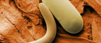
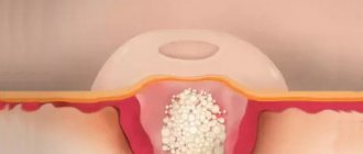
Enterobius (Oxyuris) vermicularis or pinworm is a pathogenic intestinal parasite of humans belonging to the genus Nematoda. Symptomatic pinworm infection is called enterobiasis (older term: oxyuriasis). The first description of worm eggs was made by the Swedish scientist Carl Linnaeus in 1758. It can be assumed that E. vermicularis has been successfully parasitizing the human body since ancient times. These worms have a characteristic "thread-like" appearance. Females are 9–12 mm long and approximately 0.5 mm in diameter, while males are shorter, 3–5 mm long, but visible to the naked eye due to their greater thickness. They are colored a bright whitish-beige color, have a round shape and move by energetic crawling. The extensive reproductive system of a fertilized female worm is often completely filled with eggs (>10,000 eggs per worm). Like all nematodes, E. vermicularis has a thick outer protective covering (cuticle). Their eggs are two-layered, elongated oval, asymmetrical and translucent, similar to a loaf of bread. The first larval stage in the egg can often be clearly visualized. Parasite eggs can remain viable for up to five days outside the body.
Life cycle of pinworms
Pinworms have a simple life cycle that takes place in the lumen of the gastrointestinal tract. Infection occurs through ingestion of eggs. When it enters the body's digestive system, the outer shell of the eggs softens. After passing through the pylorus of the stomach, pinworm larvae hatch in the small intestine. After a double molt, the parasitic worms copulate. The worms then penetrate the lumen of the colon, where they are found in large numbers, especially in the cecum, appendix or ascending colon. After copulation, males die. Pregnant females live up to one hundred days and migrate to the anus. The interval between ingestion of eggs and oviposition by an adult female pinworm ranges from 2 to 6 weeks. Oviposition occurs mainly when the child is resting, mainly at night when he is sleeping. In this case, the females leave the anus and attach their eggs to the tissues of the anal and perianal area using an adhesive layer. The migratory movements of the worms during this process often cause unpleasant itching, causing children to try to relieve the itching by scratching the anal area with their fingers. Breakdown of the worm's cuticle due to scratching results in massive release and spread of eggs into the underwear and surrounding areas. The infected hands of the host play an important role in maintaining the chain of infection (autoinfection). Hatched larvae may also retrogradely burrow into the anus, causing re-infection (retrograde infection). Eggs always need suitable conditions outside the gastrointestinal tract to mature. There is no intraluminal spread of eggs.
Risk factors and methods of transmission of pinworms
A number of studies have identified the main risk factors for pinworm infection: children aged 4–11 years, and sometimes adult men, are especially affected. A large percentage of sick children attend kindergarten or primary school. Close social contact, putting toys or writing utensils into the mouth, and nail biting (onychophagy/perionychophagy) play important roles in exposure to E. vermicularis at this age. Scratches in the perianal area, uncontrolled contact of the anus with fingers, poor personal hygiene, and failure to wash hands before eating are all factors associated with significantly higher rates of infection.
The significant transmission potential of E. vermicularis is due to the strength and adhesive properties of the eggs, which are particularly adhering to hands and retained under fingernails, thereby easily maintaining the chain of transmission. At room temperature, eggs cease to be infectious after 5 days. Since E. vermicularis is a purely pathogenic parasite of humans, domestic animals do not act as natural reservoirs of infection.
Decoding the results
A conclusion about the examination of the scraping is issued a day later. It can be positive or negative. If the result is positive, pinworms are found in the intestines and, depending on the severity of the parasite infestation, there are three degrees of pinworm infection:
- Weak. It is indicated when single helminth eggs are detected and is designated “+” in the certificate.
- Average. When several dozen pinworm eggs are detected. The certificate is marked with “++”.
- High. If a large number of worm larvae and eggs are detected, Fr.
In this situation, it is necessary to check for the presence of helminths in surrounding people and close relatives who are in contact with an infected patient. This is especially important when the child attends kindergarten or school.
If the result is negative, this indicates the absence of helminths in the intestines or that the collection of material was done in violation of the procedure. When collecting, a large number of factors should be taken into account, including the fact that the female pinworm has not yet laid eggs on the skin.
Children are recommended to be tested at least 3 times at weekly intervals. In this case, the reliability of the research is about 90%. Taking tests once gives results with 50% confidence. If there are no symptoms of infection, then a negative assessment is correct.
The test may show a negative result, but if symptoms of infection are present, the attending physician will usually recommend taking scrapings for enterobiasis several more times, after which he will prescribe treatment or preventive measures. However, some parents, in order to avoid having to undergo tests several times, on their own initiative give their children antihelminthic drugs for preventive purposes. This is strictly forbidden, since not all antihistamines can be used in the treatment of children due to strong toxic effects on the body. The decision to prescribe any drugs for the treatment or prevention of helminthic infestation should be made only by a qualified doctor.
Sometimes patients resort to the PCR diagnostic procedure. This method makes it possible to determine the presence of parasites with a 100% guarantee, but it is not popular due to its high cost.
Confirmation of enterobiasis allows you to begin treatment, which consists of following a special diet and using anthelmintic drugs. The course of therapy is prescribed by the doctor due to the characteristics of the body, the child’s age and body weight.
Analysis validity period
The test for enterobiasis remains effective for 3 days from the date of collection of the material. One should not forget about the expiration date, otherwise the analysis will be invalid and the patient will have to undergo the procedure again. If the scraping is overdue, it can mislead the laboratory researchers into giving an incorrect conclusion.
We should also not forget that the validity period of the scraping, or rather its results, should not exceed 10 days from the date of delivery. Otherwise, a certificate of absence of enterobiasis with a test date of more than ten days for presentation to kindergartens, schools or sports institutions will be invalid.
Symptoms and clinical picture of enterobiasis
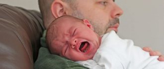
Approximately 40% of patients have few or no symptoms. If there is no constant autoinfection, then enterobiasis goes away on its own due to the short lifespan of adult worms. The main sign of infection is severe perianal itching, which occurs mainly at night when the sick child is sleeping. This can lead to sleep disturbances, childhood enuresis (up to 53% of cases) and decreased concentration of the child during the day. In some cases, developmental disorders in children are also associated with chronic enterobiasis.
Scratching in the perianal area can cause ulceration (excoriation), which can lead to bacterial superinfection. Anal dermatitis, perianal folliculitis, or ischiorectal abscess may develop. Very rarely, pinworms also migrate to the vaginal area, where they can cause vulvovaginitis, or indirectly cause urinary tract infections due to adherent enterobacteria such as Escherichia coli. The role of E. vermicularis in the pathogenesis of some cases of acute appendicitis has been the subject of debate for many years, although a causal relationship has not been proven.
In isolated cases, extraintestinal pinworm infections of the vagina, bladder, peritoneum, kidneys, liver and eyes have been described. Enterobiasis can sometimes overlap with the clinical picture of chronic inflammatory bowel disease. Along with severe itching, the disease is also characterized by severe psychosocial tension (psychological stress).
Diagnosis of enterobiasis
In addition to the typical patient history, which includes the cardinal symptom of intermittent perianal itching, examination of clothing and underwear, the anal area, and stool can provide diagnostic information. Moving, worm-like parasites can sometimes be seen on underwear, sheets, or directly on the edge of the anus. In cases of severe infestation, worms may be passed in the stool. Sometimes individual adult worms are visualized during proctoscopy or colonoscopy. A macroscopically identified worm reliably indicates infection.
Many patients do not experience visible pinworms. Because oviposition occurs on the anal folds, worm eggs, which are not visible to the naked eye, can be detected by using commercially available cellulose tape (“Scotch tape”) in the morning before bowel movements and before washing the genital area (Scotch tape test). To do this, repeatedly press the sticky side of the tape (pre-cut to size, for example, 10 × 2 cm) to the anal and perianal area with the buttocks separated. The tape is then attached to a suitable slide, sticky side down. Microscopic detection of characteristic worm eggs confirms infection. The slides do not need to be stored, prepared or preserved in any particular way. Even ready-made diagnostic kits have appeared in microbiological laboratories and pharmacies. An alternative is to clean the anal area with a cotton swab and then place it in a saline solution. The solution can then be used for microscopic examination for worm eggs (or in molecular genetic detection methods). This diagnostic method should be performed on three different days to increase its sensitivity (from approximately 50% in the case of a one-time tape test to approximately 90% if performed in three sessions). Stool microscopy is not a useful diagnostic tool because worm eggs are laid outside the intestine. Serological methods also have no diagnostic value. Likewise, neither blood eosinophilia nor elevated immunoglobulin E levels are usually expected due to the low invasiveness of the worms.
Technique for taking scrapings for enterobiasis, how they take it, how they give it
In order to find out whether there are pinworm parasites in the body, a parasitologist prescribes special tests for patients, in particular scraping for enterobiasis. Due to the research carried out, it is possible to verify the absence of helminthic infestation, or its presence.
How to properly take a scraping with a cotton swab?
To take scrapings in medicine, several objective methods are used, but the collection mainly occurs either with a cotton swab or with adhesive tape.
The first method, using a cotton swab, involves a certain procedure, and the technique of taking it from adults looks like this:
- First, gloves are put on, then the container is opened.
- Next, you should bend over and spread your buttocks.
- The stick, treated with saline or glycerin, is taken into the hands and then leisurely circular movements are made several times in the anal area.
- After this, the rod is placed in a sterile container, which is then hermetically sealed.
- And immediately the container is sent for research in the laboratory.
In some cases, the doctor has the right to prescribe additional diagnostic measures to reveal the full picture and maximum reliability. To do this, they take an enzyme-linked immunosorbent assay, which can be 100% reliable.
There is also a second, no less productive method of collecting material for enterobiasis.
Collecting material using adhesive tape
It is carried out by gluing a special adhesive tape around the anus, and can be performed on both adults and children.
The scraping procedure itself is almost no different from the previous one:
- The patient should bend over and at the same time spread his buttocks.
- Take the adhesive tape and carefully separate it from the glass.
- It should then be pressed firmly into the area near the anus.
- After this, it is slowly separated and glued to the glass again.
- The material is delivered for research.
In order for the test for enterobiasis to be more informative, the scraping should be taken immediately after waking up, since pinworms are essentially nocturnal parasites, and accordingly, eggs are laid in the dark.
There are also other, no less important recommendations. For greater information, a scraping for enterobiasis in a single case should not be considered productive if the result is negative.
There are many reasons. Perhaps the female has not laid eggs, or reinvasion is simply currently occurring. In order to obtain 100% confidence in the absence of parasitic infestation, you will need to take a similar test several more times. If, in the case of all repeated tests, the scraping for enterobiasis in adults shows a negative result, the patient can be considered healthy.
Which is the best test for Helicobacter pylori and which one is more accurate?
Treatment of enterobiasis
Mebendazole is used as a first-line drug. To prevent autoinfection, the drug is re-prescribed after two and four weeks, even when treating the first case of infection. For chronic recurrent enetrobiosis, all family members are treated with a single dose of Mebendazole at two-week intervals for 16 weeks.
In most cases, the drug is well tolerated, causing sometimes harmless and short-term side effects such as headache, abdominal pain and diarrhea.
Treatment methods such as intestinal lavage, “garlic medicine”, Carlsbad salts and appendectomy (in the absence of appendicitis) are considered outdated.
All patients undergoing treatment should also be informed about the clinical picture and modes of transmission of enterobiasis. Thorough hand washing before eating and after using the toilet, avoiding scratching in the anogenital area and contact of the anus with fingers, daily washing of the genital area (from front to back), regular change of underwear and sleeping clothes, as well as strict exclusive use of towels and diapers by the same person are necessary. . Onychophagia should be avoided.
Scraping for helminth eggs (enterobiasis)
An enterobiasis test is a microscopic examination of a fingerprint smear from the perianal area to detect eggs of the enterobiasis pathogen Enterobius vermicularis (pinworms).
Research method
Microscopy.
What biomaterial can be used for research?
Imprint from the perianal area.
How to properly prepare for research?
- In the morning, on the eve of taking the biomaterial, do not cleanse the skin in the area of the anus and buttocks.
- Considering the increased activity of the parasite at night, it is optimal to take a smear in the morning, before the patient performs the morning toilet.
- Three fingerprint swabs taken over three consecutive days are sent for research (the sensitivity of a single analysis is 50%, three - 90%, five - 99%).
General information about the study
Enterobiasis is the most common type of helminthiasis, the causative agent of which is Enterobius vermicularis (pinworm). It is especially common among children attending organized children's groups (up to 30% of children). The peak incidence occurs at 5-9 years.
The main route of transmission is fecal-oral.
Pinworm eggs enter the ileum and cecum, where they mature and mate as adults. Adult pinworms move freely in the intestinal lumen. The fertilized female migrates to the rectum, after which she lays up to 10-15,000 eggs on the surface of the skin of the perianal area (they are often called “scraping for enterobiasis”). The activity of females is maximum at night. The presence of eggs and helminths in some cases is accompanied by night itching, anxiety, sleep disturbance, and enuresis. However, more often enterobiasis is asymptomatic. From the perianal area, helminth eggs spread to the skin of the hands, clothing, toys, and household items, where they can persist under favorable conditions for up to 3 weeks, which makes the disease highly contagious. Getting eggs from the skin of the hands into the oral cavity can lead to reinfection.
Since pinworm eggs are not excreted in feces, microscopic or any other examination of stool is not used. Laboratory diagnosis of enterobiasis in children and adults is carried out using microscopy of fingerprint smears from the skin of the perianal area. Helminth eggs have a characteristic ovoid shape, smoothed on one side, their size is 25-55 microns. Microscopic examination allows for differential diagnosis of enterobiasis with another common helminthiasis - ascariasis, as well as with other non-infectious diseases accompanied by itching of the perianal area (contact dermatitis, vulvar kraurosis, diabetes mellitus). If pinworms are found on the skin of adult pinworms, they are removed.
With enterobiasis, there is no eosinophilia characteristic of invasive helminthiases.
Due to the high degree of contagiousness of helminths and the asymptomatic course of the disease, a laboratory examination of all members of the family (team) is necessary if pinworms E. vermicularis are detected in a patient.
What is the research used for?
For the diagnosis of enterobiasis.
What do the results mean?
Test norm for enterobiasis: negative.
Positive result:
- enterobiasis.
Negative result:
- absence of enterobiasis;
- incorrect taking of a smear for examination.