Congenital clubfoot is one of the most common malformations of the musculoskeletal system, in which the toes of the feet turn inward and the inner surface rises upward. In this condition, there is no complete contact of the sole with the floor. The pathology almost always affects both legs. Lack of treatment for this problem leads to dysfunction of the musculoskeletal system.
Pathology happens:
- Congenital – formed during intrauterine development.
- Acquired - occurs against the background of damage to the nervous, musculoskeletal systems and as a complication of inflammatory processes.
General information
According to statistics, congenital clubfoot occurs in three out of a thousand children. This defect occurs more often in boys than in girls, but doctors cannot yet explain this gender characteristic. Presumably, clubfoot begins to form in the 1st trimester of pregnancy under the influence of negative factors acting on the fetus. In the international classification, the disease is coded Q66. This group also includes other congenital changes of the foot. Parents often confuse pathological clubfoot with incorrect foot placement in a child who is just starting to walk. Orthopedists at the SM-Doctor clinic will help you carry out a differential diagnosis of the disease based on external objective signs and radiographic data.
Clinical picture of clubfoot
There are several main signs that characterize the clinical picture of this disease:
- The forefoot is subject to adduction.
- There is supination of the foot, forming varus.
- Equinus foot.
In advanced cases of clubfoot, another symptom is added, which is characterized by a high standing heel tubercle and internal torsion of the shin bones. Moreover, when children begin to walk, all the signs of the disease become even more noticeable.
Doctor Nikonov
With constant tension in the foot due to dysfunction of the peroneal muscles, progression of supination occurs. These events lead to the formation of callus of the skin and underlying mucous bursae.
The result of these pathologies is walking on the back of the feet with calloused skin, under which there are mucous membranes. At the same time, the soles of the feet are turned upward. The lower leg muscles undergo hypertrophy due to the inability to contract and stretch normally.
Children with signs of congenital clubfoot must be treated before they begin to walk.
Rice. 2. Healthy foot and clubfoot
Symptoms of clubfoot
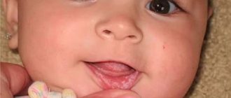
Symptoms of congenital clubfoot are noticeable after the birth of the child.
The foot bends, its outer edge sags. Clubfoot in one-year-old children is characterized by the fact that the baby turns the plantar part inward and rests on the outer part of the foot. Clubfoot in children 3-5 years old creates conditions for atrophy of the lower leg muscles. The child develops a waddling gait, it is difficult for him to run, and his motor activity is limited. With unilateral damage, the limb is shortened by 1-2 cm.
Symptoms also depend on the severity of the disease:
- Easy. Hand pressure is sufficient to correct the position of the foot. Movement in the ankle is not limited.
- Average. Resistance is felt when correcting the position of the foot. Ankle movements are slightly limited.
- Heavy. The foot and ankle are noticeably deformed. Pressing with your hand does not correct the defect.
If parents notice club feet in their baby immediately after birth, they should consult an orthopedist. The SM-Doctor clinic employs professionals who will establish an objective diagnosis in the shortest possible time.
Possible complications in the treatment of clubfoot
Doctor Nikonov
It is very irresponsible for some parents to neglect the health of the child and not make any attempts to cure the curvature of the limbs. Thinking that the symptoms will go away on their own with age, they allow clubfoot not only to develop, but also lead to more serious diseases (scoliosis, flat feet), even to disability. The following complications may also arise:
- Atrophy of entire muscle groups (disability).
- Subluxation of the bones of the feet.
- Roughness of the skin on the outside of the foot.
- Dysfunction of the knee joints (disability).
Doctor Nikonov
Incorrect walking by a child contributes to the formation of calluses and damage to the skin on the outside of the foot. The disease causes atrophy and swelling of some muscles, which results in pain. The pain prevents the child from standing and walking normally. He becomes disabled in a wheelchair.
At the same time, clubfoot is harmful not only to physical but also psychological health, and can also slow down intellectual development. At school, other children may behave badly and point out to a sick child his shortcomings. The child will feel inferior and understand how far behind his peers he is.
Clubfoot also leads to a curvature of the spine, which in turn can cause hernia, joint dysfunction, ankle sprains and complete immobility. A child suffering from this disease will have a constant headache. He won't be able to sleep properly.
Causes of clubfoot
Doctors call the main causes of clubfoot in children genetic predisposition and disruption of the formation of the child’s skeleton during intrauterine development. The influence of negative factors contributes to the development of the disease:
- mechanical pressure of amniotic fluid;
- dysfunction of the spinal cord;
- pressure of the uterine muscles due to a lack of amniotic fluid or the presence of tumors in the uterus;
- infection suffered by a woman in the 1st trimester;
- constant stress.
In medicine, there is a classification of forms of the disease according to etiology:
- Typical. Pathology develops during the laying of organs at the antenatal (intrauterine) stage. Bones and joints are deformed, and the violations are persistent and difficult to correct.
- Positional. With normal development of the osseous-ligamentous apparatus, shortening of the muscles in the lower leg and foot develops.
- Secondary. Clubfoot develops as a consequence of diseases of the muscles and nerves.
- Arthrogrypotic. The pathology is a consequence of arthrogryposis (systemic joint stiffness).
Diagnosis of clubfoot
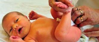
Clubfoot can be detected by performing an ultrasound of the fetus during pregnancy.
After birth, the baby is examined by a neonatologist. Sometimes a visual examination is sufficient to diagnose a disease. If pathological clubfoot is suspected, the baby is sent for consultation to an orthopedist. Before correcting a child’s clubfoot, the orthopedist conducts an examination and recommends standard tests. X-rays of the foot in various positions will help your doctor make an accurate diagnosis. The orthopedist determines the degree of the disease and the nature of the disorders, after which he determines treatment methods for clubfoot in children.
Treatment of clubfoot
Correction of congenital foot pathology begins in early preschool age. Clubfoot in children aged 6-7 years is more difficult to correct. At older ages, bones are less pliable, and proper skeletal development requires an optimal level of physical activity. Doctors can eliminate a mild form of the disease immediately after birth using conservative therapy methods:
- Exercise therapy followed by fixing the foot with bandages;
- applying a plaster bandage;
- the use of specialized shoes that allow you to fix the foot in the desired position;
- massage;
- physiotherapy (application of mud, paraffin, magnetic therapy, laser treatment).
In 90% of cases, correction of the pathological position of the limb is carried out using the Ponseti method in 3 stages:
- Application of a plaster cast. Fixes the foot in the desired position. The course of treatment includes 5-6 applications.
- Achillesotomy. The surgeon cuts the Achilles tendon, thereby helping to lengthen it.
- Wearing braces (special shoes) to maintain the correction result.
Surgical methods are used in severe cases when conservative therapy does not help. The operation is performed on ligaments and muscles, correcting their position and length. During the rehabilitation period, physiotherapy, medication, and plaster dressings are prescribed. In 90% of cases, surgery completely corrects the position of the foot. Subsequently, the child can lead an active lifestyle without any restrictions.
Rehabilitation of children with congenital clubfoot.
30.05.2017 08:13 Author: Slugin V.I., Filippova G.A.
Theories of the mechanism of development of clubfoot: - Mechanical impact on the fetus during intrauterine development. — Violation of the formation and delay in the formation of the foot and lower leg in the prenatal period. — Malformation of the nervous system. Numerous studies have shown that congenital clubfoot is based on a dysplastic process - underdevelopment of the bone, muscle and neurovascular systems. — The formation of clubfoot in the fetus can also be facilitated by unfavorable environmental conditions: electromagnetic fields, radioactive radiation, toxic-chemical influences, etc. — Viral infections at different stages of pregnancy can leave behind very serious consequences. - It may also be heredity. However, it cannot be said that this disease is inherited. Congenital adducted foot is detected either immediately after birth or during the first month. Clubfoot is most often bilateral. With unilateral localization, it is more often observed on the left (62.8%). Slightly more common in premature babies. According to statistics, this defect is most often observed in boys (57%). With normal neuromuscular development of a child, complete spontaneous correction occurs, which is observed in 80% of children under 2 years of age. However, in 16% of cases, spontaneous recovery was observed only by 5 years. The main signs of clubfoot are considered to be inward rotation of the sole with raising the inner edge of the foot and lowering the outer edge, adduction of the foot in the forefoot, plantar flexion, and significant limitation of mobility in the ankle joint. When a child begins to walk, leaning on the damaged foot, its deformation intensifies, and the shape and function of the entire leg are disrupted. This worsens posture and gait. Only at an early age, when the child’s muscles and ligaments are pliable and elastic, is there a high probability of bringing the foot into the correct position. For this purpose, a whole range of measures is used, which includes, in addition to orthopedic treatment, massage and therapeutic exercises. The diagnosis of clubfoot is made in the maternity hospital and treatment begins there: gymnastics and daily repeated dressing with a Fink-Ettinger bandage (3-4 times/day). From the 3rd week of life, correction with an “elastic foot” is used: constant force with elastic bandages (thigh-shin + flat surface on the foot). By 2 months of life, complete correction is achieved with a mild degree (Chugui E.V. et al., 2006, Tomsk). Massage. Massage of individual muscle groups of the legs and feet depends on the state of their tone and the functions they must perform: some muscles should be strengthened, while others, on the contrary, should be relaxed. Physiotherapy. Special therapeutic exercises after thermal procedures have a good effect. Paraffin (30-40 min.), ozokerite and mud (15-20 min.) applications are effective. 15-20 percent daily or h/w day. Courses can be repeated after 2 months. Electrical stimulation of the lower leg muscles, ultrasound treatment, magnetic therapy. (A.F. Krasnov. 1963, 2001). A modern and highly effective method of conservative correction of clubfoot is kinesiological taping. It is used both as an independent technique and in combination with other rehabilitation methods. Taping does not have the disadvantages that can be observed with casting: contractures of joints and muscles do not form. It ensures fixation of the joint in the anatomically correct position and at the same time does not interfere with physiologically correct movements. At the same time, favorable conditions are created for stimulating weakened muscles and relaxing spasms. This will certainly facilitate physical therapy exercises and increase the effectiveness of the activities carried out and improve the outcome of rehabilitation. Kinesio tapes are elastic bands coated with hypoallergenic glue, which is activated by body temperature. The cotton base does not interfere with skin breathing and evaporation from its surface. It is believed that the elastic properties of tapes are close to the elastic parameters of the skin. Kinesio tape can remain on the skin for 3-5 days, which allows it to be used with a continuous therapeutic effect. Only a specialist can apply the application! Most often, staged casting is used for the rehabilitation of children with clubfoot. The specialist kneads the foot, trying to bring it into a more correct position, without using force. This achieved position is secured with a plaster boot, which is applied from the foot and above the knee. After some time, the boot is removed and work is done on the foot again: the specialist achieves a more correct position compared to the previous time and again applies a plaster cast. And so on in several stages. In most cases, it is possible to bring the foot back to normal by the end of the first year of the child’s life. Surgical treatment - starting from 1 year, if conservative treatment is ineffective. If there is a severe form, surgical intervention is possible at 6 months, because severe forms of clubfoot cannot be eliminated manually. After the cast is removed, rehabilitation courses are carried out with the mandatory use of orthosis splints, braces and orthopedic shoes, the wearing of which is long-term - at least until 7-8 years of age. Maintenance therapy should be carried out continuously throughout the entire period of the child's growth. General questions of exercise therapy. 1. During sleep (if indicated), the feet are fixed (bandaged with an elastic bandage, splint) in the correction position. 2. Attach shoes to the plywood: parallel or with toes pointed out. With his feet in his shoes, the child eats, studies at the table, etc. It is recommended to perform squats on this “simulator” - many times a day. 3. Make “skis”: attach boots that fit tightly on your feet with hard sides to a board measuring 6 x 50 cm. Learn to walk with the correct (parallel) position of your feet. 4. Correcting the position of the feet while walking: - Sometimes switching shoes on the right and left foot helps. — On a strip of old linoleum, make a narrow “corridor” from slats for walking along it with parallel feet. — Nail the slats in the form of a “herringbone” — for walking with the toes of the feet apart. Walking forward and backward. - Teach the child to step on footprints (“carrots”, “cucumbers”) that are drawn (pasted) on the floor - in a parallel or apart position. Some exercises. — I.P. sitting on a chair, knees bent, heels together. Dorsal flexion of the feet while simultaneously moving them outward. Fix the heels in place. The exercise is best performed against resistance. — I.P. feet together, put a rubber ring on the toes of the feet. Pull your socks apart to stretch the elastic. The more difficult it is, the better the muscles are trained. Raising the toes of the feet can be performed with resistance from the methodologist. — I.P. standing/sitting. Flexion and extension of the toes. Pay maximum attention to extension (direction of extension upwards and outwards). The effect is greater if the exercise is performed with resistance. — I.P. stand, heels together, toes apart. Deep squat without lifting your heels off the floor. — Walking on a narrow rail with the toes of the feet apart (along the curb). - “Football”: hit the ball with the outer edge of your feet (to the side). For children who are unable to perform movements actively, perform exercises passively, at least 10 times a day. It is necessary to come up with games for the child.
To familiarize yourself with the material, you need to download the file: Rehabilitation of children with congenital clubfoot.
Prevention of clubfoot
Preventive measures that help 100% prevent congenital clubfoot in a child do not exist.
If the disease occurs in one of the relatives, the likelihood of developing pathology in the baby increases significantly. To reduce the risk of developing clubfoot in a child, even at the intrauterine stage, the mother must:
- avoid stress;
- monitor your health and promptly treat infectious diseases;
- eat foods rich in calcium;
- carry out examinations regularly.
Many parents consider congenital clubfoot to be a serious pathology. But the disease is highly treatable in infancy. If you take the time and consult a doctor, the pathology can be eliminated using traditional methods. At the SM-Doctor clinic, experienced specialists will help diagnose the disease in a timely manner and create a personalized correction program.
Acquired clubfoot
Clubfoot can develop in a child if you do not pay attention to these anomalies:
- In the area of the baby's buttocks and legs, the skin folds unequally symmetrically.
- The position of the upper limbs while walking is that the arms are raised.
- The child does not know how to sit down and stand up from his haunches without the help of his hands.
These symptoms indicate the presence of edema in the muscle tissue. It is protein swelling that prevents the muscles from contracting normally.
Signs of clubfoot in a healthy child may appear in the second or third year of life. They usually look like this:
- Incorrect gait (bearish) - while walking, the baby “rakes” with one leg.
- When a child walks, he places his feet incorrectly. This feature can be recognized by the marks left by the baby. The print of one foot is always turned towards the other.
- The kneecaps are directed inward - towards each other.
- Reduced ankle mobility.
- Violations are visible while the child is sleeping.
- The big toe looks inward: a valgus curvature, in which specific bumps appear on the inside of the foot.
The disease progresses due to increasing swelling due to the load on the muscle groups of the lower back, buttocks, and lower extremities. In children, the skeleton grows quickly, but since the muscle tissue is not able to function normally, the muscles begin to pull on the bones . Due to this, the joints cannot fully bend and extend.
Doctor Nikonov
It is for these reasons that some muscle groups are in a state of severe swelling (hypertonicity), while others are completely relaxed. All these factors manifest themselves in the form of a crooked foot.